. 2006 Jul;26(13):5155-67.
doi: 10.1128/MCB.02186-05.

Direct interaction of focal adhesion kinase (FAK) with Met is required for FAK to promote hepatocyte growth factor-induced cell invasion

Affiliations

Direct interaction of focal adhesion kinase (FAK) with Met is required for FAK to promote hepatocyte growth factor-induced cell invasion

Shu-Yi Chen et al. Mol Cell Biol. 2006 Jul.

Abstract

Focal adhesion kinase (FAK) has been implicated to be a point of convergence of integrin and growth factor signaling pathways. Here we report that FAK directly interacts with the hepatocyte growth factor receptor c-Met. Phosphorylation of c-Met at Tyr-1349 and, to a lesser extent, Tyr-1356 is required for its interaction with the band 4.1 and ezrin/radixin/moesin homology domain (FERM domain) of FAK. The F2 subdomain of the FAK FERM domain alone is sufficient for Met binding, in which a patch of basic residues (216KAKTLRK222) are critical for the interaction. Met-FAK interaction leads to FAK activation and subsequent contribution to hepatocyte growth factor-induced cell motility and cell invasion. Our results provide evidence that constitutive Met-FAK interaction may be a critical determinant for tumor cells to acquire invasive potential.


Figures
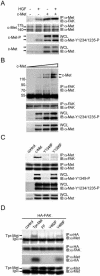
FIG. 1.
FIG. 1.
Tyr-1349 and, to a lesser extent, Tyr-1356 of Met are required for its interaction with FAK. (A) Met-FAK interaction was examined by coimmunoprecipitation (IP) of both molecules in HEK 293 cells with (+) or without (−) HGF stimulation. To measure the activation of c-Met, whole-cell lysates (WCL) were analyzed by immunoblotting (IB) with a phospho-specific antibody (anti [α]-Met-Y1234/1235-P). (B) Increasing amounts of c-Met were transiently expressed in HEK 293 cells, and its coprecipitation with FAK was examined. (C) c-Met or its mutants were transiently expressed in HEK 293 cells, and their coprecipitations with FAK were examined. Phosphorylation of c-Met at Tyr-1349 was analyzed by immunoblotting with a phospho- specific antibody (α-Met-Y1349-P). (D) HA-tagged FAK was transiently coexpressed with Tpr-Met or its mutants in HEK 293 cells, and their coprecipitations were analyzed. IgG, immunoglobulin G. Molecular size markers (in kilodaltons) are noted at the left of blots.
FIG. 2.
FIG. 2.
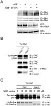
Gab1 does not mediate the interaction between Met and FAK. (A) The siRNA duplex (400 nM) for the Gab1 knockdown was delivered into HEK 293 cells by Oligofectamine. Sixty hours later, the cells were serum starved for 12 h and stimulated with or without HGF for 20 min before lysis. To examine the expression level of Gab1, whole-cell lysates (WCL) were analyzed by immunoblotting (IB) with anti-Gab1 (α-Gab1). The coimmunoprecipitation (IP) of FAK and MET was analyzed. (B) Tpr-Met was transiently expressed in Gab1+/+ cells or Gab1−/− cells, and its coprecipitation with FAK was examined. (C) HEK 293 cell lysates containing HA-tagged FAK or HA-tagged Gab1 were incubated with the lysates containing Tpr-Met in the presence or absence of a 16-amino-acid peptide containing the Met-binding sequence (MBS) of Gab1. One hour later, coprecipitation of Tpr-Met with HA-tagged FAK or Gab1 was examined. IgG, immunoglobulin G.
FIG. 3.
FIG. 3.
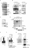
Direct interaction between Met and FAK. (A) Baculovirus-expressed recombinant FAK proteins (bac-FAK) were purified from insect cell lysates by affinity chromatography. The crude insect cell lysates, the protein A-Sepharose-bound proteins (on beads), and the eluted proteins (eluted) were fractionated by SDS-polyacrylamide gel electrophoresis and visualized by Coomassie blue staining. (B) Immobilized GST fusion proteins encoding Tpr-Met or its mutants were incubated with purified baculovirus-expressed FAK proteins, and the bound proteins were analyzed by immunoblotting (IB) with anti-FAK (α-FAK). Total tyrosine phosphorylation of GST-Tpr-Met and its specific phosphorylation at Tyr-482 (equivalent to Tyr-1349 of c-Met) were analyzed by immunoblotting with antiphosphotyrosine (α-PY) and anti-Met-Y1349-P, respectively. The GST fusion proteins on the nitrocellulose membrane were visualized by staining with Ponceau S solution. (C) Dephosphorylation of Tpr-Met decreases its association with FAK in vitro. Immobilized GST-Tpr-Met was treated with 10 U of alkaline phosphatase (AP) in the presence or absence of 2 mM sodium vanadate (Na3VO4) for 1 h and then incubated with baculovirus-expressed recombinant FAK. The bound recombinant FAK proteins were analyzed by immunoblotting with anti-FAK. The tyrosine phosphorylation of GST-Tpr-Met was analyzed by immunoblotting with anti-PY. (D) HEK 293 cell lysates containing Tpr-Met were incubated with baculovirus-expressed recombinant FAK proteins in the presence of different amounts of the phosphopeptide or its unphosphorylated counterpart. One hour later, coprecipitation of Tpr-Met and baculovirus-expressed recombinant FAK was analyzed. Molecular size markers (in kilodaltons) are noted at the left of blots. IP, immunoprecipitation; IgG, immunoglobulin G.
FIG. 4.
FIG. 4.
The FERM domain of FAK binds to Met. (A) HA-tagged full-length (FL) FAK or its FERM domain (aa 30 to 377) or COOH domain (aa 693 to 1053) was transiently coexpressed with c-Met in HEK 293 cells. Coimmunoprecipitation (IP) of HA-tagged FAK proteins and c-Met was analyzed. (B) The HA-tagged FAK FERM domain was transiently coexpressed with Tpr-Met or its mutants in HEK 293 cells, and their coprecipitation was analyzed. (C) Purified GST-Tpr-Met and the histidine-tagged NH2 domain of FAK (His-FAK-N) were analyzed by SDS-polyacrylamide gel electrophoresis and visualized by Coomassie blue staining (left). Soluble GST-Tpr-Met or its FF mutant was incubated with purified His-FAK-N. One hour later, glutathione agarose beads were added to the mixture and the bound proteins were analyzed by immunoblotting (IB) with an anti-FAK antibody (A-17) specific to the NH2 domain of FAK (α-FAK-N) (right). Molecular size markers (in kilodaltons) are noted at the left of blots. (D) HEK 293 cell lysates containing Tpr-Met and those containing the HA-tagged FAK FERM domain were mixed and incubated with (+) or without (−) 200 μM of the phosphopeptide IGEHPY1349VHVNATPY1356VNVK or its unphosphorylated peptide. One hour later, coprecipitation of Tpr-Met and the HA-tagged FAK FERM domain was analyzed. (E) The FAK FERM domain competes with the full-length FAK for Tpr-Met binding in vitro. HEK 293 cell lysates containing Tpr-Met were incubated with baculovirus-expressed FAK (bac-FAK) in the presence of increasing amounts of HEK 293 cell lysates containing the HA-tagged FAK FERM domain. Coprecipitation of Tpr-Met and HA-tagged proteins was analyzed. IgG, immunoglobulin G; WCL, whole-cell lysates.
FIG. 5.
FIG. 5.
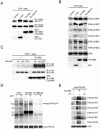
The basic residues in the F2 subdomain of the FAK FERM domain are critical for FAK to interact with Met. (A) Immobilized GST fusion proteins encoding the FAK FERM domain or its truncated mutants were incubated with HEK 293 cell lysates containing Tpr-Met. The bound proteins were analyzed by immunoblotting (IB) with anti-Met (α-Met). The GST fusion proteins on the nitrocellulose membrane were visualized by staining with Ponceau S solution. The intact GST fusion proteins on the membrane are marked by circles. (B) Immobilized GST fusion proteins encoding the F1, F2, or F3 subdomain of the FAK FERM domain were incubated with purified histidine-tagged Tpr-Met (His-Tpr-Met). The bound proteins were resolved by SDS-polyacrylamide gel electrophoresis and visualized by Coomassie blue staining (left) or subjected to immunoblotting with anti-Met (right). (C) Tpr-Met was transiently coexpressed with the HA-tagged FAK NH2 domain or its mutants in HEK 293 cells. Coprecipitation of Tpr-Met and the HA-tagged FAK NH2 domain was analyzed. (D) Immobilized GST-Tpr-Met was incubated with HEK 293 cell lysates containing the HA-tagged FAK NH2 domain or its mutants. The bound proteins were analyzed by immunoblotting with anti-HA. (E) c-Met was transiently coexpressed with HA-tagged full-length (FL) FAK or its mutants in HEK 293 cells. Coprecipitation of c-Met with HA-tagged FAK was analyzed. Molecular size markers (in kilodaltons) are noted at the left of blots. IgG, immunoglobulin G; IP, immunoprecipitation; WCL, whole-cell lysates.
FIG. 6.
FIG. 6.
Met-FAK interaction leads to FAK activation. (A) The tyrosine phosphorylation of endogenous FAK was analyzed with HEK 293 cells transiently expressing increasing amounts of c-Met. To measure the phosphorylation of c-Met, whole-cell lysates (WCL) were analyzed by immunoblotting (IB) with phospho-specific antibodies. (B) v-Src or Tpr-Met was transiently expressed in HEK 293 cells or stably expressed in NIH 3T3 cells. The tyrosine phosphorylation of FAK in those cells was analyzed. (C) HA-tagged FAK or its K222A mutant was transiently coexpressed with (+) or without (−) Tpr-Met in FAK−/− cells. The tyrosine phosphorylation of HA-tagged FAK was analyzed. (D) In vitro kinase assays for Src, Tpr-Met, and FAK, with GST-FAK (aa 378 to 406) as a substrate. Note that GST-FAK (aa 378 to 406) is phosphorylated only by FAK but not by Src, Tpr-Met, or the FAK kd mutant. (E) The kinase activity of endogenous FAK in NIH 3T3 cells stably expressing Tpr-Met or its Y482F/Y489F mutant was analyzed by an in vitro kinase assay using GST-FAK (aa 378 to 406) as a substrate. The results are the averages of two experiments. (F) The kinase activity of HA-tagged FAK or its K222A mutant expressed with or without Tpr-Met in FAK−/− cells was analyzed by an in vitro kinase assay using GST-FAK (aa 378 to 406) as a substrate. The results are the averages of two experiments. (G) Purified baculovirus-expressed FAK (bac-FAK) was incubated with soluble GST-Tpr-Met or its FF mutant in the presence of unlabeled ATP (30 μM). One hour later, the baculovirus-expressed FAK was immunoprecipitated (IP) by anti-HA (α-HA) and subjected to an in vitro kinase assay using GST-FAK (aa 378 to 406) as a substrate in the presence of [γ-32P]ATP. Molecular size markers (in kilodaltons) are noted at the left of blots. α-PY, antiphosphotyrosine; IgG, immunoglobulin G.
FIG. 7.
FIG. 7.
Met directly phosphorylates FAK. (A) The tyrosine phosphorylation of endogenous FAK in the SYF cells transiently expressing v-Src, Tpr-Met, or its mutant was examined. (B) The phosphorylation of FAK was analyzed by using antibodies specific to phosphorylated FAK at indicated tyrosine residues. (C) HA-tagged FAK or its mutants were transiently coexpressed with Tpr-Met in FAK−/− cells. The tyrosine phosphorylation of HA-tagged FAK was analyzed. (D) In vitro kinase assays for v-Src and Tpr-Met were performed using the baculovirus-expressed FAK kd mutant (bac-FAK-kd) as a substrate in the presence or absence of unlabeled ATP. The mixture of the immunocomplexes and the substrate was analyzed by immunoblotting (IB) with antiphosphotyrosine (α-PY). Molecular size markers (in kilodaltons) are noted at the left of blots. (E) The purified baculovirus-expressed FAK kd mutant was incubated with soluble GST-Tpr-Met or its FF mutant in the presence of unlabeled ATP. One hour later, the baculovirus-expressed FAK kd mutant was immunoprecipitated (IP) with anti-HA and the immunocomplexes were analyzed by immunoblotting with antibodies specific to phosphorylated FAK at indicated tyrosine residues. WCL, whole-cell lysates.
FIG. 8.
FIG. 8.
Met-FAK interaction is required for FAK to promote HGF-stimulated cell invasion. (A) MDCK cells stably overexpressing HA-tagged FAK or its K222A mutant deficient in Met binding were established. Equal amounts of cell lysates were incubated with anti-HA (α-HA), and the immunocomplexes were analyzed by immunoblotting (IB) with anti-FAK or anti-phospho-Met (α-Met-Y1234/1235-P). (B) Control MDCK cells and those overexpressing FAK or its K222A mutant were subjected to an HGF-induced scattering assay. Data (means ± standard errors) are from three experiments. formula image, P < 0.05, compared with the control MDCK cells. (C) The MDCK cells were subjected to a cell migration assay. formula image, P < 0.05, compared with the control MDCK cells in the presence of HGF. (D) The MDCK cells were subjected to a Matrigel invasion assay. formula image, P < 0.05, compared with the control MDCK cells in the presence of HGF. (E) Met-FAK interaction was analyzed with a series of lung cancer cell lines. The ability of those cells to invade through Matrigel was measured. formula image, P < 0.05, compared with A549 cells. (F) CL1-5 lung cancer cells stably expressing the FAK NH2 domain were established. The inhibitory effect of the FAK NH2 domain on Met-FAK interaction was examined. (G) CL1-5 cells and those stably expressing the FAK NH2 domain were subjected to a Matrigel invasion assay. IP, immunoprecipitation; WCL, whole-cell lysates; IgG, immunoglobulin G.
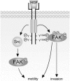
FIG. 9.
FIG. 9.
Met activates FAK through both the Met-Src-FAK pathway (our previous studies) and the Met-FAK pathway (this study). The activated FAK functions as a signaling platform to potentiate HGF-elicited signals, thereby contributing to HGF-stimulated cell motility and cell invasion. Note that the ability of FAK to promote HGF-stimulated invasion relies largely on its direct interaction with Met (see Discussion for details). P, phosphorylation.

Similar articles

Cited by

References

    1. Bardelli, A., L. Pugliese, and P. M. Comoglio. 1997. “Invasive-growth” signaling by the Met/HGF receptor: the hereditary renal carcinoma connection. Biochim. Biophys. Acta 1333:M41-M51. - PubMed
    1. Barret, C., C. Roy, P. Montcourrier, P. Mangeat, and V. Niggli. 2000. Mutagenesis of the phosphatidylinositol 4,5-bisphosphate (PIP(2)) binding site in the NH(2)-terminal domain of ezrin correlates with its altered cellular distribution. J. Cell Biol. 151:1067-1080. - PMC - PubMed
    1. Birchmeier, C., W. Birchmeier, E. Gherardi, and G. F. Vande Woude. 2003. Met, metastasis, motility and more. Nat. Rev. Mol. Cell Biol. 4:915-925. - PubMed
    1. Birchmeier, C., and E. Gherardi. 1998. Developmental roles of HGF/SF and its receptor, the c-Met tyrosine kinase. Trends Cell Biol. 8:404-410. - PubMed
    1. Bottaro, D. P., J. S. Rubin, D. L. Faletto, A. M. Chan, T. E. Kmiecik, G. F. Vande Woude, and S. A. Aaronson. 1991. Identification of the hepatocyte growth factor receptor as the c-met proto-oncogene product. Science 251:802-804. - PubMed

Publication types

MeSH terms

Substances