. 2006 Jul;26(13):4970-81.
doi: 10.1128/MCB.00308-06.

ST6Gal-I restrains CD22-dependent antigen receptor endocytosis and Shp-1 recruitment in normal and pathogenic immune signaling

Affiliations

ST6Gal-I restrains CD22-dependent antigen receptor endocytosis and Shp-1 recruitment in normal and pathogenic immune signaling

Prabhjit K Grewal et al. Mol Cell Biol. 2006 Jul.

Abstract

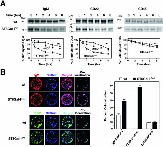
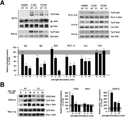
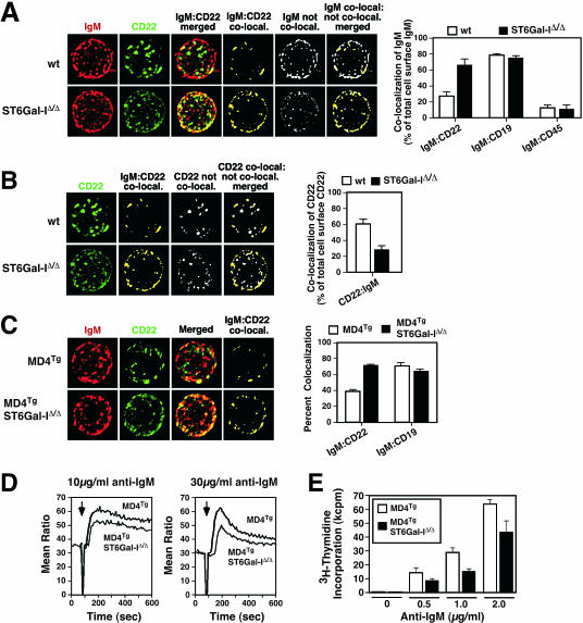
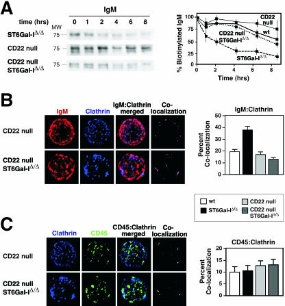
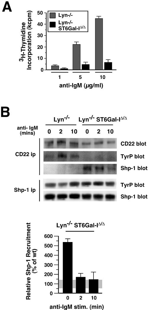
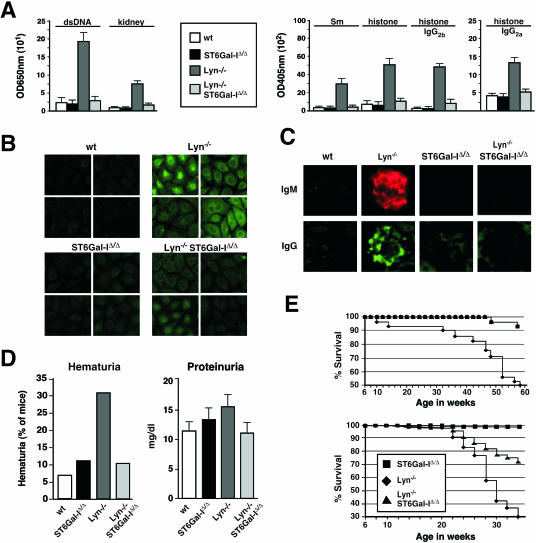
The ST6Gal-I sialyltransferase produces Siglec ligands for the B-cell-specific CD22 lectin and sustains humoral immune responses. Using multiple experimental approaches to elucidate the mechanisms involved, we report that ST6Gal-I deficiency induces immunoglobulin M (IgM) antigen receptor endocytosis in the absence of immune stimulation. This coincides with increased antigen receptor colocalization with CD22 in both clathrin-deficient and clathrin-enriched membrane microdomains concurrent with diminished tyrosine phosphorylation of Igalpha/beta, Syk, and phospholipase C-gamma2 upon immune activation. Codeficiency with CD22 restores IgM antigen receptor half-life at the cell surface in addition to reversing alterations in membrane trafficking and immune signaling. Diminished immune responses due to ST6Gal-I deficiency further correlate with constitutive recruitment of Shp-1 to CD22 in unstimulated B cells independent of Lyn tyrosine kinase activity and prevent autoimmune disease pathogenesis in the Lyn-deficient model of systemic lupus erythematosus, resulting in a significant extension of life span. Protein glycosylation by ST6Gal-I restricts access of antigen receptors and Shp-1 to CD22 and operates by a CD22-dependent mechanism that decreases the basal rate of IgM antigen receptor endocytosis in altering the threshold of B-cell immune activation.


Figures

FIG. 1.
FIG. 1.
ST6Gal-I deficiency increases IgM and CD22 endocytosis. (A) Isolated splenic and lymph node B cells were biotinylated and cultured for the indicated number of hours in the absence of immune stimulation. Biotinylated cell surface proteins were isolated and analyzed by antibody blotting to detect IgM, CD22, and CD45. Measurements (plotted) revealed glycoprotein-selective decreases in IgM and CD22 cell surface half-lives from ∼8 h among wild-type (wt) B cells to 2 h in the absence of ST6Gal-I. No effect was seen on the rate of CD45 turnover. Treatment of ST6Gal-I-deficient B-cell cultures with the endocytosis inhibitor methyl-β-cyclodextrin (MCD) increased IgM and CD22 cell surface half-lives to ∼8 h. Positions of molecular weight standards are denoted. (B) Isolated splenic and lymph node B cells were analyzed by fluorescent deconvolution microscopy. ST6Gal-I deficiency significantly increased colocalization of IgM with clathrin (P < 0.001). No significant change in colocalization of CD22 or CD45 with clathrin was observed. These data were derived from 20 individual B cells representing three separate littermate pairs 6 to 8 weeks of age.
FIG. 2.
FIG. 2.
ST6Gal-I deficiency depresses B-cell immune signal transduction coincident with inducing Shp-1 recruitment to CD22. (A) Isolated B cells were immune stimulated with 1.2 μg/ml of anti-IgM antibody for the indicated times followed by immunoprecipitation (ip) of proteins analyzed for expression level and phosphotyrosine (TyrP) accumulation by 4G10 antibody. Absence of ST6Gal-I did not significantly alter protein expression levels but significantly diminished phosphotyrosine accumulation among Igα/β, Syk, PLC-γ2 and Lyn. Results are representative of six separate experiments with 6- to 8-week-old littermates. unstim., unstimulated. (B) ST6Gal-I deficiency increased CD22 tyrosine phosphorylation and induced Shp-1 recruitment to CD22 prior to immune activation (left panel). A 2.5-fold increase in CD22 tyrosine phosphorylation was observed in ST6Gal-I deficiency prior to immune activation (middle panel). The level of Shp-1 recruitment to CD22 was elevated 6-fold prior to immune activation and remained 2.5-fold increased subsequently when compared with the levels of CD22 expression (right panel). Relative phosphorylation was calculated as ratios of phosphotyrosine to the level of protein abundance in ST6Gal-I deficiency and graphed as a percentage of wild-type (wt) measurements (gray).
FIG. 3.
FIG. 3.
Lymphocyte production and life span kinetics in ST6Gal-I deficiency. (A) Increased rate of production (pulse) and decreased half-life (chase) were noted among B cells by in vivo BrdU-labeling studies. (B) CD4+ and CD8+ T cells retained a normal rate of production and unaltered half-life in ST6Gal-I deficiency. Six mice of the indicated genotypes and 6 to 10 weeks of age were analyzed at each time point. wt, wild type.
FIG. 4.
FIG. 4.
Increased IgM colocalization with CD22, reduced Ca2+ mobilization, and attenuated proliferation in ST6Gal-I-deficient mouse B cells expressing either endogenous or MD4 transgenic (MD4Tg) IgM BCRs. (A) Intact resting B cells from the spleen or lymph nodes (spleen shown) were analyzed by fluorescent deconvolution microscopy to measure the degree of IgM and CD22 colocalization (co-local.) involving total cell surface signals. Representative deconvolution analysis of a single B cell is shown. Images are normalized for fluorescence intensity and shown as maximum projections that include all z-stacks. Cell surface IgM colocalization with cell surface CD22 was substantially increased from 30% of total IgM among wild-type (wt) B cells to 75% of total IgM in ST6Gal-I-deficient B cells (P < 0.001). Note also the reduction in total IgM signal that is not colocalized with CD22 in the absence of ST6Gal-I (white). No change in IgM colocalization occurred with either CD19 or CD45. (B) Increased cell surface dispersion of CD22 occurs in ST6Gal-I deficiency (green), identical to the corresponding panel in A. Measurements of cell surface CD22 colocalized with IgM (yellow; also identical to corresponding panel in A) compared with CD22 that was not colocalized with IgM (white) revealed that more CD22 was not colocalized in ST6Gal-I deficiency, as presented by the ratio of these measurements in the histogram to the right. (C) Increased colocalization of IgM with CD22, but not CD19, was further observed in MD4Tg mice lacking ST6Gal-I. (D) B lymphocytes from MD4Tg mice with and without ST6Gal-I were analyzed for Ca2+ mobilization in response to IgM cross-linking (arrow) at 10 μg/ml or 30 μg/ml. ST6Gal-I-deficient B cells expressing MD4Tg exhibited a reduction in the amount of cytosolic Ca2+ mobilization expressed as a ratio of bound to unbound Indo-I. (E) During the last 16 h of a 64-h assay period, MD4Tg-expressing B cells stimulated with anti-IgM at the indicated concentrations underwent reduced proliferation in the absence of ST6Gal-I. Proliferation is shown as triplicate measurements indicating mean ± standard error. Measurements in panels A to C were derived from 24 to 48 resting B cells of each genotype isolated from at least three separate donors.
FIG. 5.
FIG. 5.
CD22 is required to increase IgM endocytosis and degradation in ST6Gal-I deficiency. (A) The diminished cell surface half-life of IgM in the absence of ST6Gal-I was restored to normal among ST6Gal-I-deficient B cells further lacking CD22. Results are plotted along with those obtained from the wild-type and ST6Gal-I-deficient B cells presented in Fig. 1. CD22 deficiency slightly decreased the rate of IgM degradation compared with that in wild-type B cells. No change in the half-life of cell surface CD45 was observed. (B) Increased IgM-clathrin colocalization in ST6Gal-I deficiency was decreased to wild-type (wt) levels by further loss of CD22. (C) CD45-clathrin colocalization was not altered by ST6Gal-I deficiency (n = 20; P < 0.001).
FIG. 6.
FIG. 6.
ST6Gal-I deficiency reduces B-cell activation and promotes Shp-1 recruitment to CD22 in the absence of Lyn. (A) Isolated splenic or lymph node B cells were stimulated by anti-IgM cross-linking at various antibody concentrations and analyzed for cellular proliferation responses by thymidine incorporation. The absence of ST6Gal-I reduced Lyn-deficient B-cell proliferation responses by four- to fivefold. (B) Shp-1 recruitment to CD22 was abolished in B cells lacking Lyn, as expected. In contrast, the additional loss of ST6Gal-I restored Shp-1 recruitment to CD22 and induced CD22-Shp-1 complexes prior to immune stimulation (upper panel). Absence of both Lyn and ST6Gal-I reduced CD22 tyrosine phosphorylation (TyrP) to background levels and did not alter Shp-1 expression or tyrosine phosphorylation. The ratio of Shp-1 recruitment to the endogenous level of CD22 was elevated in the absence of both Lyn and ST6Gal-I, similar to ST6Gal-I deficiency alone (Fig. 2B), as calculated in comparison to measurements among wild-type B cells (lower panel). ip, immunoprecipitation.
FIG. 7.
FIG. 7.
Loss of ST6Gal-I activity attenuates autoimmune disease in Lyn deficiency. (A) Autoantibody titers (IgM) to dsDNA, kidney protein, Sm antigen, and histone, as well as IgG2 titers to histone induced by Lyn deficiency, were reduced or eliminated in the absence of ST6Gal-I (n = 12 sera from distinct mice of each genotype). (B) Similar findings were obtained in observing ANA titers against HepG2 cells in sera of 16-week-old littermates. Examples of ANA titers to both nuclear and cytoplasmic antigens (shown at a magnification of ×200) are representative of four littermate comparisons. (C) Absence of elevated glomerular deposition of IgM and IgG in Lyn-deficient mice lacking ST6Gal-I. Representative results are shown (×400). (D) Kidney dysfunction detected by elevated hematuria and proteinuria in Lyn deficiency was reduced to normal in the absence of ST6Gal-I. The results plotted represent 24 animals of each genotype. (E) ST6Gal-I deficiency increased the average life span of Lyn-null mice. Two separate studies were accomplished in different vivaria. Each study graphed represents results obtained involving distinct cohorts of mice comprising 40 to 50 littermates of each genotype.

Similar articles

Cited by

References

    1. Angata, K., J. M. Long, O. Bukalo, W. Lee, A. Ditayev, A. Wynshaw-Boris, M. Schachner, M. Fukuda, and J. D. Marth. 2004. Sialyltransferase ST8Sia-II assembles a subset of polysialic acid that directs hippocampal axonal targeting and promotes fear behavior. J. Biol. Chem. 279:32603-32613. - PubMed
    1. Baum, L. 2002. Developing a taste for sweets. Immunity 16:5-8. - PubMed
    1. Chan, V. W. F., F. Meng, P. Soriano, A. L. DeFranco, and C. A. Lowell. 1997. Characterization of the B lymphocyte populations in Lyn-deficient mice and the role of Lyn in signal transduction and down-regulation. Immunity 7:69-81. - PubMed
    1. Chui, D., G. Sellakumar, R. Green, M. Sutton-Smith, T. McQuistan, K. Marek, H. Morris, A. Dell, and J. D. Marth. 2001. Genetic remodeling of protein glycosylation in vivo induces autoimmune disease. Proc. Natl. Acad. Sci. USA 98:1142-1147. - PMC - PubMed
    1. Collins, B. E., O. Blixt, N. V. Bovin, C. P. Danzer, D. Chui, J. D. Marth, L. Nitschke, and J. C. Paulson. 2002. Constitutively unmasked CD22 on B cells of ST6Gal I knockout mice: novel sialoside probe for murine CD22. Glycobiology 12:563-571. - PubMed

Publication types

MeSH terms

LinkOut - more resources