. 2006 Jul;26(13):4806-17.
doi: 10.1128/MCB.02062-05.

Interaction between PCNA and diubiquitinated Mcm10 is essential for cell growth in budding yeast

Affiliations

Interaction between PCNA and diubiquitinated Mcm10 is essential for cell growth in budding yeast

Sapna Das-Bradoo et al. Mol Cell Biol. 2006 Jul.

Abstract

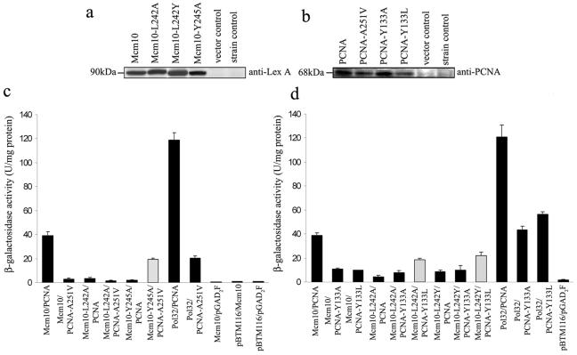
The minichromosome maintenance protein 10 (Mcm10) is an evolutionarily conserved factor that is essential for replication initiation and elongation. Mcm10 is part of the eukaryotic replication fork and interacts with a variety of proteins, including the Mcm2-7 helicase and DNA polymerase alpha/primase complexes. A motif search revealed a match to the proliferating cell nuclear antigen (PCNA)-interacting protein (PIP) box in Mcm10. Here, we demonstrate a direct interaction between Mcm10 and PCNA that is alleviated by mutations in conserved residues of the PIP box. Interestingly, only the diubiquitinated form of Mcm10 binds to PCNA. Diubiquitination of Mcm10 is cell cycle regulated; it first appears in late G(1) and persists throughout S phase. During this time, diubiquitinated Mcm10 is associated with chromatin, suggesting a direct role in DNA replication. Surprisingly, a Y245A substitution in the PIP box of Mcm10 that inhibits the interaction with PCNA abolishes cell proliferation. This severe-growth phenotype, which has not been observed for analogous mutations in other PCNA-interacting proteins, is rescued by a compensatory mutation in PCNA that restores interaction with Mcm10-Y245A. Taken together, our results suggest that diubiquitinated Mcm10 interacts with PCNA to facilitate an essential step in DNA elongation.


Figures

FIG. 1.
FIG. 1.
Mcm10 exists in two forms, and unmodified Mcm10 interacts with pol-α. (a) WCEs were prepared from asynchronous W303 cells (MCM10). Spheroplasts were lysed with 0.2% Triton X-100, and lysates were fractionated into a soluble supernatant and an insoluble pellet. Two forms of Mcm10 were detected in soluble and pellet fractions, using an anti-Mcm10 antibody. (b) ABy003 cells (MCM10-9MYC, CDC17-3HA) were synchronized in S phase with HU. WCEs were immunoprecipitated with an anti-HA antibody in the presence (+) or absence (−) of DNase I. Mcm10-9Myc was detected by an anti-Myc antibody. Protein A Sepharose beads were mock treated as a control (Mock). The asterisk indicates a nonspecific band.
FIG. 2.
FIG. 2.
Modified Mcm10 interacts with PCNA. (a) ABy025 cells (MCM10-9MYC) were synchronized in S phase with HU. WCEs were immunoprecipitated with an anti-PCNA antibody or IgG as indicated. Mcm10-9Myc (unmodified and modified) was detected with an anti-Myc antibody by Western blotting. The asterisk indicates a nonspecific band. (b) ABy025 cells (MCM10-9MYC) were synchronized in S phase with HU. WCEs were immunoprecipitated with an anti-Myc antibody or IgG. PCNA was detected by Western blotting using an anti-PCNA antibody. (c) ABy025 (MCM10-9MYC) cells were synchronized in S phase with HU. WCEs were immunoprecipitated with anti-PCNA or anti-Myc antibodies in the presence (+) or absence (−) of DNase I as indicated. PCNA was detected using an anti-PCNA antibody.
FIG. 3.
FIG. 3.
Modified Mcm10 is cell cycle regulated. ABy025 (MCM10-9MYC) cells were synchronized in G1, S, and G2/M phases of the cell cycle using α-factor, HU, and nocodazole, respectively. Myc-tagged Mcm10 was detected in TCA-precipitated proteins using an anti-Myc antibody. The asterisks indicate nonspecific bands. Another nonspecific band served as a loading control.
FIG. 4.
FIG. 4.
S. cerevisiae Mcm10 is diubiquitinated. (a) ABy025 (MCM10-9MYC) and ABy021 (nontagged) cells were synchronized in S phase. WCEs were immunoprecipitated with an anti-Myc antibody or IgG. Ubiquitinated Mcm10 was detected by an ubiquitin-specific antibody. Unmodified Mcm10 (Mcm10-9Myc) and diubiquitinated Mcm10 [Mcm10-9Myc-(Ub)2] were also detected by an anti-Myc antibody. The asterisk indicates a nonspecific band. (b) Expression of a Myc-tagged synthetic ubiquitin gene was induced in Aby078 (MCM10-3HA) and ABy104 (nontagged) cells with 100 μM copper sulfate for 4 h. Cells were allowed to grow to a cell density of 0.6 at 600 nm and then arrested in S phase by HU. WCEs were immunoprecipitated with anti-HA antibody. Mcm10-3HA (unmodified and diubiquitinated) was detected by Western blotting with an anti-HA antibody (left). Diubiquitinated Mcm10 was also detected with an anti-Myc antibody (right). (c) WCEs were prepared from asynchronous ABy003 (MCM10-9MYC, CDC17-3HA) and W303 (MCM10) cells in the presence of protease inhibitors except for N-ethylmaleimide. Mono- and diubiquitinated Mcm10 were detected in Myc-specific immunoprecipitates by a ubiquitin-specific antibody (right). Western blot analysis with the anti-Myc (9E10) antibody revealed the unmodified, mono-, and diubiquitinated forms of Mcm10 (left). All samples were fractionated on the same gel. (d) Expression of Mcm10 was analyzed in asynchronous ABy211 (GAL-MCM10-3HA CUP1-MYC-Ub), ABy035 (MCM10-3HA), and ABy021 (MCM10) cells grown overnight in 2% raffinose at room temperature. Expression of Myc-Ub was induced by addition of 100 μM copper sulfate for 4 h at a cell density of 0.4 at 600 nm. At a cell density of 0.6 at 600 nm, overexpression of Mcm10 was either induced in 2% galactose (Gal.) or repressed in 2% glucose (Gluc.) for 2 h. Unmodified, mono-, and diubiquitinated Mcm10 were detected in TCA protein precipitates using an anti-HA antibody. A nonspecific band served as a loading control. Gels in panels c and d were run much longer than those shown in panel b for higher resolution.
FIG. 5.
FIG. 5.
Mcm10 is not turned over by polyubiquitination and subsequent 26S proteasome-mediated degradation. (a) Asynchronous ABy198 (erg6Δ MCM10-9MYC) and DK1 (erg6Δ MCM10) cells were treated with either dimethyl sulfoxide (DMSO) or 100 μM MG132 (MG132) for 2 h. Unmodified and diubiquitinated Mcm10 were detected in TCA-precipitated proteins using an anti-Myc antibody. On the right, we show a darker exposure of the same gel. The number sign indicates a nonspecific band that is not identical to the one detected in W303 cells (Fig. 1b). The triangle depicts a second nonspecific band. We failed to detect any high-molecular-weight forms of Mcm10, indicative of polyubiquitination, in the top portion of the gel. Ponceau S staining served as a loading control. (b) Asynchronous ABy212 (ergΔ GAL-CLB2-HA) and DK1 (erg6Δ CLB2) cells were grown in 2% raffinose overnight at room temperature. Overexpression of Clb2 was induced by addition of 2% galactose. Samples were taken at 0 and 30 min after induction. Thirty minutes after induction, DMSO or 100 μM MG132 was added for 2 h. Clb2 was detected in TCA-precipitated proteins using an anti-HA antibody. Ponceau S staining served as a loading control. (c) Expression of untagged ubiquitin was induced with 100 μM copper sulfate in asynchronous ABy025 cells (MCM10-9MYC) transformed with plasmid coding for either wild-type ubiquitin (WT ub) (ABy177) or mutant ubiquitin ABy172 (ub-K48R), ABy174 (ub-K29R), ABy176 (ub-K63R), and ABy178 (ub-KO indicates K6R, K11R, K27R, K29R, K33R, K48R, and K63R). WCEs were immunoprecipitated with an anti-Myc antibody. Mcm10 (unmodified and diubiquitinated) was analyzed by Western blotting with an anti-Myc antibody.
FIG. 6.
FIG. 6.
Unmodified and diubiquitinated Mcm10 cycle on and off chromatin. (a) YK10MYC cells (MCM10-9MYC) were synchronized in G1 and released at 22°C. Samples were taken at the times indicated. DNA content was monitored using flow cytometry. (b) The histone association assay was performed on cells released from α-factor as shown in panel a. Immunoprecipitated proteins (Histone H3 IP) were analyzed by immunoblotting. Mcm10-9Myc, Mcm10-9Myc-(Ub)2, and histone H3 were detected. The asterisk indicates a nonspecific band. The blot for Mcm10-9Myc was overexposed to visualize diubiquitinated Mcm10. The majority of Mcm10 remained in the supernatant (not shown). Mcm10 was also detected in the cross-linked extracts (Input; 1/10 was loaded onto the gel). We could not detect diubiquitinated Mcm10 in these extracts, likely due to the cross-linking procedure. Total protein was stained with Ponceau S (Input). (c) Cultures of ABy198 (erg6Δ MCM10-9MYC) were blocked in S phase with the addition of HU. Once arrested, cells were released into yeast-peptone-dextrose containing 20 μg/ml of nocodazole at 30°C and were then treated with either 100 μM MG132 or DMSO as a control. After 90 min, cell cycle progression in the presence of the inhibitor (+MG132) or in its absence (+DMSO) was monitored using flow cytometry. (d) Samples shown in panel c were analyzed by the histone association assay for chromatin binding of Mcm10. Cross-linked extract from HU-arrested cells was mock treated as a control (Mock). Histone H3 immunoprecipitations (Histone H3 IP) and 1/10 of the input samples (Input) were analyzed by immunoblotting as described for panel b. Total protein was stained with Ponceau S (Input). Importantly, inhibition of the 26S proteasome by treatment with MG132 did not alter the capability of Mcm10 to dissociate from chromatin.
FIG. 7.
FIG. 7.
Mcm10 interacts with the C terminus of PCNA through its PIP box in yeast two-hybrid assays. Immunoblotting of TCA-precipitated proteins with anti-LexA (a) and anti-PCNA (b) antibodies confirmed the expression of bait (Mcm10, Mcm10-L242A, Mcm10-L242Y, Mcm10-Y245A) and prey (PCNA, PCNA-A251V, PCNA-Y133A, PCNA-Y133L) proteins, respectively. Empty vectors (pBTM116 and pGAD2F) and the yeast strain (L40) alone served as controls. (c) β-Galactosidase activity was measured in yeast two-hybrid strains expressing PCNA and Mcm10 wild-type and mutant alleles. The Pol32 and PCNA interaction served as a positive control. pBTM116 and pGAD2F were empty-vector controls. For each combination, three transformants were tested in duplicate, and thus the average for six measurements is shown. The error bars indicate standard deviations. The light shaded bar indicates restoration of activity in the corresponding double mutant. (d) β-Galactosidase activity was measured in yeast two-hybrid strains expressing either wild-type or IDCL mutants of PCNA and wild-type or PIP box mutants of Mcm10. The Pol32 and PCNA interaction served as a positive control. pBTM116 and pGAD2F were empty-vector controls. For each combination, three transformants were tested in duplicate, so the average for six measurements is shown. The error bars indicate standard deviations. The light shaded bars indicate restoration of activity in the corresponding double mutants.
FIG. 8.
FIG. 8.
The mcm10-Y245A allele does not support cell growth. (a) Growth phenotype of RDKY3552 (MCM10 POL30) cells that express MCM10 from plasmid pRS316 and carry integrations of either mcm10-L242A (ABy146) or mcm10-Y245A (ABy147) at the endogenous MCM10 loci. RDKY3552 cells transformed with plasmid pRS316-Mcm10 (ABy145) alone served as a control. Cells were grown for 2 to 3 days at 30°C on SC-Ura and 5′-FOA plates. (b) Successive 10-fold serial dilutions of strains RDKY3552 (MCM10 POL30), ABy142 (mcm10-L242A POL30), RDKY3556 (MCM10 pol30-A251V), ABy143 (mcm10-L242A pol30-A251V), and ABy144 (mcm10-Y245A pol30-A251V) were grown for 3 to 4 days at 30°C on yeast-peptone-dextrose (YPD) plates and YPD containing 25 mM, 50 mM, and 75 mM HU. All mutant alleles were integrated into the chromosome.
FIG. 9.
FIG. 9.
Ubiquitination is a prerequisite for Mcm10 binding to PCNA. (a) Schematic structure of protein motifs identified in Mcm10. Mcm10 is a 571-amino-acid protein that contains an OB fold from amino acid 201 to 297, a PIP domain (239 to 245), a zinc (Zn) finger domain (309 to 335) (11), and two C-terminal nuclear localization signals (NLS) (435 to 451 and 512 to 527) (7). The N-terminal half of the protein, including the OB fold domain, has previously been implicated in the interaction with pol-α (19). The PIP box Mcm10-Y245A mutant, defective in PCNA binding, is shown. (b) Mcm10-Y245A is diubiquitinated. ABy210 (MCM10-9MYC POL30) and ABy201 cells (mcm10-Y245A-9MYC POL30 pRS316.MCM10) were synchronized in S phase with HU. Unmodified and diubiquitinated Mcm10 were detected in TCA-precipitated proteins using an anti-Myc antibody. Ponceau S staining of the same extracts served as a loading control. The asterisk indicates a nonspecific band. (c) PCNA mutants express both unmodified and diubiquitinated Mcm10. ABy210 (MCM10-9MYC POL30), ABy199 (MCM10-9MYC pol30-A251V), ABy200 (mcm10-Y245A-9MYC pol30-A251V), and RDKY3552 (MCM10 POL30) cells were synchronized in S phase with HU. Unmodified and modified Mcm10 were detected in TCA-precipitated proteins using an anti-Myc antibody. The asterisk indicates a nonspecific band. Another nonspecific band served as a loading control. (d) ABy210 (MCM10-9MYC POL30), ABy199 (MCM10-9MYC pol30-A251V), ABy200 (mcm10-Y245A-9MYC pol30-A251V), and RDKY3552 (MCM10 POL30) cells were synchronized in S phase with HU. WCEs were immunoprecipitated with an anti-PCNA antibody. Diubiquitinated Mcm10 was detected by Western blotting with an anti-Myc antibody.
FIG. 10.
FIG. 10.
Diubiquitinated Mcm10 interacts with PCNA and may facilitate its recruitment to the replication fork. Unmodified Mcm10 interacts with pol-α/primase (19, 48) and stimulates the formation of a small RNA primer (rectangle) that is extended by 10 to 20 nucleotides of DNA (black line), both of which are synthesized by pol-α/primase (Pol-α). In a previous report, we estimated that approximately four Mcm10 molecules are located at a single replication fork (48). We hypothesize that ubiquitination (stars) of Mcm10 induces a conformational change, thereby exposing its normally buried PIP box and thus allowing Mcm10 to interact with PCNA (dark circle). This may facilitate the recruitment of PCNA to the primed DNA template onto which it is subsequently loaded in an RFC-dependent manner (42, 58, 63).

Similar articles

Cited by

References

    1. Amin, N. S., and C. Holm. 1996. In vivo analysis reveals that the interdomain region of the yeast proliferating cell nuclear antigen is important for DNA replication and DNA repair. Genetics 144:479-493. - PMC - PubMed
    1. Bell, S. P., and A. Dutta. 2002. DNA replication in eukaryotic cells. Annu. Rev. Biochem. 71:333-374. - PubMed
    1. Bielinsky, A. K., and S. A. Gerbi. 1999. Chromosomal ARS1 has a single leading strand start site. Mol. Cell 3:477-486. - PubMed
    1. Bielinsky, A. K., and S. A. Gerbi. 1998. Discrete start sites for DNA synthesis in the yeast ARS1 origin. Science 279:95-98. - PubMed
    1. Blow, J. J., and A. Dutta. 2005. Preventing re-replication of chromosomal DNA. Nat. Rev. Mol. Cell Biol. 6:476-486. - PMC - PubMed

Publication types

MeSH terms

LinkOut - more resources