

Cardiovascular gene delivery: The good road is awaiting
- PMID: 16769148
- PMCID: PMC3337725
- DOI: 10.1016/j.addr.2006.03.002
Cardiovascular gene delivery: The good road is awaiting
Abstract
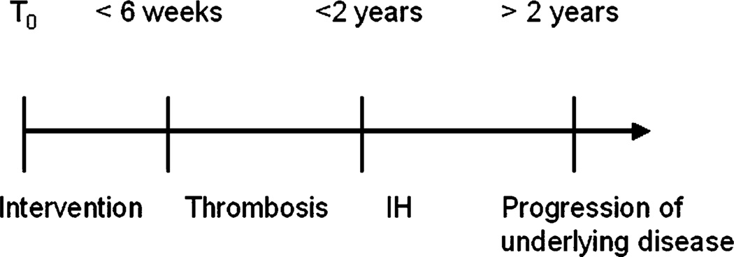
Atherosclerotic cardiovascular disease is a leading cause of death worldwide. Despite recent improvements in medical, operative, and endovascular treatments, the number of interventions performed annually continues to increase. Unfortunately, the durability of these interventions is limited acutely by thrombotic complications and later by myointimal hyperplasia followed by progression of atherosclerotic disease over time. Despite improving medical management of patients with atherosclerotic disease, these complications appear to be persisting. Cardiovascular gene therapy has the potential to make significant clinical inroads to limit these complications. This article will review the technical aspects of cardiovascular gene therapy; its application for promoting a functional endothelium, smooth muscle cell growth inhibition, therapeutic angiogenesis, tissue engineered vascular conduits, and discuss the current status of various applicable clinical trials.
Figures

Similar articles
-
Therapeutic angiogenesis and wound healing--IBC's International Conference. 15-16 November 2004, Cambridge, MA, USA.IDrugs. 2005 Feb;8(2):94-6. IDrugs. 2005. PMID: 15696404 No abstract available.
-
Angiogenic Growth Factors for Coronary Artery Disease: Current Status and Prospects.J Cardiovasc Pharmacol Ther. 2018 Mar;23(2):130-141. doi: 10.1177/1074248417735399. Epub 2017 Oct 12. J Cardiovasc Pharmacol Ther. 2018. PMID: 29025278 Review.
-
Gene therapy for atherosclerotic cardiovascular disease: a time for optimism and caution.Mayo Clin Proc. 2000 Aug;75(8):831-4. doi: 10.4065/75.8.831. Mayo Clin Proc. 2000. PMID: 10943238 Review.
-
The emerging role of gene therapy in the treatment of cardiovascular diseases.Crit Rev Clin Lab Sci. 2003 Oct;40(5):499-545. Crit Rev Clin Lab Sci. 2003. PMID: 14653356 Review.
-
Identifying and overcoming obstacles in angiogenic gene therapy for myocardial ischemia.J Cardiovasc Pharmacol. 2014 Aug;64(2):109-19. doi: 10.1097/FJC.0000000000000107. J Cardiovasc Pharmacol. 2014. PMID: 24736636 Review.
Cited by
-
Delivery of large biopharmaceuticals from cardiovascular stents: a review.Biomacromolecules. 2007 Nov;8(11):3281-93. doi: 10.1021/bm700540p. Epub 2007 Oct 12. Biomacromolecules. 2007. PMID: 17929968 Free PMC article. Review.
-
Reducible poly(amido ethylenediamine) for hypoxia-inducible VEGF delivery.J Control Release. 2007 Apr 2;118(2):254-61. doi: 10.1016/j.jconrel.2006.12.018. Epub 2006 Dec 28. J Control Release. 2007. PMID: 17276536 Free PMC article.
-
Electroporation-mediated delivery of a naked DNA plasmid expressing VEGF to the porcine heart enhances protein expression.Gene Ther. 2010 Mar;17(3):419-23. doi: 10.1038/gt.2009.153. Epub 2009 Dec 3. Gene Ther. 2010. PMID: 19956270 Free PMC article.
-
The grafts modified by heparinization and catalytic nitric oxide generation used for vascular implantation in rats.Regen Biomater. 2018 Mar;5(2):105-114. doi: 10.1093/rb/rby003. Epub 2018 Mar 6. Regen Biomater. 2018. PMID: 29644092 Free PMC article.
-
Radionuclide reporter gene imaging for cardiac gene therapy.Eur J Nucl Med Mol Imaging. 2007 Jun;34 Suppl 1:S27-33. doi: 10.1007/s00259-007-0438-x. Eur J Nucl Med Mol Imaging. 2007. PMID: 17464505 Review.
References
-
- Murray CJ, Lopez AD. Mortality by cause for eight regions of the world: Global Burden of Disease Study. Lancet. 1997;349:1269–1276. - PubMed
-
- World Health Organization. [cited 11/10/05]; Available from: www.who.int.
-
- American Heart Association. [cited 11/10/05]; Available from: www.americanheart.org.
-
- Tassiopoulos AK, Greisler HP. Angiogenic mechanisms of endothelialization of cardiovascular implants: a review of recent investigative strategies. J. Biomater. Sci., Polym. Ed. 2000;11:1275–1284. - PubMed
-
- Martin GH, Allen RC, Noel BL, Talkington CM, Garrett WV, Smith BL, Pearl GJ, Thompson JE. Carotid endarterectomy in patients less than 50 years old. J. Vasc. Surg. 1997;26:447–454. (discussion 454–445). - PubMed
Publication types
MeSH terms
Grants and funding
LinkOut - more resources
Full Text Sources
Other Literature Sources
Medical
