. 2006 Aug 1;296(1):104-18.
doi: 10.1016/j.ydbio.2006.04.442.

Dose-dependent Smad1, Smad5 and Smad8 signaling in the early mouse embryo

Affiliations

Dose-dependent Smad1, Smad5 and Smad8 signaling in the early mouse embryo

Sebastian J Arnold et al. Dev Biol. .

Abstract

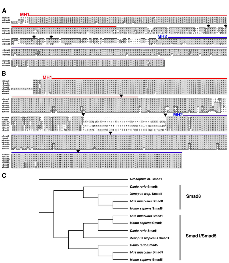
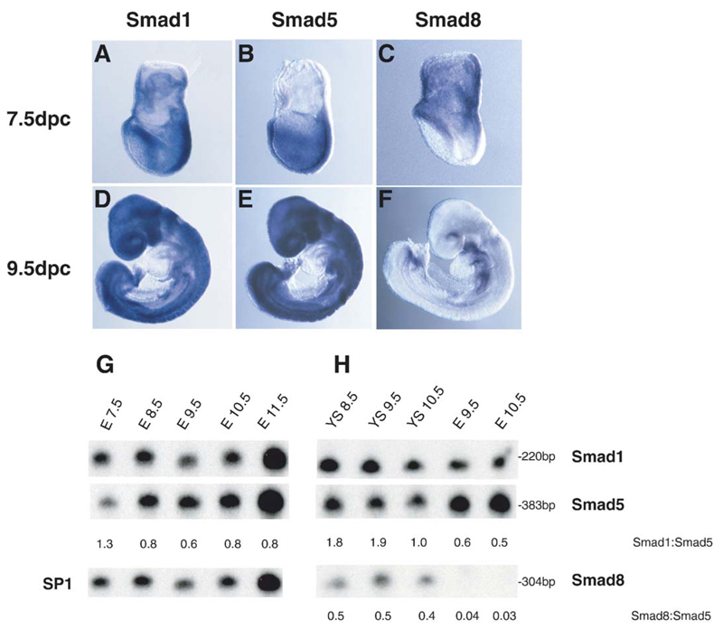
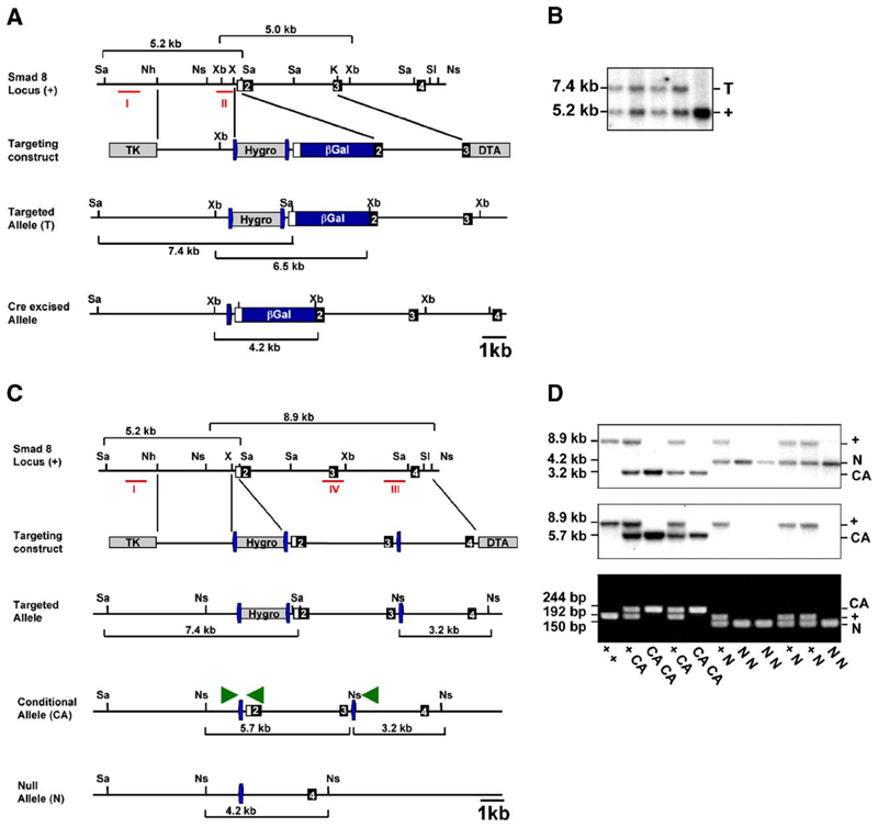
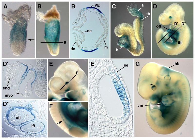
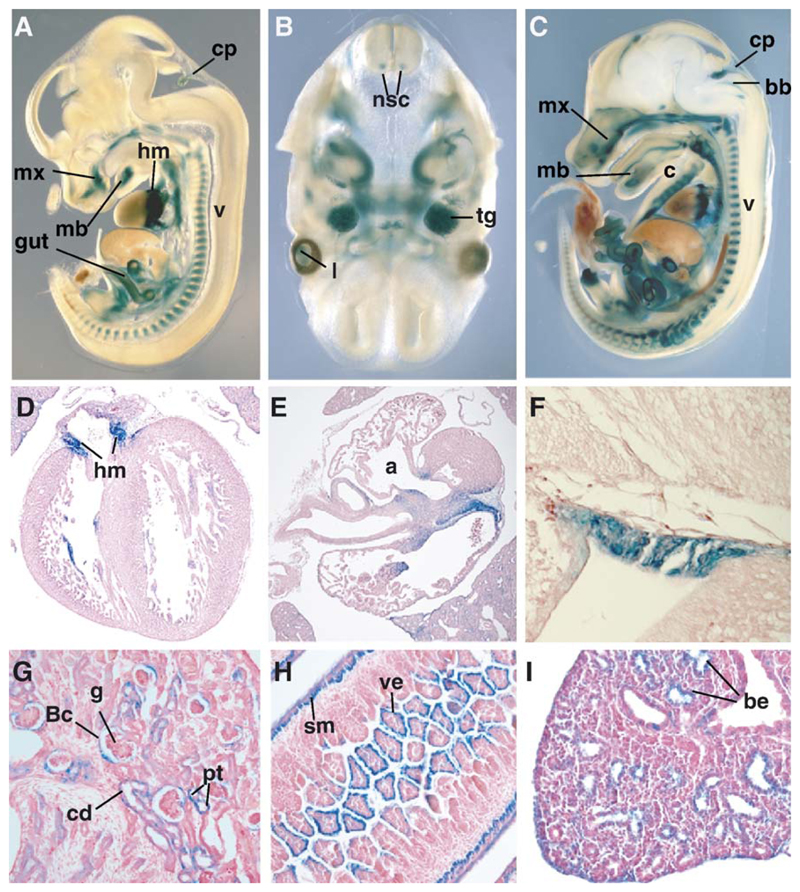
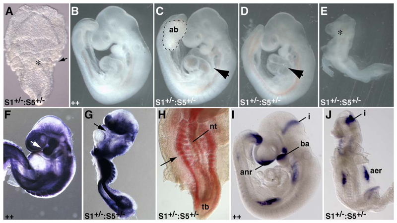
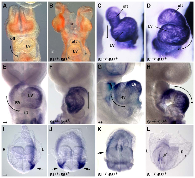
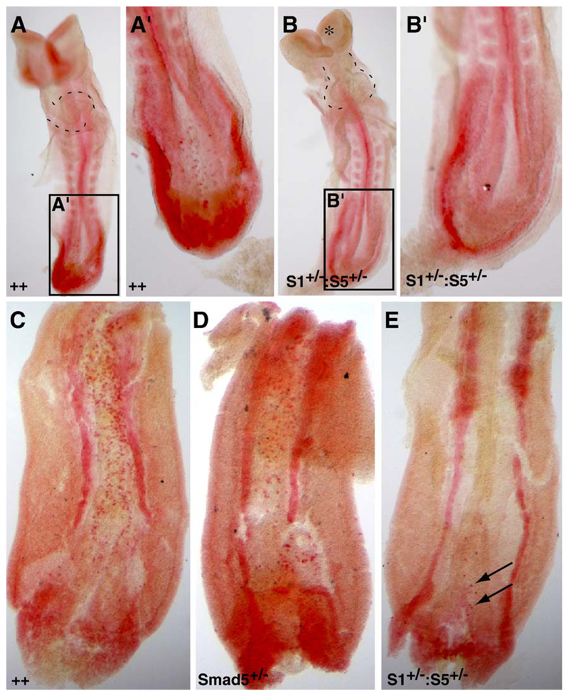
Three closely related mammalian R-Smads, namely Smad1, Smad5 and Smad8, are activated by BMP receptors. Here we have taken a genetic approach to further dissect their possibly unique and/or shared roles during early mouse development. A Smad8.LacZ reporter allele was created to visualize Smad8 expression domains. Smad8 is initially expressed only in the visceral yolk sac (VYS) endoderm and shows a highly restricted pattern of expression in the embryo proper at later stages. In addition, Smad8 conditional and null alleles were engineered. All alleles clearly demonstrate that adult Smad8 homozygous mutants are viable and fertile. To elucidate gene dosage effects, we manipulated expression ratios of the three BMP R-Smads. Smad8 homozygotes also lacking one copy of Smad1 or Smad5 did not exhibit overt phenotypes, and the tissue disturbances seen in Smad1 or Smad5 null embryos were not exacerbated in the absence of Smad8. However, we discovered a profound genetic interaction between Smad1 and Smad5. Thus, as for Smad1 and Smad5 mutant embryos, Smad1+/-:Smad5+/- double heterozygotes die by E10.5 and display defects in allantois morphogenesis, cardiac looping and primordial germ cell (PGC) specification. These experiments demonstrate for the first time that Smad1 and Smad5 function cooperatively to govern BMP target gene expression in the early mammalian embryo.


Figures

Fig. 1
Fig. 1. Smad1, Smad5 and Smad8 sequence alignments.
(A) Comparison of the Smad1, Smad5 and Smad8 amino acid sequences in mouse. Boxes show the conserved amino acids. The dots indicate serine residues within Erk-consensus motifs (PXSP). The presumptive boundaries of the MH1and MH2 domains are indicated. (B) The mouse Smad8 (mSmad8) sequence is aligned with those of rat (r), human (h), frog (x) and zebrafish (z). Human and Xenopus longer isoforms are indicated as hSmad8L and xSmad8L, respectively. Boxes show conserved amino acids and exon boundaries are denoted by the arrow heads. Sequence alignments were performed using the Macvector software package (Accelrys.com). (C) A phylogenetic tree of BMP R-Smads shows early branching of Smad8 from Smad1 and 5 genes. Fruitfly (Drosophila melanogaster), zebrafish (Danio rerio), frog (Xenopus tropicalis), mouse (Mus musculus) and human (Homo sapiens). Phylogenetic alignment was generated using Macvector (Accelrys.com).
Fig. 2
Fig. 2. Partially overlapping Smad1, Smad5 and Smad8 expression domains in the early mouse embryo.
(A–F) Whole-mount in situ hybridization analysis of Smad1, Smad5 and Smad8 expression. (A and B) At the early head fold stage (E7.75), Smad1 and Smad5 are robustly expressed throughout the extra-embryonic and embryonic regions. (D and E) Similarly at E9.5 both transcripts are widely expressed throughout the embryo proper. (C) In contrast, Smad8 is exclusively expressed in the extra-embryonic region at E7.75, specifically in the endoderm of the visceral yolk sac (VYS). (F) At E9.5 Smad8 expression in the embryo is restricted to a subregion of the heart tube and surrounding mesenchyme, the eye and the tail bud. (G and H) Ribonuclease protection assays analyzing Smad1, Smad5 and Smad8 expression ratios. Total embryonic (E) and yolk sac (YS) RNA at the various stages indicated were tested. The numbers to the right hand side indicate the sizes of the protected fragments. The expression ratios shown as numbers at the bottom of the lanes were calculated by scanning the gels in a PhosphorImager, measuring the amount of radioactivity in each band and adjusting for the CTP content of the antisense probes. Smad1 and Smad5 are co-expressed in the embryo and yolk sac, whereas in contrast Smad8 transcripts are predominantly localized to the yolk sac. Data shown are representative of 4 independent experiments testing several embryonic and yolk sac total RNA samples.
Fig. 3
Fig. 3. Generation of Smad8.LacZ, conditional and null alleles.
(A) A β-galactosidase-polyA cassette was introduced into the ATG-containing exon 2 of the Smad8 locus. (B) Hygromycin-resistant ES cell clones were screened by Southern blot analysis of SacI-digested DNA using a 5′ external probe (probe I) that distinguishes 5.2-kb wild-type (+) and 7.4-kb targeted (T) alleles. Correctly targeted clones were subjected to in vitro Cre-mediated excision to remove the loxP (blue arrows)-flanked hygromycin resistance cassette and verified by Southern blot analysis using an internal probe (probe II) on XbaI-digested DNA. (C) Targeting strategy for generation of Smad8 conditional and null alleles. A loxP (blue arrows)-flanked hygromycin resistance cassette was inserted 5′ of exon 2 and a single loxP site placed 3′ of exon 3. Hygromycin-resistant clones were screened by Southern blot analysis using a 5′ external probe (probe I). Cre-mediated recombination yielded ES cell subclones carrying the conditional (CA) and null (N) alleles. (D) Genotypes of offspring from heterozygous intercross matings. Southern blot analysis of NsiI-digested genomic DNA using a 3′ internal probe (probe III in C) distinguishes 8.9-kb wild-type (+), 3.2-kb conditional (CA) and 4.2-kb null alleles (N). The 3′ internal probe (probe IV in panel C) confirms excision of exons 2 and 3 in homozygous null animals. Confirmatory multiplex PCR genotyping analysis using the indicated primers (green arrowheads in C) distinguishes wt (192 bp), conditional (244 bp) and null allele (150 bp). K, KpnI; Nh, NheI; Ns, NsiI; Sa, SacI; Sl, SalI; X, XhoI; Xb, XbaI.
Fig. 4
Fig. 4. Highly restricted Smad8.LacZ expression during early mouse development.
Whole-mount X-Gal staining of Smad8.LacZ/+ embryos at 5.5 dpc (A), 7.5 dpc (B, B′), 8.5 dpc (C), 9.5 dpc (D, D′, D″), 10.5 dpc (E, E′, F) and 12.5 dpc (G). (A, B, B′) Smad8.LacZ expression is initially detected in the extra-embryonic visceral endoderm(VE) from day 5.5, but not in the definitive endoderm (de), neuroectoderm (ne) or mesoderm (m) of the embryo proper. (C) LacZ expression in the embryo is first detected at 8.5 dpc in the mesenchyme surrounding the forming heart (h) and at lower levels in the tail bud region (tb) and allantois (a). (D, D′, D″) At 9.5 dpc, embryonic expression is strongly seen within the myocardial layer (myo) of the cardiac outflow tract (oft) and inflow tract (ift) but is absent from the forming ventricular regions and endocardium (end). (E, E′) Expression is also initiated within the forming eye and by 10.5 dpc expression is confined to the inner retinal layer of the optic vesicle exclusively in the rostral dorsal half. No expression is seen within the surface ectoderm (se). (F) From E10.5 expression is detectable in the sclerotomal component (arrow) of the somites. (G) At E11.5 X-Gal staining is seen in presumptive areas of chondrogenesis throughout the forming cranial, axial and appendicular skeleton. LacZ staining also marks ventral mesenchyme of the thoracic and abdominal regions (vm). Low levels of expression are also seen in the neuroepithelium of the hindbrain (hb) and eye (asterisk).
Fig. 5
Fig. 5. The Smad8.LacZ reporter allele reveals a dynamic pattern of expression during organogenesis.
(A) Sagittal vibratome section of an E14.5 embryo. LacZ expression is detected in the choroid plexus (cp), forming vertebra (v), in a band of mesenchyme located in the atrioventricular regions of the developing heart (hm) and in the developing gut. (B) Transverse section of the brain of a 16.5 dpc embryo showing LacZ expression in a subset of motorneurons in the anterior spinal cord (nsc), the trigeminal ganglion (tg) and the lens (l). (C) Sagittal vibratome section of an E16.5 embryo. LacZ-positive sites include all of the cartilage primordia. In this particular section, cartilage primordia of the vertebrae (v), ribs (c), basioccipital bone (bb) and forming maxilla (mx) and mandible (mb) are clearly stained. (D and E) Transverse and coronal sections of 16.5 dpc heart showing expression of the Smad8 reporter allele in the mesenchyme tissue (hm) separating the atria (a) from the ventricles. (F) Close-up of the choroid plexus. (G) Sagittal section of an E16.5 kidney reveals LacZ expression in the epithelium of the collecting ducts (cd) and proximal tubules (pt) as well as in the juxtaglomerular apparatus and Bowman’s capsule (Bc) of the glomeruli (g). (H) Transverse section of the intestine of a 16.5 dpc embryo. Stained cells correspond to the epithelium layer of the villi (ve). The outer smooth muscle layer (sm) is also positive for LacZ expression. (I) At E16.5, within the lung LacZ expression is detectable in the epithelial cells of the bronchiole (be).
Fig. 6
Fig. 6. Smad1 +/– :Smad5 +/– double heterozygous embryos display pleiotropic tissue abnormalities.
(A) The earliest abnormality seen in a proportion of embryos at E7.5 is ruffling of the visceral yolk sac (asterisk). The arrow marks the boundary between the embryo and extra-embryonic regions. At E9.5, in contrast to wild-type litter mates (B) where the allantois has fused to the chorion, Smad1 +/−:Smad5 +/− double heterozygotes (C) display an unfused allantoic bud (ab, outlined with dashed line). In this example, the remainder of the embryo is grossly normal including the heart (arrow). (D) Approximately two thirds of mutant embryos show abnormalities in heart morphogenesis including heart looping (arrow in panel D) and patterning defects. (E) Approximately one third of mutant embryos arrest at embryonic turning and show severe defects including lack of anterior neural structures (asterisk). (F, G) In situ hybridization for Twist expression in wild-type (F) and mutant embryos (G) shows that severely affected embryos have a paucity of mesoderm and lack branchial arches (arrows in panels F and G). (H) Ventral view of the caudal region of a severely affected embryo shows that the somites are disorganized and fragmented (arrow). (I, J) Analysis of Fgf8 expression in wild-type (I) and severely affected (J) embryos documents the absence of anterior-most neural tissue whereas the midbrain/hindbrain isthmus (i) is specified. The developing limb buds form but are disorganized. (ab, allantoic bud; aer, apical ectodermal ridge; anr, anterior neural ridge; ba, branchial arch; nt, neural tube; i, isthmus; tb, tail bud).
Fig. 7
Fig. 7. L/R and heart patterning defects in Smad1 +/− :Smad5 +/− double heterozygous embryos.
(A, B) Frontal views of E9.5 wild-type and mutant embryos. The arrows indicate the direction of cardiac looping. Normally, the heart tube is looped to the right and the left ventricle (LV) is readily seen. In contrast, in a proportion of Smad1 +/− :Smad5 +/− mutants, the heart fails to loop and the forming left ventricle remains caudal. The asterisk marks the unfused anterior neural folds in mutants. (C) Staining of hearts with myosin light chain V (MLCV) highlights the disturbances to heart tube morphogenesis and (D) reversal in the direction of heart looping. (E, G) eHand expression in E9.5 wild-type embryos delineates the outer curvature of the left ventricle. Staining is largely absent from the forming right ventricle. (F, H) In Smad1 +/− :Smad5 +/− embryos, the domain of eHand expression has expanded to encompass the length of the heart tube. (I) At E8.5 Nodal is normally expressed in the lateral plate mesoderm on the left side of the axis. Smad1 +/− :Smad5 +/− embryos show bilateral (J), right-sided (K) Nodal expression or in the majority of cases fail to activate asymmetric expression, whereas in the node, Nodal is expressed appropriately (L, posterior view) (ift, inflow tract; L, left side of embryo; LV, left ventricle; oft, outflow tract; R, right side of embryo; RV right ventricle).
Fig. 8
Fig. 8. Dose-dependent Smad1/Smad5 activities are essential for specification of primordial germ cells.
(A, A′, B, B′) Fast red alkaline phosphatase staining of primordial germ cells (PGCs) in E8.5 (A, A′) wild-type and (B, B′) Smad1 +/−:Smad5+ /- double heterozygous mutant embryos. The mutants display a significant reduction in number of primary germ cells within the hindgut region (compare panel A′ to B′) and additional tissue defects as indicated (heart morphogenesis, dashed line; unfused anterior neural folds, asterisk). (C, D, E) At E9.5, germ cells defects become more pronounced (compare panel C to E, arrows indicating few detectable PGCs). Smad5 −/− heterozygous embryos display PGCs at intermediate numbers (D).

Similar articles

Cited by

References

    1. Aoki H, Fujii M, Imamura T, Yagi K, Takehara K, Kato M, Miyazono K. Synergistic effects of different bone morphogenetic protein type I receptors on alkaline phosphatase induction. J Cell Sci. 2001;114:1483–1489. - PubMed
    1. Aubin J, Davy A, Soriano P. In vivo convergence of BMP and MAPK signaling pathways: impact of differential Smad1 phosphorylation on development and homeostasis. Genes Dev. 2004;18:1482–1494. - PMC - PubMed
    1. Biben C, Harvey RP. Homeodomain factor Nkx2-5 controls left/right asymmetric expression of bHLH gene eHand during murine heart development. Genes Dev. 1997;11:1357–1369. - PubMed
    1. Brennan J, Lu CC, Norris DP, Rodriguez TA, Beddington RS, Robertson EJ. Nodal signalling in the epiblast patterns the early mouse embryo. Nature. 2001;411:965–969. - PubMed
    1. Chang H, Matzuk MM. Smad5 is required for mouse primordial germ cell development. Mech Dev. 2001;104:61–67. - PubMed

Publication types

LinkOut - more resources