The E delta enhancer controls the generation of CD4- CD8- alphabetaTCR-expressing T cells that can give rise to different lineages of alphabeta T cells
- PMID: 16754716
- PMCID: PMC2118313
- DOI: 10.1084/jem.20051711
The E delta enhancer controls the generation of CD4- CD8- alphabetaTCR-expressing T cells that can give rise to different lineages of alphabeta T cells
Erratum in
- J Exp Med. 2006 Jul 10;203(7):1831
Abstract
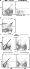
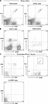
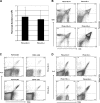
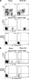
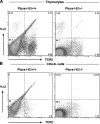
It is well established that the pre-T cell receptor for antigen (TCR) is responsible for efficient expansion and differentiation of thymocytes with productive TCRbeta rearrangements. However, Ptcra- as well as Tcra-targeting experiments have suggested that the early expression of Tcra in CD4- CD8- cells can partially rescue the development of alphabeta CD4+ CD8+ cells in Ptcra-deficient mice. In this study, we show that the TCR E delta but not E alpha enhancer function is required for the cell surface expression of alphabetaTCR on immature CD4- CD8- T cell precursors, which play a crucial role in promoting alphabeta T cell development in the absence of pre-TCR. Thus, alphabetaTCR expression by CD4- CD8- thymocytes not only represents a transgenic artifact but occurs under physiological conditions.
Figures


Similar articles
-
Impact of early expression of TCR alpha chain on thymocyte development.Eur J Immunol. 2004 Jun;34(6):1532-41. doi: 10.1002/eji.200424870. Eur J Immunol. 2004. PMID: 15162422
-
The CD3gamma chain is essential for development of both the TCRalphabeta and TCRgammadelta lineages.EMBO J. 1998 Apr 1;17(7):1871-82. doi: 10.1093/emboj/17.7.1871. EMBO J. 1998. PMID: 9524111 Free PMC article.
-
T lymphocyte development in the absence of Fc epsilon receptor I gamma subunit: analysis of thymic-dependent and independent alpha beta and gamma delta pathways.Eur J Immunol. 1996 Aug;26(8):1935-43. doi: 10.1002/eji.1830260839. Eur J Immunol. 1996. PMID: 8765042
-
CD4/CD8 lineage commitment in T cell receptor transgenic mice: evidence for precommitment of CD4+ CD8+ thymocytes.Semin Immunol. 1994 Aug;6(4):249-56. doi: 10.1006/smim.1994.1032. Semin Immunol. 1994. PMID: 8000034 Review.
-
Thymic selection revisited: how essential is it?Immunol Rev. 2003 Feb;191:62-78. doi: 10.1034/j.1600-065x.2003.00010.x. Immunol Rev. 2003. PMID: 12614352 Review.
Cited by
-
αβ versus γδ fate choice: counting the T-cell lineages at the branch point.Immunol Rev. 2010 Nov;238(1):169-81. doi: 10.1111/j.1600-065X.2010.00947.x. Immunol Rev. 2010. PMID: 20969592 Free PMC article. Review.
-
The thymus chapter in the life of gut-specific intra epithelial lymphocytes.Curr Opin Immunol. 2008 Apr;20(2):185-91. doi: 10.1016/j.coi.2008.03.009. Epub 2008 May 2. Curr Opin Immunol. 2008. PMID: 18456487 Free PMC article. Review.
-
Early expression of mature αβ TCR in CD4-CD8- T cell progenitors enables MHC to drive development of T-ALL bearing NOTCH mutations.Proc Natl Acad Sci U S A. 2022 Jul 5;119(27):e2118529119. doi: 10.1073/pnas.2118529119. Epub 2022 Jun 29. Proc Natl Acad Sci U S A. 2022. PMID: 35767640 Free PMC article.
-
Identification of CD4(-)CD8(-) double-negative natural killer T cell precursors in the thymus.PLoS One. 2008;3(11):e3688. doi: 10.1371/journal.pone.0003688. Epub 2008 Nov 10. PLoS One. 2008. PMID: 18997862 Free PMC article.
-
Regulation of T-cell Receptor Gene Expression by Three-Dimensional Locus Conformation and Enhancer Function.Int J Mol Sci. 2020 Nov 11;21(22):8478. doi: 10.3390/ijms21228478. Int J Mol Sci. 2020. PMID: 33187197 Free PMC article. Review.
References
-
- Godfrey, D.I., and A. Zlotnik. 1993. Control points in early T-cell development. Immunol. Today. 14:547–553. - PubMed
-
- Rodewald, H.R., and H.J. Fehling. 1998. Molecular and cellular events in early thymocyte development. Adv. Immunol. 69:1–112. - PubMed
-
- Groettrup, M., K. Ungewiss, O. Azogui, R. Palacios, M.J. Owen, A.C. Hayday, and H. von Boehmer. 1993. A novel disulfide-linked heterodimer on pre-T cells consists of the T cell receptor beta chain and a 33 kd glycoprotein. Cell. 75:283–294. - PubMed
-
- Saint-Ruf, C., K. Ungewiss, M. Groettrup, L. Bruno, H.J. Fehling, and H. von Boehmer. 1994. Analysis and expression of a cloned pre-T cell receptor gene. Science. 266:1208–1212. - PubMed
Publication types
MeSH terms
Substances
Grants and funding
LinkOut - more resources
Full Text Sources
Other Literature Sources
Molecular Biology Databases
Research Materials
Miscellaneous

