. 2006 Jun 5;173(5):645-50.
doi: 10.1083/jcb.200512079. Epub 2006 May 30.

Regulation of mitochondrial fusion by the F-box protein Mdm30 involves proteasome-independent turnover of Fzo1

Affiliations

Regulation of mitochondrial fusion by the F-box protein Mdm30 involves proteasome-independent turnover of Fzo1

Mafalda Escobar-Henriques et al. J Cell Biol. .

Abstract

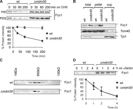
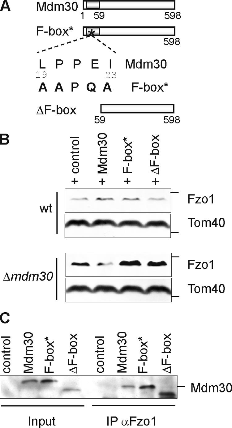
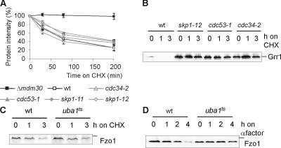
Mitochondrial morphology depends on balanced fusion and fission events. A central component of the mitochondrial fusion apparatus is the conserved GTPase Fzo1 in the outer membrane of mitochondria. Mdm30, an F-box protein required for mitochondrial fusion in vegetatively growing cells, affects the cellular Fzo1 concentration in an unknown manner. We demonstrate that mitochondrial fusion requires a tight control of Fzo1 levels, which is ensured by Fzo1 turnover. Mdm30 binds to Fzo1 and, dependent on its F-box, mediates proteolysis of Fzo1. Unexpectedly, degradation occurs along a novel proteolytic pathway not involving ubiquitylation, Skp1-Cdc53-F-box (SCF) E3 ubiquitin ligase complexes, or 26S proteasomes, indicating a novel function of an F-box protein. This contrasts to the ubiquitin- and proteasome-dependent turnover of Fzo1 in alpha-factor-arrested yeast cells. Our results therefore reveal not only a critical role of Fzo1 degradation for mitochondrial fusion in vegetatively growing cells but also the existence of two distinct proteolytic pathways for the turnover of mitochondrial outer membrane proteins.


Figures

Figure 1.
Figure 1.
Fzo1 stability is controlled by two independent proteolytic pathways. (A, top) The stability of Fzo1 in exponentially growing (exp) or post–diauxic-shift cultures (PDS) after adding cycloheximide (CHX) was monitored by SDS-PAGE and immunoblotting. (bottom) A quantification including standard deviation of three independent experiments. (B) Cellular subfractionation. Exponentially growing wild-type (wt) and Δmdm30 cells were split by differential centrifugation into a mitochondrial (pellet) and a cytosolic (sup) fraction as described previously (Rapaport et al., 1998). The fractions were analyzed by SDS-PAGE and immunoblotting for the presence of Fzo1. The mitochondrial outer membrane protein Tom40 and the cytosolic protein Tpi1 were used as marker proteins to validate the fractionation. (C) Complex formation of Fzo1 in the mitochondrial outer membrane. Mitochondrial membranes of wild-type and Δmdm30 cells were solubilized in Triton X-100 and fractionated by Superose 6 gel filtration chromatography. Eluate fractions were analyzed by immunoblotting using Fzo1-specific antibodies. (D) Fzo1 degradation upon α-factor– induced cell cycle arrest. Fzo1 stability in cell cycle–arrested wild-type and Δmdm30 cells was analyzed as in A. The position of standard molecular mass markers during electrophoresis corresponding to 116, 43, and 27 kD are indicated by the small bars in panels showing Fzo1, Tom40, or Tpi1, respectively.
Figure 2.
Figure 2.
The constitutive Fzo1 turnover depends on the F-box motif of its interacting partner, Mdm30. (A) Schematic representation of Mdm30 variants. The Mdm30 derivative F-box* was obtained, replacing amino acid residues 19, 20, 22, and 23 within consensus residues of the F-box motif of Mdm30 as shown. The variant ΔF-box was generated by deleting amino acid residues 1–58 of Mdm30. (B) Mitochondrial extracts of wild-type (wt) and Δmdm30 strains expressing Mdm30 variants or transformed with the vector control were analyzed by immunoblotting using Fzo1-specific and, as a loading control, Tom40-specific antibodies. (C) Mitochondria derived from wild-type cells expressing several Mdm30 variants harboring an NH2-terminal Flag epitope or cells transformed with the vector control were solubilized in digitonin and subjected to immunoprecipitation using Fzo1-specific antibodies. Total extracts (input; 5%) and immunoprecipitates (IP; 100%) were analyzed by immunoblotting with antibodies directed against the Flag epitope of the Mdm30 proteins. The position of standard molecular mass markers during electrophoresis corresponding to 116, 43, and 66 kD are indicated by the small bars in panels showing Fzo1, Tom40, or Mdm30, respectively.
Figure 3.
Figure 3.
Differential requirement of ubiquitylation for constitutive and induced Fzo1 degradation. (A–C) The stability of Fzo1 and Grr1-myc was analyzed after inhibition of cytosolic protein synthesis with cycloheximide (CHX) in vegetatively growing wild-type (wt), Δmdm30, cdc34-2, cdc53-1, skp1-11, skp1-12, and uba1ts cells at 37°C, as described in Fig. 1 A. (D) Proteolysis of Fzo1 in the presence of α-factor was monitored at 37°C in wild-type and uba1ts cells. The position of standard molecular mass markers during electrophoresis corresponding to 158 and 116 kD are indicated by the small bars in panels showing Grr1 or Fzo1, respectively.
Figure 4.
Figure 4.
Fzo1 degradation is mediated by a novel proteolytic system. (A) The stability of Fzo1 was monitored in wild-type (wt), Δmdm30, pre1-1, cim5-1, Δump1, Δyme1, and Δpep4 cells after addition of cycloheximide (CHX) as in Fig. 1 A. (B) Fzo1 stability upon α-factor–induced cell cycle arrest was examined in wild-type, pre1-1, and cim5-1 cells, which also lacked BAR1. (C) Exponentially growing wild-type cells were treated with cycloheximide (CHX) and additionally with sodium azide (NaN3) or CCCP as indicated. The stability of Fzo1 was determined as in Fig. 1 A. The position of the standard molecular mass marker during electrophoresis corresponding to 116 kD is indicated by the small bars.
Figure 5.
Figure 5.
Control of mitochondrial fusion by Mdm30-dependent proteolysis of Fzo1. (A) Cellular (Nomarski) and mitochondrial (GFP) morphology was visualized by fluorescence microscopy after expressing mtGFP in wild-type (wt), Δfzo1, and Δmdm30 cells or in wild-type cells expressing Fzo1 from a multicopy plasmid under the control of the CUP1 promoter in the presence of added copper (50 μM; wt + Fzo1). (B) Steady-state concentrations of Fzo1 in extracts of the indicated strains were analyzed by immunoblotting using Fzo1-specific and, as a loading control, Tom40-specific antibodies. (C) Stability of Fzo1 present at different levels in wild-type and Δmdm30 cells. Protein synthesis shut-off experiments were performed as in Fig. 1 A using exponentially growing wild-type or Δmdm30 cells expressing HA-Fzo1 from either a centromeric (cen) or a multicopy (2μ) plasmid under the control of the endogenous FZO1 promoter. (D) Wild-type and Δmdm30 cells expressing mtGFP and harboring a chromosomal integration of the GAL1 promoter upstream of the FZO1 coding region were grown on glucose-, raffinose-, or galactose-containing medium. Cellular (Nomarski) and mitochondrial (GFP) morphology was visualized after expression of mtGFP by fluorescence microscopy. (E) Wild-type cells expressing mtGFP and harboring a chromosomal integration of the GAL1 promoter upstream of the FZO1 coding region were grown on glucose- or galactose-containing medium. Cells were expressing MDM30 from a multicopy plasmid under the control of the CUP1 promoter when indicated. Cellular (Nomarski) and mitochondrial (GFP) morphology was visualized by fluorescence microscopy. The position of standard molecular mass markers during electrophoresis corresponding to 116 and 43 kD are indicated by the small bars in panels showing Fzo1 or Tom40, respectively. Bars, 5 μm.

Similar articles

Cited by

References

    1. Altmann, K., and B. Westermann. 2005. Role of essential genes in mitochondrial morphogenesis in Saccharomyces cerevisiae. Mol. Biol. Cell. 16:5410–5417. - PMC - PubMed
    1. Chen, H., and D.C. Chan. 2005. Emerging functions of mammalian mitochondrial fusion and fission. Hum. Mol. Genet. 14:R283–R289. - PubMed
    1. Dimmer, K.S., S. Fritz, F. Fuchs, M. Messerschmitt, N. Weinbach, W. Neupert, and B. Westermann. 2002. Genetic basis of mitochondrial function and morphology in Saccharomyces cerevisiae. Mol. Biol. Cell. 13:847–853. - PMC - PubMed
    1. Fritz, S., N. Weinbach, and B. Westermann. 2003. Mdm30 is an F-box protein required for maintenance of fusion-competent mitochondria in yeast. Mol. Biol. Cell. 14:2303–2313. - PMC - PubMed
    1. Galan, J.M., and M. Peter. 1999. Ubiquitin-dependent degradation of multiple F-box proteins by an autocatalytic mechanism. Proc. Natl. Acad. Sci. USA. 96:9124–9129. - PMC - PubMed

Publication types

MeSH terms