Comparative Study
. 2006 May;2(5):e50.
doi: 10.1371/journal.ppat.0020050. Epub 2006 May 26.

The malarial host-targeting signal is conserved in the Irish potato famine pathogen

Affiliations
Comparative Study

The malarial host-targeting signal is conserved in the Irish potato famine pathogen

Souvik Bhattacharjee et al. PLoS Pathog. 2006 May.

Abstract

Animal and plant eukaryotic pathogens, such as the human malaria parasite Plasmodium falciparum and the potato late blight agent Phytophthora infestans, are widely divergent eukaryotic microbes. Yet they both produce secretory virulence and pathogenic proteins that alter host cell functions. In P. falciparum, export of parasite proteins to the host erythrocyte is mediated by leader sequences shown to contain a host-targeting (HT) motif centered on an RxLx (E, D, or Q) core: this motif appears to signify a major pathogenic export pathway with hundreds of putative effectors. Here we show that a secretory protein of P. infestans, which is perceived by plant disease resistance proteins and induces hypersensitive plant cell death, contains a leader sequence that is equivalent to the Plasmodium HT-leader in its ability to export fusion of green fluorescent protein (GFP) from the P. falciparum parasite to the host erythrocyte. This export is dependent on an RxLR sequence conserved in P. infestans leaders, as well as in leaders of all ten secretory oomycete proteins shown to function inside plant cells. The RxLR motif is also detected in hundreds of secretory proteins of P. infestans, Phytophthora sojae, and Phytophthora ramorum and has high value in predicting host-targeted leaders. A consensus motif further reveals E/D residues enriched within approximately 25 amino acids downstream of the RxLR, which are also needed for export. Together the data suggest that in these plant pathogenic oomycetes, a consensus HT motif may reside in an extended sequence of approximately 25-30 amino acids, rather than in a short linear sequence. Evidence is presented that although the consensus is much shorter in P. falciparum, information sufficient for vacuolar export is contained in a region of approximately 30 amino acids, which includes sequences flanking the HT core. Finally, positional conservation between Phytophthora RxLR and P. falciparum RxLx (E, D, Q) is consistent with the idea that the context of their presentation is constrained. These studies provide the first evidence to our knowledge that eukaryotic microbes share equivalent pathogenic HT signals and thus conserved mechanisms to access host cells across plant and animal kingdoms that may present unique targets for prophylaxis across divergent pathogens.


Conflict of interest statement

Competing interests. The authors have declared that no competing interests exist.

Figures

Figure 1
Figure 1. Schematic of Intracellular Infection of Plasmodium and Phytophthora Parasites
(A) A human erythrocyte (pink) infected by P. falciparum (blue). Invasion by the extracellular merozoite stage leads to formation of a host-derived PVM within which the parasite resides. Proteins (brown squares) secreted by the parasite must cross the PVM to reach and mediate virulence and structural changes in the erythrocyte. (B) Plant cells (green) infected by P. infestans (blue). P. infestans parasite colonizes the host intracellular spaces and forms haustoria (yellow). The host-derived haustorial membrane must be crossed by pathogenic effectors (brown squares) released into cells to mediate virulence and plant hypersensitive responses.
Figure 2
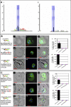
Figure 2. Conserved, Plant Pathogenic Oomycete Motif Functions as a Host-Targeting Signal in P. falciparum–Infected Erythrocytes
(A) Sequence alignment of six effectors from P. falciparum HT-secretome (upper panel) and five Avr proteins from the oomycete pathogens Phytophthora and Hyaloperonospora (lower panel). Each sequence contains an 11 amino acid region (black bar) centered on RxL residues (blue). Negatively charged residues (E or D) downstream of the RxL are colored red. Seven residues upstream of the RxL are underlined purple and boxed in the case of PfHRPII. (B) Live cells expressing secretory GFP chimeras of AVR3a (residues 21 to 69) with no change (i–iii), or where RRLLRK was replaced by AASTAI (iv–vi), where (ii) and (v) indicate fluorescence images, (i) and (iv) corresponding brightfield images, and (iii) and (vi) the respective merges. Constructs contain SS (black) followed by indicated sequences of AVR3a (orange) and GFP (green). For quantitative analyses, two hundred fluorescent images were analyzed as described in Materials and Methods. Fraction of GFP exported to the erythrocyte cytosol is indicated in (vii). As expected, all parasitized cells express and export the transgene (viii). Standard deviations are shown in pink. In all cases: p, parasite; e, erythrocyte; nucleus is Hoechst-stained (blue); scale bar represents 2 μm.
Figure 3
Figure 3. HT Motif Has High Predictive Value for Phytophthora Effectors
(A) Sequence logos derived from 59 predicted P. infestans secretory proteins and the P. falciparum HT-secretome (boxed inset). Amino acids are represented by one-letter abbreviations and color-coded as follows: blue, basic; red, acidic; black, hydrophobic; and green, polar. Height of amino acids indicates their frequency at that position. (B) Live cells expressing secretory GFP chimeras of PH001D5 (residues 19 to 88) with no change (i–iii) or where DRQLRGF was replaced with ISAATAI (iv–vi), where (ii) and (v) indicate fluorescence images, (i and iv) corresponding brightfield images, and (iii and vi) show respective merges. The asterisk (*) in panel (ii) indicates intraerythrocytic loop structure that excluded GFP. Constructs contain SS (black), followed by indicated sequences of PH001D5 (orange) and GFP (green). For quantitative analyses, 220 fluorescent images were analyzed as described in Materials and Methods. Fraction of GFP exported to the erythrocyte cytosol is indicated in (vii) in a culture where all parasites are transformed (as expected) and export the transgene (viii). Standard deviations are shown in pink. Export of green fluorescence to erythrocyte is quantitatively blocked on replacement of the P. infestans motif (as indicated in the bar chart in vii [standard deviation show in pink]). p, parasite; e, erythrocyte; nucleus is Hoechst-stained (blue), scale bar is 2 μm.
Figure 4
Figure 4. Comparative Analyses of Secretory and Cytosolic RxLR–Containing Sequences in Phytophthora: In Silico and Functional Evidence for the Requirement of Downstream E, D Residue Sequences in HT Consensus
(Ai) Secretory: logos generated from sequences containing RxLR in the first 100 residues after the SS cleavage site for predicted secretory proteins (~10% of the SS set). (Aii) Cytosolic: logos from sequences containing RxLR in the first 100 residues of predicted cytosolic proteins (~5% of the cytosolic set). (B) Live cells expressing secretory GFP chimeras of Avr3a (residues 21–69) with no change (i–iii), replacement of D/E residues downstream of RxLR with hydrophobic residues (iv–vi), replacement of downstream sequence KNEENEETSEERAPNFNLANLN by NQYSHTALVKIQGLTDKKDYLGK found downstream of RxLR from a non-secretory Phytophthora protein AAY43422.1 (vii–ix). (C) Live cells expressing secretory GFP chimeras of PH001D5 (residues 19–88) with no change (i–iii), replacement of D/E residues downstream of RxLR with hydrophobic residues (iv–vi), replacement of downstream sequence GFYATENTDPVNNQDTAHEDGEERV by VGPAGGAAAVGTGSGNAASNTAPHAG found downstream of RxLR from a non-secretory Phytophthora protein 83742 (vii–ix). (B and C) Panels (ii, v, and viii) indicate fluorescence micrographs; (i, iv, and vii), brightfield images; (iii, vi, and ix), respective merges. Constructs contain SS (black) followed by indicated AVR3a or PH001D5 wild-type and modified sequences (orange) and GFP (green). Fraction of GFP exported to the erythrocyte cytosol is indicated in Bx and Cx. Standard deviations are shown in pink. Export of green fluorescence to erythrocyte is quantitatively blocked on replacement of the regions downstream of the Avr3a and PH001D5 motifs, as indicated in the bar charts in (xi). Standard deviations are shown in pink. p, parasite; e, erythrocyte; nucleus is Hoechst-stained (blue); scale bar is 2 μm. In both (ii) panels, the asterisk (*) indicates intraerythrocytic loop structure that excluded GFP. Note, in the case of PH001D5 (Cviii), a small amount of GFP is detected in tubovesicular elements in the erythrocyte. However, export to the erythrocyte cytoplasm or periphery is never detected.
Figure 5
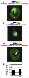
Figure 5. Requirement of Sequences Downstream of HT Motif in Protein Export to Erythrocyte Cytosol
(i) Images of live cells exporting a secretory GFP chimera containing the five amino acid HT core (blue) followed by 16 amino acids downstream sequence from PfHRPII (red). (ii) Removal of the terminal nine amino acids (VHHAHHADV) blocked export to the erythrocyte. (iii) Replacement of VHHAHHADV with VGMMSMMDV restored export of GFP to the erythrocyte. For quantitative analyses, two hundred fluorescent images were analyzed as described in Materials and Methods. (iv) Fraction of GFP exported to the erythrocyte cytosol is indicated, and all parasitized cells export the transgene (as expected for stable transfections; unpublished data). Standard deviations are as shown. Constructs contain SS (black), upstream region (purple), followed by sequence containing core host-targeting motif (blue), the downstream spacer region (red), and GFP (green). In all cases: p, parasite; e, erythrocyte; nucleus is Hoechst-stained (blue); scale bar is 2 μm.
Figure 6
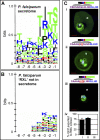
Figure 6. Analysis of Upstream Region of RxL Motifs in the P. falciparum HT-Secretome
Logos derived from eight positions upstream of the P. falciparum HT core. (A) Logo was derived from sequences in the predicted HT-secretome. (B) Logo was derived from all proteins containing an SS and RxL alone, but excluded from the HT-secretome. Sequences were aligned without gaps using the RxL motif as an anchor. Amino acids are represented by one-letter abbreviations and color-coded as follows: blue, basic; red, acidic; black, hydrophobic; and green, polar. Height of amino acids indicates their relative frequency at that position. (C) Live cells expressing secretory GFP chimeras of wild-type (i), and mutations in the region upstream of HT core of PfHRPIIGFP IGDN (ii), IVDI (iii). For quantitative analyses, 230 fluorescent images were analyzed as described in Materials and Methods. Fraction of GFP exported to the erythrocyte cytosol is indicated in (iv), and all parasitized cells export the transgene (unpublished data). Standard deviations are as shown. Constructs contain SS (black), upstream region (purple), with indicated point mutations (pink), followed by sequence containing HT core motif (in blue), downstream region (red), and GFP (green). p, parasite; e, erythrocyte; nucleus is Hoechst-stained (blue), scale bar is 2 μm.
Figure 7
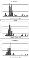
Figure 7. Positional Equivalence of HT Motifs in Phytophthora and P. falciparum
Histograms plotting the lengths of the upstream regions of proteins in the HT secretomes of P. ramorum (i, 147 sequences), P. sojae (ii, 176 sequences), and P. falciparum (iii, 112 sequences). Sequence distribution data from P. infestans is not shown because the complete sequencing of this genome is still under way.

Similar articles

Cited by

References

    1. Christie PJ, Atmakuri K, Krishnamoorthy V, Jakubowski S, Cascales E. Biogenesis, architecture, and function of bacterial type iv secretion systems. Annu Rev Microbiol. 2005;59:451–485. - PMC - PubMed
    1. Lammertyn E, Anne J. Protein secretion in Legionella pneumophila and its relation to virulence. FEMS Microbiol Lett. 2004;238:273–279. - PubMed
    1. Preston GM, Studholme DJ, Caldelari I. Profiling the secretomes of plant pathogenic Proteobacteria. FEMS Microbiol Rev. 2005;29:331–360. - PubMed
    1. Journet L, Hughes KT, Cornelis GR. Type III secretion: A secretory pathway serving both motility and virulence (review) Mol Membr Biol. 2005;22:41–50. - PubMed
    1. Hiller NL, Bhattacharjee S, van Ooij C, Liolios K, Harrison T, et al. A host-targeting signal in virulence proteins reveals a secretome in malarial infection. Science. 2004;306:1934–1937. - PubMed

Publication types

Substances