

Mycobacterium marinum Erp is a virulence determinant required for cell wall integrity and intracellular survival
- PMID: 16714540
- PMCID: PMC1479242
- DOI: 10.1128/IAI.02061-05
Mycobacterium marinum Erp is a virulence determinant required for cell wall integrity and intracellular survival
Abstract
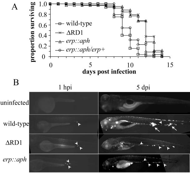
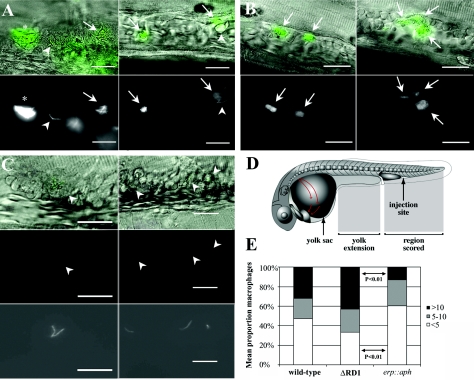
The Mycobacterium tuberculosis exported repetitive protein (Erp) is a virulence determinant required for growth in cultured macrophages and in vivo. To better understand the role of Erp in Mycobacterium pathogenesis, we generated a mutation in the erp homologue of Mycobacterium marinum, a close genetic relative of M. tuberculosis. erp-deficient M. marinum was growth attenuated in cultured macrophage monolayers and during chronic granulomatous infection of leopard frogs, suggesting that Erp function is similarly required for the virulence of both M. tuberculosis and M. marinum. To pinpoint the step in infection at which Erp is required, we utilized a zebrafish embryo infection model that allows M. marinum infections to be visualized in real-time, comparing the erp-deficient strain to a DeltaRD1 mutant whose stage of attenuation was previously characterized in zebrafish embryos. A detailed microscopic examination of infected embryos revealed that bacteria lacking Erp were compromised very early in infection, failing to grow and/or survive upon phagocytosis by host macrophages. In contrast, DeltaRD1 mutant bacteria grow normally in macrophages but fail to induce host macrophage aggregation and subsequent cell-to-cell spread. Consistent with these in vivo findings, erp-deficient but not RD1-deficient bacteria exhibited permeability defects in vitro, which may be responsible for their specific failure to survive in host macrophages.
Figures


Similar articles
-
Mycobacterium marinum infection of adult zebrafish causes caseating granulomatous tuberculosis and is moderated by adaptive immunity.Infect Immun. 2006 Nov;74(11):6108-17. doi: 10.1128/IAI.00887-06. Infect Immun. 2006. PMID: 17057088 Free PMC article.
-
A mycobacterial virulence gene cluster extending RD1 is required for cytolysis, bacterial spreading and ESAT-6 secretion.Mol Microbiol. 2004 Sep;53(6):1677-93. doi: 10.1111/j.1365-2958.2004.04261.x. Mol Microbiol. 2004. PMID: 15341647
-
A mycobacterial phosphoribosyltransferase promotes bacillary survival by inhibiting oxidative stress and autophagy pathways in macrophages and zebrafish.J Biol Chem. 2015 May 22;290(21):13321-43. doi: 10.1074/jbc.M114.598482. Epub 2015 Mar 30. J Biol Chem. 2015. PMID: 25825498 Free PMC article.
-
[Insight into tuberculosis pathogenic mechanism from the zebra fish-Mycobacterium marinum model--a review].Wei Sheng Wu Xue Bao. 2010 Jan;50(1):15-22. Wei Sheng Wu Xue Bao. 2010. PMID: 20344935 Review. Chinese.
-
Comparative pathogenesis of Mycobacterium marinum and Mycobacterium tuberculosis.Cell Microbiol. 2008 May;10(5):1027-39. doi: 10.1111/j.1462-5822.2008.01133.x. Epub 2008 Feb 20. Cell Microbiol. 2008. PMID: 18298637 Review.
Cited by
-
An enzyme that inactivates the inflammatory mediator leukotriene b4 restricts mycobacterial infection.PLoS One. 2013 Jul 11;8(7):e67828. doi: 10.1371/journal.pone.0067828. Print 2013. PLoS One. 2013. PMID: 23874453 Free PMC article.
-
Mycobacterium marinum infection of adult zebrafish causes caseating granulomatous tuberculosis and is moderated by adaptive immunity.Infect Immun. 2006 Nov;74(11):6108-17. doi: 10.1128/IAI.00887-06. Infect Immun. 2006. PMID: 17057088 Free PMC article.
-
Insights into early mycobacterial pathogenesis from the zebrafish.Curr Opin Microbiol. 2008 Jun;11(3):277-83. doi: 10.1016/j.mib.2008.05.013. Epub 2008 Jun 19. Curr Opin Microbiol. 2008. PMID: 18571973 Free PMC article. Review.
-
Insights from the complete genome sequence of Mycobacterium marinum on the evolution of Mycobacterium tuberculosis.Genome Res. 2008 May;18(5):729-41. doi: 10.1101/gr.075069.107. Epub 2008 Apr 10. Genome Res. 2008. PMID: 18403782 Free PMC article.
-
Mycobacterium abscessus cording prevents phagocytosis and promotes abscess formation.Proc Natl Acad Sci U S A. 2014 Mar 11;111(10):E943-52. doi: 10.1073/pnas.1321390111. Epub 2014 Feb 24. Proc Natl Acad Sci U S A. 2014. PMID: 24567393 Free PMC article.
References
-
- Aronson, J. D. 1926. Spontaneous tuberculosis in salt water fish. J. Infect. Dis. 39:315-320.
-
- Banu, S., N. Honore, B. Saint-Joanis, D. Philpott, M. C. Prevost, and S. T. Cole. 2002. Are the PE-PGRS proteins of Mycobacterium tuberculosis variable surface antigens? Mol. Microbiol. 44:9-19. - PubMed
-
- Berthet, F. X., M. Lagranderie, P. Gounon, C. Lauren-Winter, D. Ensergueix, P. Chavarot, F. Thouron, E. Maranghi, V. Pelicic, D. Portnoi, G. Marchal, and B. Gicquel. 1998. Attenuation of virulence by disruption of the Mycobacterium tuberculosis erp gene. Science 282:759-762. - PubMed
-
- Bigi, F., A. Gioffre, L. Klepp, M. P. Santangelo, C. A. Velicovsky, G. H. Giambartolomei, C. A. Fossati, M. I. Romano, T. Mendum, J. J. McFadden, and A. Cataldi. 2005. Mutation in the P36 gene of Mycobacterium bovis provokes attenuation of the bacillus in a mouse model. Tuberculosis 85:221-226. - PubMed
Publication types
MeSH terms
Substances
Grants and funding
LinkOut - more resources
Full Text Sources
Research Materials
