. 2006 Jul;141(3):977-87.
doi: 10.1104/pp.106.076604. Epub 2006 May 19.

AINTEGUMENTA contributes to organ polarity and regulates growth of lateral organs in combination with YABBY genes

Affiliations

AINTEGUMENTA contributes to organ polarity and regulates growth of lateral organs in combination with YABBY genes

Staci Nole-Wilson et al. Plant Physiol. 2006 Jul.

Abstract

Lateral organs in flowering plants display polarity along their adaxial-abaxial axis with distinct cell types forming at different positions along this axis. Members of three classes of transcription factors in Arabidopsis (Arabidopsis thaliana; the Class III homeodomain/leucine zipper [HD-ZIP] proteins, KANADI proteins, and YABBY proteins) are expressed in either the adaxial or abaxial domain of organ primordia where they confer these respective identities. Little is known about the factors that act upstream of these polarity-determining genes to regulate their expression. We have investigated the relationship between AINTEGUMENTA (ANT), a gene that promotes initiation and growth of lateral organ primordia, and polarity genes. Although ant single mutants do not display any obvious defects in organ polarity, loss of ANT activity in combination with mutations in one or more YABBY genes results in polarity defects greater than those observed in the yabby mutants alone. Our results suggest that ANT acts in combination with the YABBY gene FILAMENTOUS FLOWER (FIL) to promote organ polarity by up-regulating the expression of the adaxial-specifying HD-ZIP gene PHABULOSA. Furthermore, we show that ANT acts with FIL to up-regulate expression of the floral homeotic gene APETALA3. Our work defines new roles for ANT in the development of lateral organs.


Figures

Figure 1.
Figure 1.
ANT binds to a sequence within the FIL and YAB3 promoters. A, Alignment of a conserved element within the 5′ regulatory region of FIL and YAB3 to the ANT consensus site. The putative ANT binding site is underlined. Nucleotides shared between the ANT consensus binding site and the FIL and YAB3 promoters are shown in bold. A putative Kruppel binding site (−1,748 to −1,737), required for repression of FIL in the adaxial domain, is boxed. Numbers indicate positions relative to the start codons. B, Gel shift showing binding of ANT to the consensus binding site (BS15), the YAB3 promoter site, and the FIL promoter site. The YAB3 promoter fragment is a 106-bp sequence corresponding to nucleotides −1,561 to −1,456 and the FIL promoter fragment is a 131-bp sequence corresponding to nucleotides −1,763 to −1,633. Lanes 2 and 5 contain no protein. Increasing amounts of ANT protein are shown in lanes 3 and 4 (and 6 and 7). The same amount of ANT protein was used in lanes 1, 4, and 7.
Figure 2.
Figure 2.
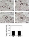
FIL and YAB3 expression in wild-type and ant-4 plants. A, FIL mRNA is present in floral primordia and sepal primordia in this transverse Ler inflorescence section. B, FIL mRNA in an ant-4 inflorescence. C, FIL expression in a stage 8 Ler flower. D, FIL expression in a stage 8 ant-4 flower. Size bars correspond to 50 μm in A to D. Bottom section, Relative expression levels (compared to ACTIN2) of YAB3 in Ler and ant-4 inflorescences. The average of two experiments is shown. The bars show sd.
Figure 3.
Figure 3.
Wild-type, fil-8 yab3-2, fil-8 ant-4, and fil-8 yab3-2 ant-4 leaves. A to D, Mature rosettes just prior to bolting: Ler (A), fil-8 yab3-2 (B), fil-8 ant-4 (C), and fil-8 yab3-2 ant-4 (D). The pictures in A to D are taken at the same magnification. Insets in B and D show closer views. E to H, Fully expanded leaves from Ler (E), fil-8 yab3-2 (F), fil-8 ant-4 (G), and fil-8 yab3-2 ant-4 (H) plants. The pictures in E to H are taken at the same magnification. I to L, Vascular patterns of Ler (I), fil-8 yab3-2 (J), fil-8 ant-4 (K), and fil-8 yab3-2 ant-4 (L) fully expanded leaves. The pictures in I to L are taken at the same magnification. Leaves shown in E to L were from position 5 or 6 of the rosette. Size bars correspond to 1 mm in I to L.
Figure 4.
Figure 4.
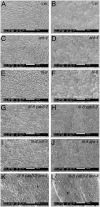
Adaxial and abaxial surfaces of leaves from wild-type, ant-4, fil-8, fil-8 yab3-2, fil-8 ant-4, and fil-8 yab3-2 ant-4 plants. Shown are adaxial leaf surfaces of Ler (A), ant-4 (C), fil-8 (E), fil-8 yab3-2 (G), fil-8 ant-4 (I), and fil-8 yab3-2 ant-4 (K). Also shown are abaxial leaf surfaces of Ler (B), ant-4 (D), fil-8 (F), fil-8 yab3-2 (H), fil-8 ant-4 (J), and fil-8 yab3-2 ant-4 (L). Two cells with similar morphologies in K and L are noted with *. Size bars correspond to 100 μm.
Figure 5.
Figure 5.
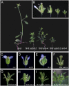
Flowers from wild-type, ant-4, fil-8, fil-8 yab3-2, fil-8 ant-4, and fil-8 yab3-2 ant-4 plants. A, Side view of fil-8, fil-8 yab3-2, fil-8 ant-4, and fil-8 yab3-2 ant-4 plants of approximately 3 weeks of age. B, A single image showing the side views of fil-8, fil-8 yab3-2, fil-8 ant-4, and fil-8 yab3-2 ant-4 flowers (from left to right) and the relative sizes of the flowers. C, Ler flower. D, ant-4 flower. E, fil-8 flower. F, fil-8 yab3-2 flower. G, fil-8 ant-4 flower. H, Early arising fil-8 ant-4 flower. Arrow points to a staminoid organ and arrowhead points to a white petaloid organ. I, fil-8 yab3-2 ant-4 flower. J, fil-8 yab3-2 ant-4 inflorescence meristem that has initiated filament production. Arrow points to the inflorescence meristem.
Figure 6.
Figure 6.
SEM analyses of fil-8 ant-4 flowers. A, fil-8 ant-4 inflorescence meristem producing a mixture of flowers (white arrow) and filamentous structures (black arrow). B, fil-8 ant-4 flower. C, fil-8 ant-4 flower. Stigmatic papillae are indicated with an arrow. D, Stage 7 fil-8 ant-4 flower showing three whorls of organ primordia. E, fil-8 ant-4 flower with two visible whorls of organs. The inner whorl contains three unfused organ primordia. F, Stage 8 fil-8 ant-4 flower with two visible whorls of organs. A black arrow points to a region of fusion between the inner whorl organ primordia. G, An outer whorl organ with sepal-like cells. H, An outer whorl organs with leaf-like (Le) and sepal-like (Se) cells. I, An outer whorl filamentous organ with sepal-like (Se) cells. J, An inner whorl filamentous organ (arrow). K, An inner whorl carpel-like organ with ovary valve-like (Ov) cells at the base and style-like (Sy) cells at the top. L, Close-up of organ shown in K. M, An early arising fil-8 ant-4 flower. A black arrow indicates a petal-like organ. N, Close-up of the petal-like organ in M showing cells with petal morphologies. O, Close-up of the organ in M. Petal cells with adaxial (black arrows) and abaxial (white arrows) morphologies are present on the abaxial surface of this fil-8 ant-4 organ. The adaxial petal epidermal cells are conical in shape with epicuticular thickenings oriented along the cone axis. The abaxial epidermal petal cells are flatter with more zigzagged epicuticular thickenings. P, Staminoid organ (black arrow) present in an early arising fil-8 ant-4 flower. The white arrow points to adaxial carpel tissue. Q, Close-up of the anther-like region of the staminoid organ in P. Size bars correspond to 20 μm in H, L, O, and Q; 50 μm in D to G, I, J, and N; 100 μm in A and K; 200 μm in M and P; and 500 μm in B and C.
Figure 7.
Figure 7.
AP3 and AG expression in wild-type and fil-8 ant-4 flowers. AP3 expression is shown in A to H, and AG expression is shown in I to P. Size bars correspond to 50 μm in A to P. A, AP3 mRNA in a Ler stage 3 flower. B, AP3 mRNA in a stage 3 fil-8 ant-4 flower. C, Arrow points to region of AP3 expression in a stage 3 fil-8 ant-4 flower. D, No AP3 mRNA was detected in this fil-8 ant-4 flower. E, AP3 mRNA is detected in the second and third whorls of stage 5 (left) and stage 8 (right) flowers. F, AP3 mRNA is detected in a stamen-like organ of a fil-8 ant-4 flower. G, AP3 mRNA is present in a few cells located between the outer and inner whorl in older fil-8 ant-4 flowers. H, AP3 mRNA was rarely detected in filaments (arrow) initiated by the inflorescence meristem of fil-8 ant-4 plants. I, AG expression is first detected in a stage 3 flower in wild type. J, AG is expressed in the inflorescence meristem of this fil-8 ant-4 plant. K, AG mRNA is detected in the central part of the floral meristem in a stage 3 Ler flower. L, AG expression in a stage 3 fil-8 ant-4 flower. M, AG expression in the outer organ primordia (arrow) of a stage 4 fil-8 ant-4 flower. N, AG is expressed in the stamens and carpels of a stage 8 Ler flower. O, AG mRNA is detected on the adaxial surface of the central carpel-like organs in a fil-8 ant-4 flower. P, AG mRNA is detected in the central region of filamentous structures (arrow) produced by a fil-8 ant-4 inflorescence meristem.
Figure 8.
Figure 8.
PHB expression in wild-type, fil-8, ant-4, and fil-8 ant-4 flowers. Size bars correspond to 50 μm in A to H. A, PHB expression in a wild-type inflorescence. B, PHB is expressed in the adaxial region of sepal primordia (arrows) and in the floral meristem dome in a stage 4 Ler flower. C, PHB expression in a fil-8 inflorescence. D, PHB expression in a stage 3 fil-8 flower. E, PHB expression in an ant-4 inflorescence. F, PHB expression in a stage 4 ant-4 flower. G, PHB mRNA is present at reduced levels in fil-8 ant-4 inflorescences. H, PHB is expressed in the floral meristem dome but not in the outer whorl organ primordia of this stage 3 fil-8 ant-4 flower. St 2/4, Stage 2 and stage 4 flowers; IM, inflorescence stem.

Similar articles

Cited by

References

    1. Baker SC, Robinson-Beers K, Villanueva JM, Gaiser JC, Gasser CS (1997) Interactions among genes regulating ovule development in Arabidopsis thaliana. Genetics 145: 1109–1124 - PMC - PubMed
    1. Bowman JL (2000) The YABBY gene family and abaxial cell fate. Curr Opin Plant Biol 3: 17–22 - PubMed
    1. Bowman JL, Eshed Y, Baum SF (2002) Establishment of polarity in angiosperm lateral organs. Trends Genet 18: 134–141 - PubMed
    1. Bowman JL, Smyth DR (1999) CRABS CLAW, a gene that regulates carpel and nectary development in Arabidopsis, encodes a novel protein with zinc finger and helix-loop-helix domains. Development 126: 2387–2396 - PubMed
    1. Chen Q, Atkinson A, Otsuga D, Christensen T, Reynolds L, Drews GN (1999) The Arabidopsis FILAMENTOUS FLOWER gene is required for flower formation. Development 126: 2715–2726 - PubMed

Publication types

MeSH terms

LinkOut - more resources