. 2006 Jun 7;25(11):2575-83.
doi: 10.1038/sj.emboj.7601142. Epub 2006 May 18.

Topoisomerase II, not topoisomerase I, is the proficient relaxase of nucleosomal DNA

Affiliations

Topoisomerase II, not topoisomerase I, is the proficient relaxase of nucleosomal DNA

Javier Salceda et al. EMBO J. .

Abstract

Eukaryotic topoisomerases I and II efficiently remove helical tension in naked DNA molecules. However, this activity has not been examined in nucleosomal DNA, their natural substrate. Here, we obtained yeast minichromosomes holding DNA under (+) helical tension, and incubated them with topoisomerases. We show that DNA supercoiling density can rise above +0.04 without displacement of the histones and that the typical nucleosome topology is restored upon DNA relaxation. However, in contrast to what is observed in naked DNA, topoisomerase II relaxes nucleosomal DNA much faster than topoisomerase I. The same effect occurs in cell extracts containing physiological dosages of topoisomeraseI and II. Apparently, the DNA strand-rotation mechanism of topoisomerase I does not efficiently relax chromatin, which imposes barriers for DNA twist diffusion. Conversely, the DNA cross-inversion mechanism of topoisomerase II is facilitated in chromatin, which favor the juxtaposition of DNA segments. We conclude that topoisomerase II is the main modulator of DNA topology in chromatin fibers. The nonessential topoisomerase I then assists DNA relaxation where chromatin structure impairs DNA juxtaposition but allows twist diffusion.


Figures
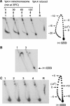
Figure 1
Figure 1
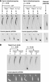
DNA supercoiling density in yeast circular minichromosomes. (A) Lanes1–4, DNA topology in Yp4.4 minichromosomes extracted from JCW28 cells that expressed of E. coli topo I, and were then shifted to 35°C to inactivate topo II for the indicated times (min). Lane R, Yp4.4 plasmid relaxed in vitro at 35°C with topoisomerase I. Two-dimensional electrophoresis of DNA was carried at 25°C in a 0.8% agarose gel in TBE buffer at 50 V for 14 h in the first dimension (top to bottom), and TBE buffer plus 2 μg/ml of chloroquine, at 60 V for 8 h in the second dimension (left to right). The gel-blot was probed for Yp4.4. The scheme depicts gel migrations of Yp4.4 topoisomers, indicating ΔLk values relative to relaxed DNA. (B) DNA samples (lanes 1 and 3 as in A) analyzed like above but in TBE buffer plus 3 μg/ml of chloroquine in the first dimension, and TBE buffer plus 15 μg/ml of chloroquine in the second dimension. (C) DNA samples (all as in A) analyzed by two-dimensional electrophoresis at 4°C in a 0.8% agarose gel in TBE buffer plus magnesium acetate 5 mM at 33 V for 40 h in the first dimension (top to bottom), and TBE buffer alone at 60 V for 4 h in the second dimension (left to right). The scheme depicts gel migrations of Yp4.4 topoisomers, indicating ΔLk values relative to relaxed DNA.
Figure 2
Figure 2
Relaxation of yeast minichromosomes by DNA topoisomerases. (A) Yeast minichromosomes, for which σ∼−0.05 (lane 1) and for which σ∼>+0.04 (lane 5) were incubated at 30°C for 30 min with catalytic amounts of vaccinia virus topo I (lanes 2 and 6), S. cerevisiae topo II (lanes 3 and 7) or no enzyme (7 and 8). Plasmids Yp4.4 and pHC624 were also relaxed in vitro (lane R). Two-dimensional electrophoresis of DNA was carried out at 25°C in a 0.8% agarose gel in TBE buffer plus 0.6 μg/ml of chloroquine at 50 V for 14 h in the first dimension (top to bottom), and TBE buffer plus 3 μg/ml of chloroquine, at 60 V for 8 h in the second dimension (left to right). Gel-blots were probed for Yp4.4 (upper panel) and for the control plasmid pHC624 (lower panel). (B) Yeast minichromosomes, for which σ∼−0.05 (lane 1) and for which σ>+0.04 (lane 4) were supplemented with an excess of control plasmid pHC624 (1 mg/ml), and incubated with catalytic amounts of S. cerevisiae topo I (lanes 2 and 5) or S. cerevisiae topo II (lanes 3 and 6) at 30°C for 30 min. After two-dimensional electrophoresis of DNA, gels were blotted and probed for the yeast 2-μm circle (upper panel) or ethidium stained to visualize pHC624 (lower panel).
Figure 3
Figure 3
Chromatin structure upon relaxation of yeast minichromosomes. Preferential DNA cleavage sites by micrococcal nuclease (MN) digestion along the TRP1-ARS1 region of Yp4.4 were mapped on the following substrates: Yp4.4 plasmid (A), Yp4.4 minichromosome for which σ∼−0.05 (B), Yp4.4 minichromosomes for which σ>+0.04 (C), and after their relaxation by S. cerevisiae topo I (D) or S. cerevisiae topo II (E). Digested DNA samples were restricted with endonuclease EcoRI, separated on a 1% agarose gel, blotted and probed with the radio-labeled 186 bp EcoRI–XbaI TRP1 fragment (p). Lane 1, XbaI and HindIII site markers. Arrowheads denote main DNA cleavage sites visible in (B, C, D, E). Asterisks denote additional DNA cleavage sites visible in (C). The scheme depicts nucleosome positions determined by Thoma et al (1984) along the TRP1-ARS1 yeast DNA segment.
Figure 4
Figure 4
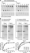
Relaxation kinetics of supercoiled chromatin. (A) Positively supercoiled Yp4.4 minichromosomes and their accompanying control plasmid pHC624 were incubated with catalytic amounts of topoisomerase I or topoisomerase II (as indicated). Reactions were quenched at the indicated periods (min). Following DNA electrophoresis, the gel-blots were probed for Yp4.4 (up) and for pHC624 (down). Note that topoisomerase I and II activities were adjusted to relax the control plasmid at comparable rates. (B) Mixtures containing (+) supercoiled Yp4.4 plasmid plus (−) supercoiled pHC624 plasmid, or containing (+) supercoiled Yp4.4 minichromosome plus (−) supercoiled pHC624 plasmid were relaxed with catalytic amounts of topoisomerase I or topoisomerase II (as indicated). Reactions were quenched at the indicated periods (min). DNA electrophoresis was carried at 25°C in a 0.9% agarose gel in TBE buffer at 40 V for 14 h. Gel-blots were probed for Yp4.4 plus pHC624. DNA relaxation rates, by topoisomerases I and II, for supercoiled minichromosomes (CHR S(+)) and supercoiled plasmids (DNA S(−), DNA S(+)) were determined by measuring the gain of relaxed topoisomers excluding nicked ones. Graphs represent the average of four experiments with error bars indicating s.d.'s from the mean.
Figure 5
Figure 5
Relative relaxase activities of intracellular topoisomerases I and II. (A) Positively supercoiled Yp4.4 minichromosomes (CHR) and their accompanying control plasmid pHC624 (DNA) were supplemented with extracts from Δtop1 TOP2 or TOP1 TOP2 yeast cells, as indicated. Incubations proceeded at 30°C in the absence or presence of ATP, and were quenched at the indicated time periods (min). Two-dimensional electrophoresis of DNA was as in Figure 2A and the gel blots were probed for Yp4.4 and pHC624. (B) Right, rates of chromatin and DNA relaxation, in the presence and the absence of ATP, determined by measuring the gain of relaxed topoisomers excluding nicked ones. Left, relative specific activities of topo I (ATP-independent rate) and topo II (ATP- dependent minus ATP-independent rate). Error bars denote the s.d. from the mean of five different experiments.
Figure 6
Figure 6
Plausible chromatin conformations that accommodate positive helical tension of DNA. The topology of DNA in the canon nucleosome and in models a, b, and c is explained in the text. To calculate approximate σ values supported in each model, the ratio of the change in writhe to the change in twist was fixed at ∼2.6:1 in histone-free DNA regions (Boles et al, 1990). For σ over +0.03, the twist regime tends to saturate and deformation occurs mainly by the writhe regime (Koster et al, 2005). We then took ΔTw values no higher than 1-U/200 bp and ΔWr values by averaging (−) and (+) crossings in several planar projections of a given conformation. For simplicity, we depicted histone octamers as spheres, although partial unfolding of octamers may occur in models b and c.
Figure 7
Figure 7
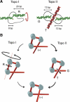
Comparative mechanics of topoisomerases I and II in relaxing nucleosomal DNA. (A) DNA segment lengths required for the function of eukaryotic topoisomerases are represented according to structural and biochemical data. Topo I clamps around 8 bp of DNA up to the cleavage site. Then, about 20 bp of duplex (R) should be free to rotate without colliding with the protruding domains of the enzyme (Stewart et al, 1998). Topo II interacts with the gated duplex (G) along 26 bp of DNA. The transported duplex (T) must cross the 50-Å wide dimer interface of the enzyme (Fass et al, 1999). (B) In nucleosomal DNA, bending of the duplex slows twist diffusion, and rotation of entire nucleosomes implies high viscous friction. Consequently, driving torque is small and a duplex cleaved by topo I comes up against kinetic limitations to complete axial rotations. Conversely, chromatin folding may favor the juxtaposition of DNA segments. Then, the cross-inversion mechanism of topo II involves a short translocation of the T-segment across the G-segment.

Similar articles

Cited by

References

    1. Adachi Y, Luke M, Laemmli UK (1991) Chromosome assembly in vitro: topoisomerase II is required for condensation. Cell 64: 137–148 - PubMed
    1. Almouzni G, Mechali M (1988) Assembly of spaced chromatin involvement of ATP and DNA topoisomerase activity. EMBO J 7: 4355–4365 - PMC - PubMed
    1. Bednar J, Furrer P, Stasiak A, Dubochet J, Egelman EH, Bates AD (1994) The twist, writhe and overall shape of supercoiled DNA change during counterion-induced transition from a loosely to a tightly interwound superhelix. J Mol Biol 235: 825–847 - PubMed
    1. Bjornsti MA, Fertala J (1999) Overexpression and purification of DNA topoisomerase I from yeast. Methods Mol Biol 94: 179–186 - PubMed
    1. Boles TC, White JH, Cozzarelli NR (1990) Structure of plectonemically supercoiled DNA. J Mol Biol 213: 931–951 - PubMed

Publication types

MeSH terms