Identification of a tumour suppressor network opposing nuclear Akt function
- PMID: 16680151
- PMCID: PMC1976603
- DOI: 10.1038/nature04809
Identification of a tumour suppressor network opposing nuclear Akt function
Abstract
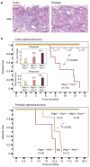
The proto-oncogene AKT (also known as PKB) is activated in many human cancers, mostly owing to loss of the PTEN tumour suppressor. In such tumours, AKT becomes enriched at cell membranes where it is activated by phosphorylation. Yet many targets inhibited by phosphorylated AKT (for example, the FOXO transcription factors) are nuclear; it has remained unclear how relevant nuclear phosphorylated AKT (pAKT) function is for tumorigenesis. Here we show that the PMLtumour suppressor prevents cancer by inactivating pAKT inside the nucleus. We find in a mouse model that Pml loss markedly accelerates tumour onset, incidence and progression in Pten-heterozygous mutants, and leads to female sterility with features that recapitulate the phenotype of Foxo3a knockout mice. We show that Pml deficiency on its own leads to tumorigenesis in the prostate, a tissue that is exquisitely sensitive to pAkt levels, and demonstrate that Pml specifically recruits the Akt phosphatase PP2a as well as pAkt into Pml nuclear bodies. Notably, we find that Pml-null cells are impaired in PP2a phosphatase activity towards Akt, and thus accumulate nuclear pAkt. As a consequence, the progressive reduction in Pml dose leads to inactivation of Foxo3a-mediated transcription of proapoptotic Bim and the cell cycle inhibitor p27(kip1). Our results demonstrate that Pml orchestrates a nuclear tumour suppressor network for inactivation of nuclear pAkt, and thus highlight the importance of AKT compartmentalization in human cancer pathogenesis and treatment.
Figures


Similar articles
-
Activation of protein kinase CK2 attenuates FOXO3a functioning in a PML-dependent manner: implications in human prostate cancer.Cell Death Dis. 2013 Mar 14;4(3):e543. doi: 10.1038/cddis.2013.63. Cell Death Dis. 2013. PMID: 23492774 Free PMC article.
-
The deubiquitinylation and localization of PTEN are regulated by a HAUSP-PML network.Nature. 2008 Oct 9;455(7214):813-7. doi: 10.1038/nature07290. Epub 2008 Aug 20. Nature. 2008. PMID: 18716620 Free PMC article.
-
PML inhibits HIF-1alpha translation and neoangiogenesis through repression of mTOR.Nature. 2006 Aug 17;442(7104):779-85. doi: 10.1038/nature05029. Nature. 2006. PMID: 16915281
-
PML: a tumor suppressor that regulates cell fate in mammary gland.Cell Cycle. 2009 Sep 1;8(17):2711-7. doi: 10.4161/cc.8.17.9462. Epub 2009 Sep 8. Cell Cycle. 2009. PMID: 19652541 Review.
-
Pondering the puzzle of PML (promyelocytic leukemia) nuclear bodies: can we fit the pieces together using an RNA regulon?Biochim Biophys Acta. 2008 Nov;1783(11):2145-54. doi: 10.1016/j.bbamcr.2008.06.005. Epub 2008 Jun 18. Biochim Biophys Acta. 2008. PMID: 18616965 Free PMC article. Review.
Cited by
-
Polymorphisms in microRNA target sites of forkhead box O genes are associated with hepatocellular carcinoma.PLoS One. 2015 Mar 4;10(3):e0119210. doi: 10.1371/journal.pone.0119210. eCollection 2015. PLoS One. 2015. PMID: 25739100 Free PMC article.
-
PML Surfs into HIPPO Tumor Suppressor Pathway.Front Oncol. 2013 Mar 1;3:36. doi: 10.3389/fonc.2013.00036. eCollection 2013. Front Oncol. 2013. PMID: 23459691 Free PMC article.
-
Regulation of NF-E2-related factor 2 signaling for cancer chemoprevention: antioxidant coupled with antiinflammatory.Antioxid Redox Signal. 2010 Dec 1;13(11):1679-98. doi: 10.1089/ars.2010.3276. Epub 2010 Aug 17. Antioxid Redox Signal. 2010. PMID: 20486765 Free PMC article. Review.
-
Regional hippocampal differences in AKT survival signaling across the lifespan: implications for CA1 vulnerability with aging.Cell Death Differ. 2009 Mar;16(3):439-48. doi: 10.1038/cdd.2008.171. Epub 2008 Nov 28. Cell Death Differ. 2009. PMID: 19039330 Free PMC article.
-
Posttranslational regulation of Akt in human cancer.Cell Biosci. 2014 Oct 1;4(1):59. doi: 10.1186/2045-3701-4-59. eCollection 2014. Cell Biosci. 2014. PMID: 25309720 Free PMC article. Review.
References
-
- Luo J, Manning BD, Cantley LC. Targeting the PI3K–Akt pathway in human cancer: rationale and promise. Cancer Cell. 2003;4:257–262. - PubMed
-
- Brenkman AB, Burgering BM. FoxO3a eggs on fertility and aging. Trends Mol Med. 2003;9:464–467. - PubMed
-
- Salomoni P, Pandolfi PP. The role of PML in tumor suppression. Cell. 2002;108:165–170. - PubMed
-
- Gurrieri C, et al. Loss of the tumour suppressor PML in human cancers of multiple histologic origins. J Natl Cancer Inst. 2004;96:269–279. - PubMed
-
- Goel A, et al. Frequent inactivation of PTEN by promoter hypermethylation in microsatellite instability-high sporadic colorectal cancers. Cancer Res. 2004;64:3014–3021. - PubMed
MeSH terms
Substances
Grants and funding
LinkOut - more resources
Full Text Sources
Other Literature Sources
Molecular Biology Databases
Research Materials
Miscellaneous

