Comparative Study
. 2006 May 3;26(18):4870-9.
doi: 10.1523/JNEUROSCI.4527-05.2006.

Regulation of nuclear factor kappaB in the hippocampus by group I metabotropic glutamate receptors

Affiliations
Comparative Study

Regulation of nuclear factor kappaB in the hippocampus by group I metabotropic glutamate receptors

Kenneth J O'Riordan et al. J Neurosci. .

Abstract

An increasing amount of evidence suggests that the family of nuclear factor kappaB (NF-kappaB) transcription factors plays an important role in synaptic plasticity and long-term memory formation. The present study investigated the regulation of NF-kappaB family members p50, p65/RelA, and c-Rel in the hippocampus in response to metabotropic glutamate receptor (mGluR) signaling. Activation of group I metabotropic glutamate receptors (GpI-mGluRs) with the agonist (S)-3,5-dihydroxyphenylglycine (DHPG) resulted in a time-dependent increase in DNA binding activity of p50, p65, and c-Rel in area CA1 of the hippocampus. An antagonist of mGluR5, 2-Methyl-6-(phenylethynyl)pyridine, inhibited the DHPG-induced activation of NF-kappaB, whereas an antagonist of mGluR1, (S)-(+)-alpha-amino-4-carboxy-2-methylbenzeneacetic acid, did not. Using a series of inhibitors, we investigated the signaling pathways necessary for DHPG-induced activation of NF-kappaB and found that they included the phosphatidyl inositol 3-kinase, protein kinase C, mitogen-activated protein kinase kinase, and p38-mitogen-activated protein kinase pathways. To determine the functional significance of mGluR-induced regulation of NF-kappaB, we measured long-term depression (LTD) of Schaffer-collateral synapses in the hippocampus of c-Rel knock-out mice. Early phase LTD was normal in c-rel(-/-) mice. However, late-phase LTD (>90 min) was impaired in c-rel(-/-) mice. The observations of this deficit in hippocampal synaptic plasticity prompted us to further investigate long-term memory formation in c-rel(-/-) mice. c-rel(-/-) mice exhibited impaired performance in a long-term passive avoidance task, providing additional evidence for c-Rel in long-term memory formation. These results demonstrate that the NF-kappaB transcription factor family is regulated by GpI-mGluRs in the hippocampus and that the c-Rel transcription factor is necessary for long-term maintenance of LTD and formation of long-term memory.


Figures
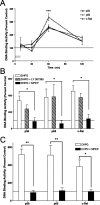
Figure 1.
Figure 1.
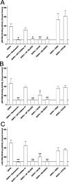
mGluR5 can activate NF-κB in area CA1 of the hippocampus. Nuclear DNA binding activity of NF-κB subunits p50, p65, and c-Rel was measured in area CA1 of transverse hippocampal slices (400 μm) in response to activation of GpI-mGluRs. A, Hippocampal slices were treated with DHPG (50 μm; 10 min), and DNA binding activity was measured in area CA1 of the hippocampus at various times after treatment. DHPG induced significant, time-dependent increases in nuclear DNA binding activity for p50 (F(3,13) = 34; p < 0.0001), p65 (F(3,13) = 11; p < 0.002), and c-Rel (F(3,13) = 16; p < 0.0005). The asterisk indicates significant difference (*p < 0.01) relative to the first time point as determined by Dunnett’s post hoc test. B, Slices were pretreated with either an mGluR1 antagonist (LY 367385, 30 μm) or an mGluR5 antagonist (MPEP, 10 μm) before treatment with DHPG (50 μm; 10 min). Nuclear DNA binding activity was measured 50 min after the end of DHPG treatment. LY 367385 had no effect on DHPG-induced increases in NF-κB DNA binding activity. MPEP significantly decreased DHPG-induced activation of NF-κB (F(4,53) = 11; p < 0.0001). The asterisk indicates significant difference (*p < 0.05) relative to DHPG-treated slices as determined by post hoc Bonferroni comparisons. C, To further confirm a role for mGluR5 in activation of NF-κB, slices were treated with the mGluR5 agonist CHPG (1 mm; 10 min). Significant activation of p50, p65, and c-Rel were observed 50 min after the end of treatment (F(2,23) = 38; p < 0.001). Pretreatment with MPEP (10 μm) blocked CHPG-induced increases in p50 (p < 0.01), p65 (p < 0.01), and c-Rel (p < 0.05). Asterisks indicate significant differences as determined by post hoc Bonferroni comparisons. For all panels, error bars indicate SEM. Percentage change was calculated based on DNA binding activity measured from matched control slices treated with vehicle.
Figure 2.
Figure 2.
GpI-mGluR-mediated signaling activates PDK1, AKT, Raf-1, ERK2, and p38-MAPK. The signaling pathways that were activated by DHPG in area CA1 of the hippocampus were determined by Western blotting using phospho-specific antibodies. Equal loading of protein was confirmed by blotting for total protein and actin (data not shown). A, Levels of phospho-PDK1, AKT, Raf-1, ERK2, and p38-MAPK were significantly increased (p < 0.05) immediately after treatment with DHPG (50 μm; 10 min). Representative Western blots are shown above summary densitometry. C indicates samples from matched control slices receiving vehicle; E indicates slices receiving DHPG. Arrows indicate a band specific for a particular signaling molecule. Each bar represents the average of 11 replicates. B, No significant differences in levels of phospho-PDK1, AKT, Raf-1, ERK2, or p38-MAPK were observed 50 min after DHPG treatment. Each bar represents the average of nine replicates. C, DHPG-induced activation of ERK2, but not p38 MAPK, was inhibited by pretreatment with the PKC inhibitor GF 109,203× (1 μm; F(1,8) = 11; p < 0.05). Each bar represents the average of four to six replicates. D, DHPG-induced activation of ERK2, but not p38 MAPK, was inhibited by pretreatment with the PI3-K inhibitor Wortmannin (50 nm; F(1,8) = 7.8; p < 0.05). Each bar represents the average of four to six replicates. In A and B, asterisks indicate significant difference from vehicle controls as determined by one-sample t test. In C and D, asterisks indicate significant difference between groups as determined by two-way ANOVA with repeated measures. Error bars indicate SEM.
Figure 3.
Figure 3.
PI3-K, PKC, MEK, and p38-MAPK are necessary for DHPG-induced activation of NF-κB. To determine which signaling pathways were necessary for GpI-mGluR-dependent activation of NF-κB, slices were pretreated with various inhibitors of signaling pathways before treatment with DHPG. The panels represent summary data for p50 (F(7,43) = 9; p < 0.0001) (A), p65 (F(7,43) = 6; p < 0.0001) (B), or c-Rel (F(7,43) = 7; p < 0.0001) (C). KN-93 (CaMKII; 10 μm), KT5720 (PKA, 1 μm), and AKT inhibitor IV (AKT, 3 μm) had no significant effect on DHPG-induced activation of NF-κB. Wortmannin (PI3-K, 50 nm), GF 109,203× (PKC, 1 μm), U0126 (MEK, 20 μm), and SB 203580 (p38-MAPK, 1 μm) significantly inhibited DHPG-induced activation of NF-κB. In each panel, bars represent the mean of four to eight replicates. Error bars are SEM. Asterisks indicate significant difference from DHPG alone (*p < 0.05; **p < 0.01) as determined by Dunnett’s multiple comparison test.
Figure 4.
Figure 4.
c-rel−/− mice have normal levels of p50 and p65 mRNA and protein. Area CA1 and the dentate gyrus of c-rel+/+, c-rel+/−, and c-rel−/− animals was isolated from each hemisphere and processed for RNA or protein extraction. A, No significant differences in expression of p50 or p65 mRNA were observed using real-time reverse transcription-QPCR in area CA1 (p50 and p65 c-rel+/+ vs c-rel−/−; p > 0.05 as determined by Bonferroni posttests). Expression of c-Rel mRNA in c-rel+/− animals was ∼50% of c-rel+/+ animals and was significantly decreased in c-rel−/− animals (F(2,14) = 4; p < 0.05). B, No significant differences in expression of p50 or p65 mRNA were observed using real-time reverse transcription-QPCR in the dentate gyrus (p50 and p65 c-rel+/+ vs c-rel−/−, p > 0.05 as determined by Bonferroni posttests). Expression of c-Rel mRNA in c-rel+/− animals was ∼50% of c-rel+/+ animals and was significantly decreased in c-rel−/− animals (F(2,14) = 8; p < 0.001). C, Analysis of tissue derived from the same animals used in A revealed a significant difference in expression of c-Rel between c-rel+/+ and c-rel−/− animals (F(2,64) = 5; p < 0.05). No significant differences were observed in expression of p50 or p65 protein (p > 0.05; post hoc Bonferroni analysis). D, Analysis of tissue derived from the same animals used in B revealed a significant difference in expression of c-Rel between c-rel+/+ and c-rel−/− animals (F(2,58) = 4; p < 0.05). No significant differences were observed in expression of p50 or p65 protein (p > 0.05; post hoc Bonferroni analysis). In C and D, representative Western blots are shown above summary densitometry. Arrows next to blots indicate immunoreactive band specific for p50, p65, or c-Rel, and c-Rel genotype is indicated by either +/+ (wild type), +/− (heterozygous), or −/− (null). In all panels, error bars indicate SEM. The asterisk indicates significant difference (*p < 0.05) from c-rel+/+ as determined by post hoc Bonferroni comparisons.
Figure 5.
Figure 5.
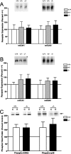
Expression of GpI-mGluRs and induction of MAPK is normal in c-rel−/− animals. Levels of GpI-mGluRs and activation of MAPK was measured in the hippocampus from c-rel−/− animals. A, No significant differences in expression of mGluR1 or mGluR5 were observed in area CA1 (F(2,40) = 0.3; p < 0.8). B, No significant differences in expression of mGluR1 or mGluR5 were observed in the dentate gyrus (F(2,38) = 0.8; p < 0.5). C, No significant differences in DHPG-induced activation of ERK2 or p38 MAPK were observed in area CA1 (F(1,16) = 0.003; p < 1.0). In all panels, representative Western blots are shown above summary densitometry. Genotype and experimental treatment (C, control; E, DHPG) are indicated above blots. Error bars indicate SEM.
Figure 6.
Figure 6.
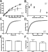
Loss of c-Rel on hippocampal synaptic transmission. Synaptic transmission was assessed at Schaffer-collateral synapses of c-rel+/+ and c-rel−/− littermate animals. A, Synaptic transmission as determined by the ratio of fEPSP slope (postsynaptic depolarization) versus fiber volley slope (presynaptic depolarization). Animals lacking c-Rel had significantly reduced synaptic transmission relative to wild-type littermates (F(2,28) = 34; p < 0.0001). B, No significant differences were seen in paired-pulse facilitation measured at several interstimulus intervals (20, 50, 100, 200, 300 ms) between c-rel+/+ and c-rel−/− littermates (F(1,62) = 0.3; p < 0.7). C, Minianalysis of Schaffer-collateral synapses revealed no significant difference in spontaneous event amplitude (D = 0.02; n = 560; p < 1.0). D, Minianalysis of Schaffer-collateral synapses revealed no significant difference in spontaneous interevent interval (D = 0.05; n = 719; p < 0.1). E, No significant differences were observed in resting membrane potential of pyramidal neurons in area CA1 of the hippocampus (t = 1.5; df = 16; p < 0.2). F, No significant differences were observed in input resistance of pyramidal neurons in area CA1 of the hippocampus (t = 0.7; df = 16; p < 0.6). In all panels, error bars indicate SEM.
Figure 7.
Figure 7.
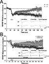
Impaired hippocampal long-term depression in c-rel−/− animals. mGluR-LTD was induced at Schaffer-collateral synapses by treatment with DHPG (50 μm, 10 min; gray bar). A, Acute synaptic depression was identical between slices from c-rel+/+ and c-rel−/− littermates. LTD was similar between c-rel+/+ and c-rel−/− animals for ∼90 min, when synaptic depression in slices from c-rel−/− animals decayed to basal levels of synaptic efficacy (F(1,89) = 340; p < 0.0001). Representative traces 2 min before, 10 min after, and 150 min after DHPG treatment are shown below summary electrophysiology. Calibration: 1 mV, 5 ms. B, Pretreatment of slices with the transcription inhibitor DRB (90 μm; 20 min) attenuated induction of mGluR-LTD (F(1,19) = 214; p < 0.001). In all panels, error bars indicate SEM.
Figure 8.
Figure 8.
Impaired performance in a passive avoidance task in c-rel−/− animals. Passive avoidance was assessed in a two-chamber shuttle box. During the training day, c-rel+/+ and c-rel−/− animals performed equally, moving into the dark side of the chamber within 15 s of placement into the shuttle box. When tested 24 h after training, c-rel−/− animals exhibited a significantly lower latency to enter the dark side compared with c-rel+/+ littermates (U = 159; p < 0.05; n+/+ = 15; n−/− = 36), indicating reduced capacity for formation of long-term passive avoidance memory.

Similar articles

Cited by

References

    1. Albensi BC, Mattson MP (2000). Evidence for the involvement of TNF and NF-kappaB in hippocampal synaptic plasticity. Synapse 35:151–159. - PubMed
    1. Ambrogi Lorenzini CG, Baldi E, Bucherelli C, Sacchetti B, Tassoni G (1997). Analysis of mnemonic processing by means of totally reversible neural inactivations. Brain Res Brain Res Protoc 1:391–398. - PubMed
    1. Baeuerle PA, Henkel T (1994). Function and activation of NF-kappa B in the immune system. Annu Rev Immunol 12:141–179. - PubMed
    1. Balschun D, Manahan-Vaughan D, Wagner T, Behnisch T, Reymann KG, Wetzel W (1999). A specific role for group I mGluRs in hippocampal LTP and hippocampus-dependent spatial learning. Learn Mem 6:138–152. - PMC - PubMed
    1. Bolshakov VY, Siegelbaum SA (1994). Postsynaptic induction and presynaptic expression of hippocampal long-term depression. Science 264:1148–1152. - PubMed

Publication types

MeSH terms