Epidermal growth factor receptor variant III mutations in lung tumorigenesis and sensitivity to tyrosine kinase inhibitors
- PMID: 16672372
- PMCID: PMC1456806
- DOI: 10.1073/pnas.0510284103
Epidermal growth factor receptor variant III mutations in lung tumorigenesis and sensitivity to tyrosine kinase inhibitors
Abstract
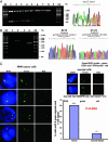
The tyrosine kinase inhibitors gefitinib (Iressa) and erlotinib (Tarceva) have shown anti-tumor activity in the treatment of non-small cell lung cancer (NSCLC). Dramatic and durable responses have occurred in NSCLC tumors with mutations in the tyrosine kinase domain of the epidermal growth factor receptor (EGFR). In contrast, these inhibitors have shown limited efficacy in glioblastoma, where a distinct EGFR mutation, the variant III (vIII) in-frame deletion of exons 2-7, is commonly found. In this study, we determined that EGFRvIII mutation was present in 5% (3/56) of analyzed human lung squamous cell carcinoma (SCC) but was not present in human lung adenocarcinoma (0/123). We analyzed the role of the EGFRvIII mutation in lung tumorigenesis and its response to tyrosine kinase inhibition. Tissue-specific expression of EGFRvIII in the murine lung led to the development of NSCLC. Most importantly, these lung tumors depend on EGFRvIII expression for maintenance. Treatment with an irreversible EGFR inhibitor, HKI-272, dramatically reduced the size of these EGFRvIII-driven murine tumors in 1 week. Similarly, Ba/F3 cells transformed with the EGFRvIII mutant were relatively resistant to gefitinib and erlotinib in vitro but proved sensitive to HKI-272. These findings suggest a therapeutic strategy for cancers harboring the EGFRvIII mutation.
Conflict of interest statement
Conflict of interest statement: No conflicts declared.
Figures


Similar articles
-
Irreversible inhibitors of the EGF receptor may circumvent acquired resistance to gefitinib.Proc Natl Acad Sci U S A. 2005 May 24;102(21):7665-70. doi: 10.1073/pnas.0502860102. Epub 2005 May 16. Proc Natl Acad Sci U S A. 2005. PMID: 15897464 Free PMC article.
-
The T790M "gatekeeper" mutation in EGFR mediates resistance to low concentrations of an irreversible EGFR inhibitor.Mol Cancer Ther. 2008 Apr;7(4):874-9. doi: 10.1158/1535-7163.MCT-07-2387. Mol Cancer Ther. 2008. PMID: 18413800
-
Clinical impact of switching to a second EGFR-TKI after a severe AE related to a first EGFR-TKI in EGFR-mutated NSCLC.Jpn J Clin Oncol. 2012 Jun;42(6):528-33. doi: 10.1093/jjco/hys042. Epub 2012 Mar 28. Jpn J Clin Oncol. 2012. PMID: 22457323
-
First-line treatment of advanced epidermal growth factor receptor (EGFR) mutation positive non-squamous non-small cell lung cancer.Cochrane Database Syst Rev. 2016 May 25;(5):CD010383. doi: 10.1002/14651858.CD010383.pub2. Cochrane Database Syst Rev. 2016. Update in: Cochrane Database Syst Rev. 2021 Mar 18;3:CD010383. doi: 10.1002/14651858.CD010383.pub3 PMID: 27223332 Updated. Review.
-
Chasing targets for EGFR tyrosine kinase inhibitors in non-small-cell lung cancer: Asian perspectives.Expert Rev Mol Diagn. 2007 Nov;7(6):821-36. doi: 10.1586/14737159.7.6.821. Expert Rev Mol Diagn. 2007. PMID: 18020911 Review.
Cited by
-
Specific and sensitive hydrolysis probe-based real-time PCR detection of epidermal growth factor receptor variant III in oral squamous cell carcinoma.PLoS One. 2012;7(2):e31723. doi: 10.1371/journal.pone.0031723. Epub 2012 Feb 16. PLoS One. 2012. PMID: 22359620 Free PMC article.
-
Epidermal growth factor receptor in non-small cell lung cancer.Transl Lung Cancer Res. 2015 Apr;4(2):110-8. doi: 10.3978/j.issn.2218-6751.2015.01.01. Transl Lung Cancer Res. 2015. PMID: 25870793 Free PMC article. Review.
-
Epidermal growth factor receptor variant III mutation in Chinese patients with squamous cell cancer of the lung.Thorac Cancer. 2015 May;6(3):319-26. doi: 10.1111/1759-7714.12204. Epub 2015 Jan 15. Thorac Cancer. 2015. PMID: 26273378 Free PMC article.
-
Pan-cancer molecular analysis of EGFR large fragment deletion in the Asian population.Cancer Med. 2023 Apr;12(7):8083-8088. doi: 10.1002/cam4.5603. Epub 2023 Jan 9. Cancer Med. 2023. PMID: 36622089 Free PMC article.
-
Targeting EGFR resistance networks in head and neck cancer.Cell Signal. 2009 Aug;21(8):1255-68. doi: 10.1016/j.cellsig.2009.02.021. Epub 2009 Mar 1. Cell Signal. 2009. PMID: 19258037 Free PMC article. Review.
References
-
- Hynes N. E., Lane H. A. Nat. Rev. Cancer. 2005;5:341–354. - PubMed
-
- Arteaga C. L. Exp. Cell Res. 2003;284:122–130. - PubMed
-
- Mendelsohn J., Baselga J. Oncogene. 2000;19:6550–6565. - PubMed
-
- Paez J. G., Janne P. A., Lee J. C., Tracy S., Greulich H., Gabriel S., Herman P., Kaye F. J., Lindeman N., Boggon T. J., et al. Science. 2004;304:497–500. - PubMed
-
- Lynch T. J., Bell D. W., Sordella R., Gurubhagavatula S., Okimoto R. A., Brannigan B. W., Harris P. L., Haserlat S. M., Supko J. G., Haluska F. G., et al. N. Engl. J. Med. 2004;350:2129–2139. - PubMed
Publication types
MeSH terms
Substances
Grants and funding
LinkOut - more resources
Full Text Sources
Other Literature Sources
Medical
Molecular Biology Databases
Research Materials
Miscellaneous

