Generation of measles virus with a segmented RNA genome
- PMID: 16611883
- PMCID: PMC1472037
- DOI: 10.1128/JVI.80.9.4242-4248.2006
Generation of measles virus with a segmented RNA genome
Abstract
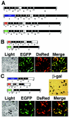
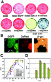
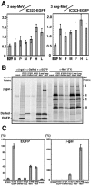
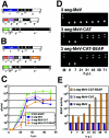
Viruses classified in the order Mononegavirales have a single nonsegmented RNA molecule as the genome and employ similar strategies for genome replication and gene expression. Infectious particles of Measles virus (MeV), a member of the family Paramyxoviridae in the order Mononegavirales, with two or three RNA genome segments (2 seg- or 3 seg-MeV) were generated using a highly efficient reverse genetics system. All RNA segments of the viruses were designed to have authentic 3' and 5' self-complementary termini, similar to those of negative-stranded RNA viruses that intrinsically have multiple RNA genome segments. The 2 seg- and 3 seg-MeV were viable and replicated well in cultured cells. 3 seg-MeV could accommodate up to six additional transcriptional units, five of which were shown to be capable of expressing foreign proteins efficiently. These data indicate that the MeV genome can be segmented, providing an experimental insight into the divergence of the negative-stranded RNA viruses with nonsegmented or segmented RNA genomes. They also illustrate a new strategy to develop mononegavirus-derived vectors harboring multiple additional transcriptional units.
Figures


Similar articles
-
Long untranslated regions of the measles virus M and F genes control virus replication and cytopathogenicity.J Virol. 2005 Nov;79(22):14346-54. doi: 10.1128/JVI.79.22.14346-14354.2005. J Virol. 2005. PMID: 16254369 Free PMC article.
-
The C Protein Is Recruited to Measles Virus Ribonucleocapsids by the Phosphoprotein.J Virol. 2020 Jan 31;94(4):e01733-19. doi: 10.1128/JVI.01733-19. Print 2020 Jan 31. J Virol. 2020. PMID: 31748390 Free PMC article.
-
Polyploid measles virus with hexameric genome length.EMBO J. 2002 May 15;21(10):2364-72. doi: 10.1093/emboj/21.10.2364. EMBO J. 2002. PMID: 12006489 Free PMC article.
-
Transcription and replication of nonsegmented negative-strand RNA viruses.Curr Top Microbiol Immunol. 2004;283:61-119. doi: 10.1007/978-3-662-06099-5_3. Curr Top Microbiol Immunol. 2004. PMID: 15298168 Review.
-
Sendai Virus and a Unified Model of Mononegavirus RNA Synthesis.Viruses. 2021 Dec 9;13(12):2466. doi: 10.3390/v13122466. Viruses. 2021. PMID: 34960735 Free PMC article. Review.
Cited by
-
Morbillivirus infections: an introduction.Viruses. 2015 Feb 12;7(2):699-706. doi: 10.3390/v7020699. Viruses. 2015. PMID: 25685949 Free PMC article. Review.
-
Non-transmissible MV Vector with Segmented RNA Genome Establishes Different Types of iPSCs from Hematopoietic Cells.Mol Ther. 2020 Jan 8;28(1):129-141. doi: 10.1016/j.ymthe.2019.09.007. Epub 2019 Sep 12. Mol Ther. 2020. PMID: 31677955 Free PMC article.
-
Measles virus infection of SLAM (CD150) knockin mice reproduces tropism and immunosuppression in human infection.J Virol. 2007 Feb;81(4):1650-9. doi: 10.1128/JVI.02134-06. Epub 2006 Nov 29. J Virol. 2007. PMID: 17135325 Free PMC article.
-
Expression of transgenes from newcastle disease virus with a segmented genome.J Virol. 2008 Mar;82(6):2692-8. doi: 10.1128/JVI.02341-07. Epub 2008 Jan 16. J Virol. 2008. PMID: 18199643 Free PMC article.
-
Peste des petits ruminants.Vet Microbiol. 2015 Dec 14;181(1-2):90-106. doi: 10.1016/j.vetmic.2015.08.009. Epub 2015 Sep 5. Vet Microbiol. 2015. PMID: 26443889 Free PMC article. Review.
References
-
- Biacchesi, S., M. H. Skiadopoulos, K. C. Tran, B. R. Murphy, P. L. Collins, and U. J. Buchholz. 2004. Recovery of human metapneumovirus from cDNA: optimization of growth in vitro and expression of additional genes. Virology 321:247-259. - PubMed
-
- Bitzer, M., S. Armeanu, U. M. Lauer, and W. J. Neubert. 2003. Sendai virus vectors as an emerging negative-strand RNA viral vector system. J. Gene Med. 5:543-553. - PubMed
-
- Dahlberg, J. E., and E. H. Simon. 1969. Physical and genetic studies of Newcastle disease virus: evidence for multiploid particles. Virology 38:666-678. - PubMed
-
- Fielding, A. K. 2005. Measles as a potential oncolytic virus. Rev. Med. Virol. 15:135-142. - PubMed
Publication types
MeSH terms
Substances
LinkOut - more resources
Full Text Sources
Other Literature Sources

