Histone H3 variants and their potential role in indexing mammalian genomes: the "H3 barcode hypothesis"
- PMID: 16571659
- PMCID: PMC1564199
- DOI: 10.1073/pnas.0600803103
Histone H3 variants and their potential role in indexing mammalian genomes: the "H3 barcode hypothesis"
Abstract
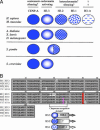
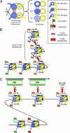
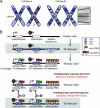
In the history of science, provocative but, at times, controversial ideas have been put forward to explain basic problems that confront and intrigue the scientific community. These hypotheses, although often not correct in every detail, lead to increased discussion that ultimately guides experimental tests of the principal concepts and produce valuable insights into long-standing questions. Here, we present a hypothesis, the "H3 barcode hypothesis." Hopefully, our ideas will evoke critical discussion and new experimental approaches that bear on general topics, such as nuclear architecture, epigenetic memory, and cell-fate choice. Our hypothesis rests on the central concept that mammalian histone H3 variants (H3.1, H3.2, and H3.3), although remarkably similar in amino acid sequence, exhibit distinct posttranslational "signatures" that create different chromosomal domains or territories, which, in turn, influence epigenetic states during cellular differentiation and development. Although we restrict our comments to H3 variants in mammals, we expect that the more general concepts presented here will apply to other histone variant families in organisms that employ them.
Conflict of interest statement
Conflict of interest statement: No conflicts declared.
Figures


Similar articles
-
Epigenetic and replacement roles of histone variant H3.3 in reproduction and development.Int J Dev Biol. 2009;53(2-3):231-43. doi: 10.1387/ijdb.082653go. Int J Dev Biol. 2009. PMID: 19412883 Review.
-
In and out: histone variant exchange in chromatin.Trends Biochem Sci. 2005 Dec;30(12):680-7. doi: 10.1016/j.tibs.2005.10.003. Epub 2005 Oct 28. Trends Biochem Sci. 2005. PMID: 16257529 Review.
-
Histone H3 variants and their chaperones during development and disease: contributing to epigenetic control.Annu Rev Cell Dev Biol. 2014;30:615-46. doi: 10.1146/annurev-cellbio-100913-013311. Annu Rev Cell Dev Biol. 2014. PMID: 25288118 Review.
-
Histone variants in development and diseases.J Genet Genomics. 2013 Jul 20;40(7):355-65. doi: 10.1016/j.jgg.2013.05.001. Epub 2013 May 20. J Genet Genomics. 2013. PMID: 23876776 Review.
-
Genome-wide identification, evolutionary, and expression analyses of histone H3 variants in plants.Biomed Res Int. 2015;2015:341598. doi: 10.1155/2015/341598. Epub 2015 Feb 26. Biomed Res Int. 2015. PMID: 25815311 Free PMC article.
Cited by
-
Critical Role of Histone Turnover in Neuronal Transcription and Plasticity.Neuron. 2015 Jul 1;87(1):77-94. doi: 10.1016/j.neuron.2015.06.014. Neuron. 2015. PMID: 26139371 Free PMC article.
-
Combinatorial Histone H3 Modifications Are Dynamically Altered in Distinct Cell Cycle Phases.J Am Soc Mass Spectrom. 2021 Jun 2;32(6):1300-1311. doi: 10.1021/jasms.0c00451. Epub 2021 Apr 5. J Am Soc Mass Spectrom. 2021. PMID: 33818074 Free PMC article.
-
EvoChromo: towards a synthesis of chromatin biology and evolution.Development. 2019 Sep 26;146(19):dev178962. doi: 10.1242/dev.178962. Development. 2019. PMID: 31558570 Free PMC article. Review.
-
The evolutionary history of histone H3 suggests a deep eukaryotic root of chromatin modifying mechanisms.BMC Evol Biol. 2010 Aug 25;10:259. doi: 10.1186/1471-2148-10-259. BMC Evol Biol. 2010. PMID: 20738881 Free PMC article.
-
Stable transmission of reversible modifications: maintenance of epigenetic information through the cell cycle.Cell Mol Life Sci. 2011 Jan;68(1):27-44. doi: 10.1007/s00018-010-0505-5. Epub 2010 Aug 27. Cell Mol Life Sci. 2011. PMID: 20799050 Free PMC article. Review.
References
-
- Strahl B. D., Allis C. D. Nature. 2000;403:41–45. - PubMed
-
- Fischle W., Wang Y., Allis C. D. Curr. Opin. Cell Biol. 2003;15:172–183. - PubMed
-
- Fischle W., Wang Y., Allis C. D. Nature. 2003;425:475–479. - PubMed
-
- Fischle W., Tseng B. S., Dormann H. L., Ueberheide B. M., Garcia B. A., Shabanowitz J., Hunt D. F., Funabiki H., Allis C. D. Nature. 2005;438:1116–1122. - PubMed
-
- Hirota T., Lipp J. J., Toh B. H., Peters J. M. Nature. 2005;438:1176–1180. - PubMed
Publication types
MeSH terms
Substances
Grants and funding
LinkOut - more resources
Full Text Sources
Other Literature Sources

