. 2006 Apr;80(7):3189-204.
doi: 10.1128/JVI.80.7.3189-3204.2006.

Acetylated Tat regulates human immunodeficiency virus type 1 splicing through its interaction with the splicing regulator p32

Affiliations

Acetylated Tat regulates human immunodeficiency virus type 1 splicing through its interaction with the splicing regulator p32

Reem Berro et al. J Virol. 2006 Apr.

Abstract

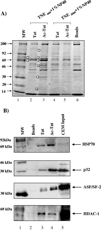
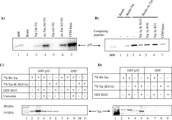
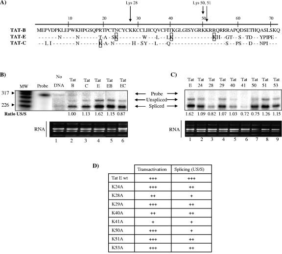
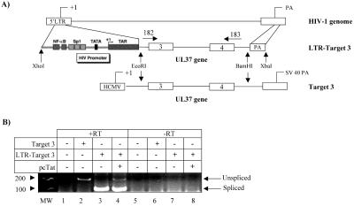
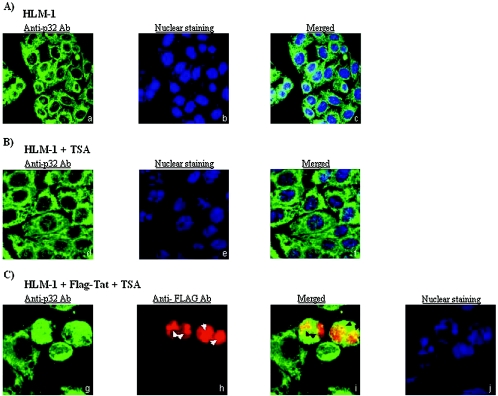
The human immunodeficiency virus type 1 (HIV-1) potent transactivator Tat protein mediates pleiotropic effects on various cell functions. Posttranslational modification of Tat affects its activity during viral transcription. Tat binds to TAR and subsequently becomes acetylated on lysine residues by histone acetyltransferases. Novel protein-protein interaction domains on acetylated Tat are then established, which are necessary for both sustained transcriptional activation of the HIV-1 promoter and viral transcription elongation. In this study, we investigated the identity of proteins that preferentially bound acetylated Tat. Using a proteomic approach, we identified a number of proteins that preferentially bound AcTat, among which p32, a cofactor of splicing factor ASF/SF-2, was identified. We found that p32 was recruited to the HIV-1 genome, suggesting a mechanism by which acetylation of Tat may inhibit HIV-1 splicing needed for the production of full-length transcripts. Using Tat from different clades, harboring a different number of acetylation sites, as well as Tat mutated at lysine residues, we demonstrated that Tat acetylation affected splicing in vivo. Finally, using confocal microscopy, we found that p32 and Tat colocalize in vivo in HIV-1-infected cells.


Figures

FIG. 1.
FIG. 1.
Genomic structure of HIV-1. (A) Structure of HIV-1 proviral genome and location of the HIV-1 exons and splice donor (D) and splice acceptor (A) sites. (B) The structure of the alternatively spliced mRNAs (lines) and open reading frames (ORFs) (filled boxes).
FIG. 2.
FIG. 2.
Identification of cellular Tat-binding proteins. (A) Uninfected CEM extracts (2 mg) were incubated with 100 μg of biotinylated unmodified or acetylated Tat (AcTat) peptides (aa 36 to 54) for 2 h at 4°C, pulled down with streptavidin beads, and washed with TNE600 or TNE2000 plus 1% NP-40 (0.6 M and 2 M NaCl, respectively). As a negative control, whole-cell lysates were incubated with streptavidin beads alone (Beads). Bound proteins were separated on a 4 to 20% Tris-glycine gel and stained with Coomassie blue. Eight bands were chosen for mass spectrometric analysis (numbered circles). (B) Western analysis of Tat-interacting proteins. Samples were prepared as for panel A and washed with TNE600 plus 1% NP-40. Bound proteins were separated on a 4 to 20% Tris-glycine gel, transferred overnight to an Immobilon P membrane, and Western blotted with antibodies against p32, HSP-70, ASF/SF-2, and HDAC1. MW, DNA marker.
FIG. 3.
FIG. 3.
p32 preferentially interacts with acetylated Tat in vitro. (A) Biotin-labeled Tat (aa 36 to 53) unmodified (lane 3) or acetylated at positions 41, 50, and 51 (lane 4) and biotin-labeled Tat (aa 42 to 54) unmodified (lane 5) or acetylated at lysine 50 and 51 (lane 6) were incubated with CEM cell extracts. Bound proteins were separated on 4 to 20% Tris-glycine gels, transferred overnight to an Immobilon P membrane, and Western blotted with antibodies against p32. (B) Three competing peptides (aa 42 to 54) were added to the biotin-labeled AcTat pull down: Tat Ac K50 and 51, Tat Ac K50, or Tat Ac K51, and the same experiment described for panel A was performed. (C, D) TNT-synthesized 32P-labeled full-length WT Tat, mutant Tat (K50, 51A) (C), and Tat (K41A) (D) were incubated with acetyl coenzyme A in the presence (+) or absence (−) of GST-p300 (HAT domain) for 2 h and then incubated with GST-p32 or GST overnight. In panel C, curcumin (100 μM) was added to inhibit HAT activity. Bound complexes were washed with TNE600 plus 1% NP-40, and proteins were separated on a 4 to 20% Tris-glycine polyacrylamide gel, dried, and exposed to a PhosphorImager cassette. MW, DNA marker.
FIG. 4.
FIG. 4.
TSA treatment increases the presence of p32 and acetylated Tat at the HIV-1 promoter. (A) OM10.1 cells were treated with (+) or without (−) 450 nM TSA to induce Tat acetylation followed by ChIP assays. Equal amounts of treated and untreated cells were harvested after 24 h, cross-linked, and lysed using an SDS-containing buffer. The cross-linked chromatin was sonicated, and transcription complexes were immunoprecipitated with 5 μg of antibodies to anti-Tab172 (Control Ab), anti-Pol II (C-21), anti-p32, cdk-9, ASF/SF-2, or anti-AcTat 50/51 (AcTat), and recovered DNA was used for PCR amplification of the HIV-1 LTR to determine the occupancy of the immunoprecipitated protein on the HIV-1 promoter (upper panel) and on the actin gene (lower panel). (B) Specificity of the AcTat antibody. One microgram of either GST (lane 1), GST-Tat acetylated with p300 HAT domain (lane 2; as described in legend to Fig. 3), or unmodified GST-Tat (lane 3) were separated by 4 to 20% SDS-PAGE, transferred, and Western blotted (WB) with anti-Tat (α-Tat) antibody (NIH AIDS reagent) or anti-AcTat (α-AcTat) antibody (rabbit polyclonal antibody raised against the Tat peptide 36 to 70 acetylated at positions 50 and 51). (C) Western blot for ASF/SF-2 and actin (as a loading control) of OM10.1 cell extracts before and after treatment with TSA. The circled P indicates phosphorylation.
FIG. 5.
FIG. 5.
Effect of TSA on HIV-1 singly and doubly spliced mRNA. (A) The proviral plasmid pNL-CAT contains the CAT reporter gene cloned into the env open reading frame (ORF) (32), which is expressed from singly spliced (4-kb) viral transcripts. The proviral plasmid pNL-Luc contains the firefly luciferase gene cloned into the nef ORF (89), and luciferase is expressed from the doubly spliced (2-kb) viral transcripts. (B) HeLa cells were infected with MLV-pseudotyped pNL-CAT or pNL-Luc and treated with 450 nM TSA 24 h later. After 48 h, cells were lysed and CAT and luciferase activities were determined. The average of results from three independent experiments is shown. (C) Western blot for actin (Santa Cruz) and cyclin E (Santa Cruz) on cells transfected with pNL-CAT and pNL-Luc treated or untreated with 450 nM TSA. +, present; −, absent.
FIG. 6.
FIG. 6.
Effect of WT-Tat and mutant Tat on the pattern of HIV-1 splicing. (A) Total RNA was extracted from HLM-1 (HIV-1+/Tat) cells transfected with WT Tat or mutant (K50A, K51A) Tat and analyzed by RPA after 48 h. Upper panel: RPA. Lanes 3 and 4, untreated; lanes 5 and 6, treatment with 200 nM TSA for 4 h. The free probe (312 nucleotides) and two protected fragments from unspliced (262 nucleotides) and spliced (213 nucleotides) transcripts are indicated by arrows. MW, RNA marker. Lower panel: RNA from the different samples was run on 1% agarose gel and stained with ethidium bromide. (B) RNA from HLM-1 transfected with pcTat alone, pcTat with ASF/SF-2, or pcTat with p32 was extracted and analyzed by RPA 48 h after transfection. The US/S ratio is calculated and normalized to the ratio of pcTat which is set as 1. (C) Ratios of unspliced to spliced mRNA in panel A are shown as bars and correspond to the averages of results from three independent transfections. +,present; −, absent.
FIG. 7.
FIG. 7.
Effect of Tat from different clades and Tat acetylation mutants on the pattern of HIV-1 splicing. (A) Sequence variation in subtype-specific Tat proteins. Differences in amino acid residues (1 to 72 aa) in Tat B, C, and E are shown. Lys 28, 50, and 51, which are targets for acetylation by P/CAF and p300, are conserved (indicated by arrows). Additional lysine residues of Tat E and C that may be targets of acetylation are boxed. (B and C) Total RNA was extracted from HLM-1 cells transfected with plasmids encoding Tat from clades B, C, and E, and chimeric Tat BE and EC (B) or Tat E and Tat E mutants (Tat E K 24A, K28A, K29A, K40A, K41A, K50A, K51A, and K53A) (C) and analyzed by RPA after 48 h as described for Fig. 6 (upper panel). RNA from the different samples was run on a 1% agarose gel (lower panel). Ratios of US/S transcripts normalized according to the Tat B ratio (set at 1) are indicated at the bottom. (D) Table representing the transactivation efficiency and the effect on splicing of all the Tat E lysine mutants. Transactivation efficiency was calculated as the sum of unspliced and spliced transcript intensity as measured by Image Quant. +++, no significant effect on transactivation; ++, modest decrease in transactivation; +, complete disruption of transactivation. The effect on splicing was measured as the US/S ratio. US/S ratios were classified as follows: high ratio, +++; moderate ratio, ++; low ratio, +.
FIG. 8.
FIG. 8.
Effect of Tat acetylation on the splicing of the LTR-Target 3 reporter minigene. (A) The HIV-1 LTR was cloned from HIV-1 (pLAI strain) and inserted in place of the HCMV IE promoter of the Target 3 reporter minigene (Target 3) and is referred to as LTR-Target 3. The polyadenylation signal from HIV-1 (pLAI) was cloned downstream of Target 3. The localization of the forward (182) and reverse (183) primers used in the RT-PCR assay are indicated (87). (B) 293T cells were cotransfected with no DNA (lanes 1 and 5), Target 3 (lanes 2 and 6), or LTR-Target 3 with (+) (lanes 4 and 8) or without (−) (lanes 3 and 7) pcTat. Total RNA was isolated, reverse transcribed, and PCR amplified using primers 182 and 183 to detect spliced and unspliced messages. PCR products were separated by electrophoresis through a 1% agarose gel and stained with ethidium bromide. Control reactions did not contain RT (−) (lanes 5 to 8). Arrows indicate the positions of unspliced (202 bp) and spliced (99 bp) cDNA products. MW, DNA marker.
FIG. 9.
FIG. 9.
Colocalization of Tat and p32 in HIV-1-infected cells. Confocal optical sections (z = 1.0 μm) are shown in all panels. Localization of p32 in HLM-1 untreated cells (A) or cells treated with 450 nM TSA for 2 h (B). p32 staining as detected by rabbit polyclonal anti-p32 primary antibody and Alexa Fluor 488 secondary antibody is shown in panels a and d. Nuclear staining detected utilizing TOTO-3, a dimeric cyanine nucleic acid stain, is shown in panels b and e. Merged images of p32 and nuclear staining are shown in panels c and f. (C) HLM-1 cells transfected with Flag-Tat and treated with 450 nM TSA for 2 h. p32 staining as detected by rabbit polyclonal anti-p32 primary antibody and Alexa Fluor 488 secondary antibody is shown in panel g. Tat staining detected by mouse monoclonal anti-Flag primary antibody and Alexa Fluor 568 secondary antibody is shown in panel h. White arrows indicate nucleolar localization of Tat. A merged image of p32 and Tat staining is shown in panel i. Black arrows indicate points where colocalization is occurring, shown as yellow coloring when the two images are merged. Nuclear staining detected utilizing TOTO-3, a dimeric cyanine nucleic acid stain, is shown in panel j.
FIG. 10.
FIG. 10.
Proposed model for AcTat regulation of HIV-1 splicing. (A) Low amounts of Tat, p32, and hyperphysphorylated ASF/SF-2 are bound to the HIV-1 LTR before virus activation. (B) Upon treatment with TSA, Tat becomes acetylated and recruits more p32 to the LTR, which in turn acts on ASF/SF-2 and decreases its phosphorylation, impairing its binding to RNA and subsequently leading to a decrease in HIV-1 splicing. (C) AcTat/p32/SF-2 form a complex and affect splicing, probably sequestering ASF/SF-2 away from the HIV-1 mRNA, which may occur in the absence of HIV-1 LTR DNA.

Similar articles

Cited by

References

    1. Adair, R., G. W. Liebisch, Y. Su, and A. M. Colberg-Poley. 2004. Alteration of cellular RNA splicing and polyadenylation machineries during productive human cytomegalovirus infection. J. Gen. Virol. 85:3541-3553. - PubMed
    1. Balasubramanyam, K., R. Varier, M. Altaf, V. Swaminathan, N. Siddappa, U. Ranga, and T. Kundu. 2004. Curcumin, a novel p300/CREB-binding protein-specific inhibitor of acetyltransferase, represses the acetylation of histone/nonhistone proteins and histone acetyltransferase-dependent chromatin transcription. J. Biol. Chem. 279:51163-51171. - PubMed
    1. Benkirane, M., R. F. Chun, H. Xiao, V. V. Ogryzko, B. H. Howard, Y. Nakatani, and K. T. Jeang. 1998. Activation of integrated provirus requires histone acetyltransferase. p300 and P/CAF are coactivators for HIV-1 Tat. J. Biol. Chem. 273:24898-24905. - PubMed
    1. Bieniasz, P., and B. Cullen. 2000. Multiple blocks to human immunodeficiency virus type 1 replication in rodent cells. J. Virol. 74:9868-9877. - PMC - PubMed
    1. Bieniasz, P. D., T. A. Grdina, H. P. Bogerd, and B. R. Cullen. 1998. Recruitment of a protein complex containing Tat and cyclin T1 to TAR governs the species specificity of HIV-1 Tat. EMBO J. 17:7056-7065. - PMC - PubMed

Publication types

MeSH terms