. 2006 May 1;496(1):13-26.
doi: 10.1002/cne.20885.

Functional effectiveness of the blood-brain barrier to small water-soluble molecules in developing and adult opossum (Monodelphis domestica)

Affiliations

Functional effectiveness of the blood-brain barrier to small water-soluble molecules in developing and adult opossum (Monodelphis domestica)

Carl Joakim Ek et al. J Comp Neurol. .

Abstract

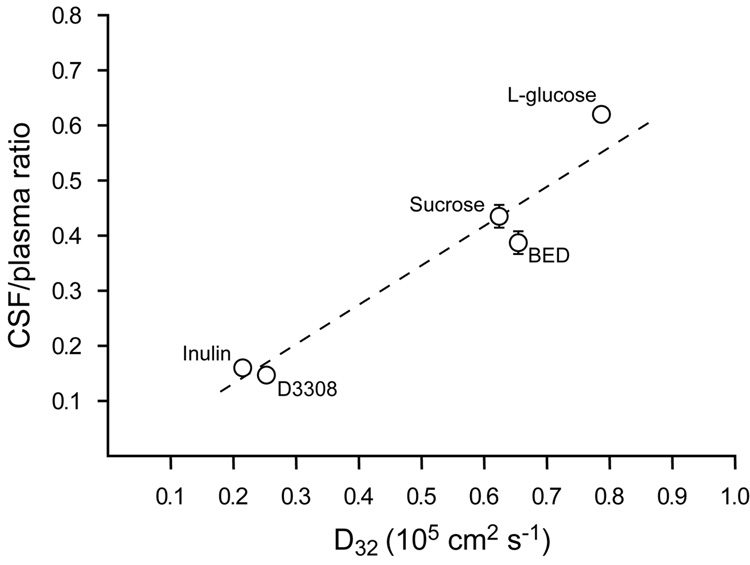
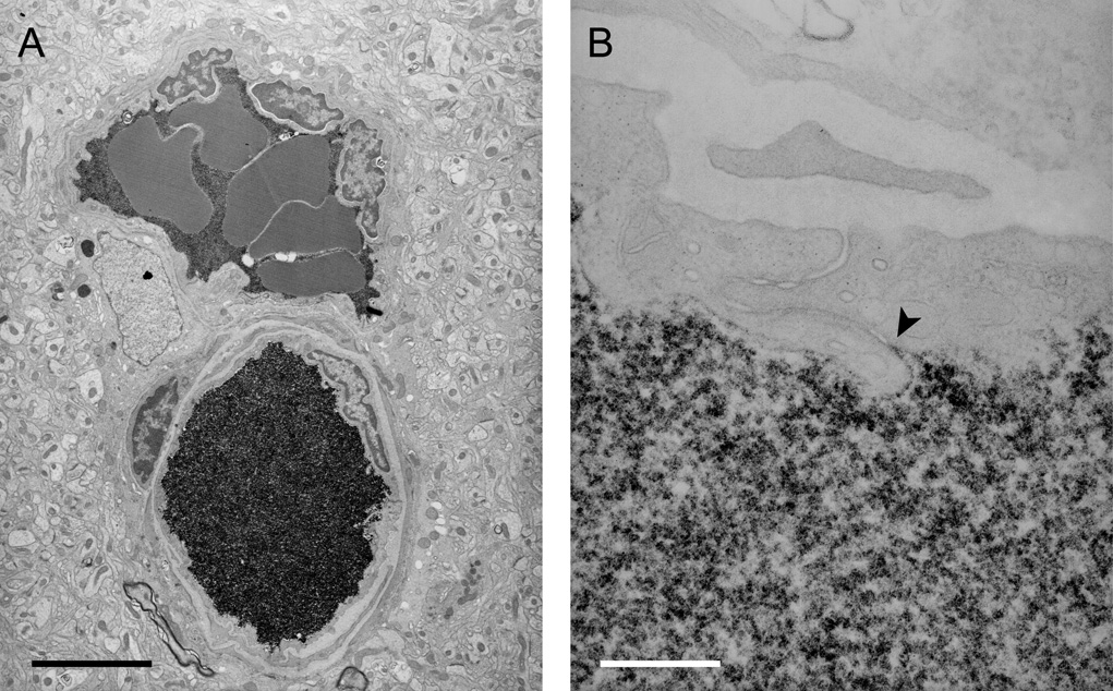
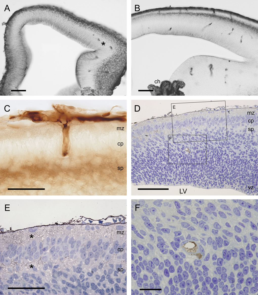
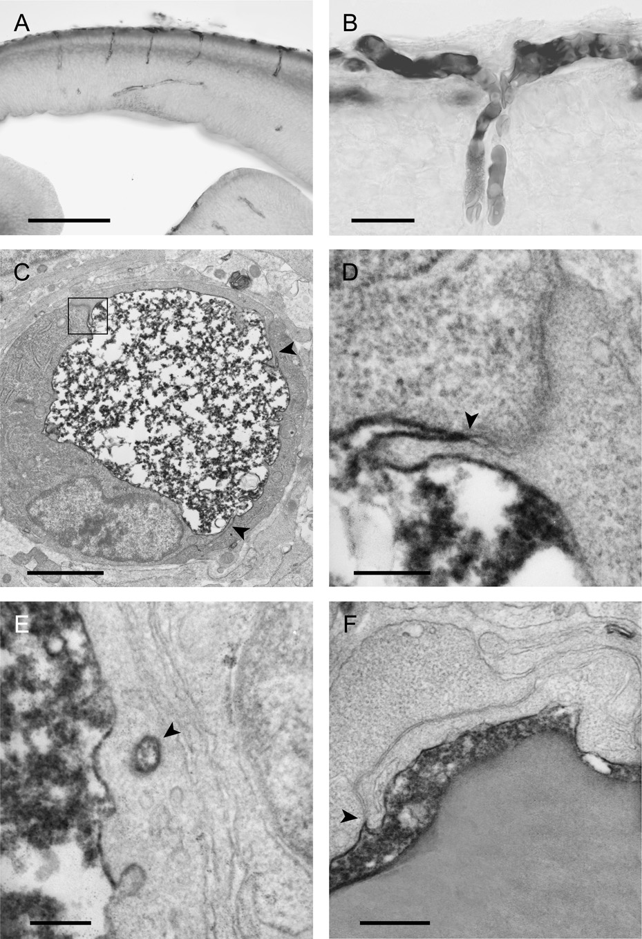
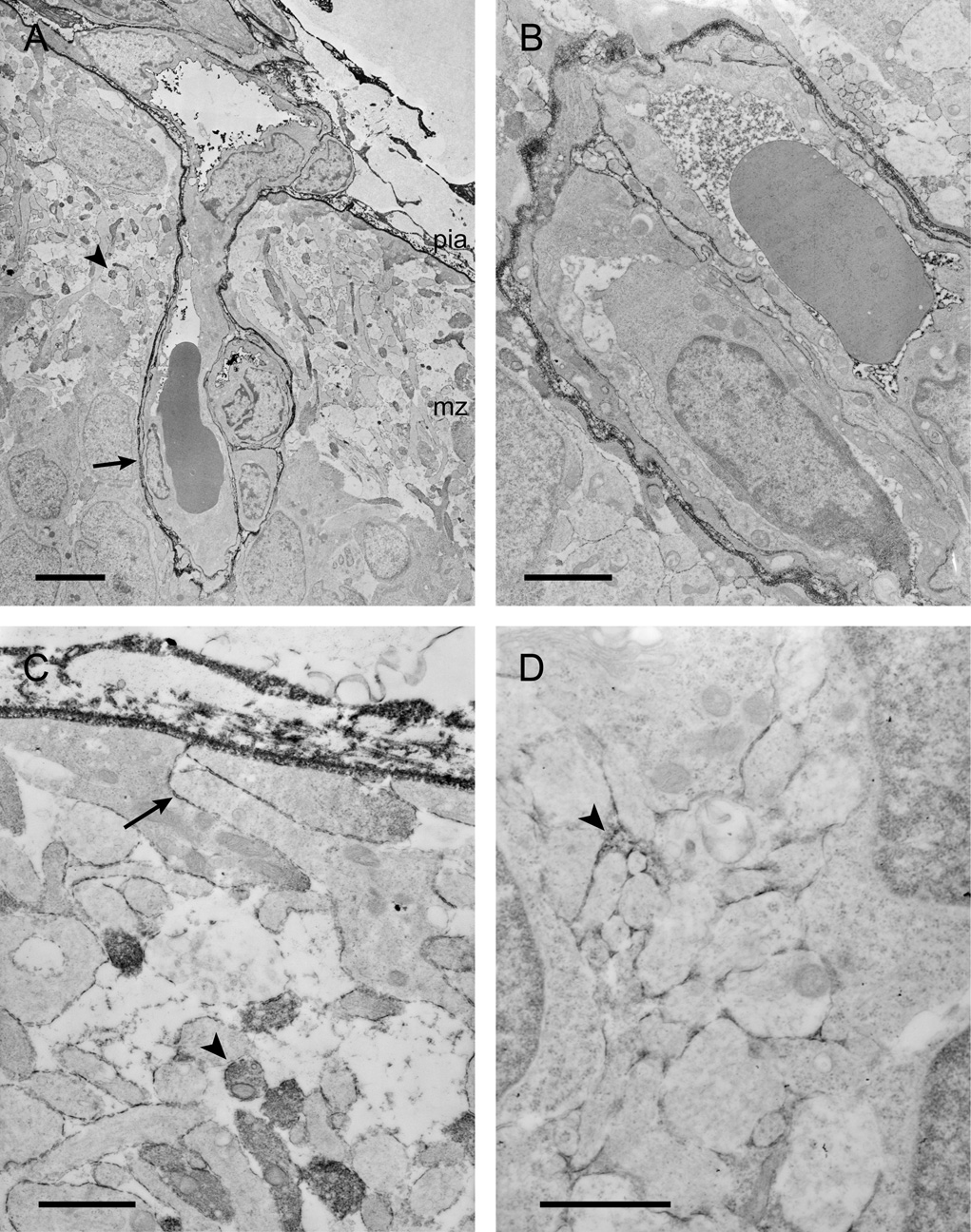
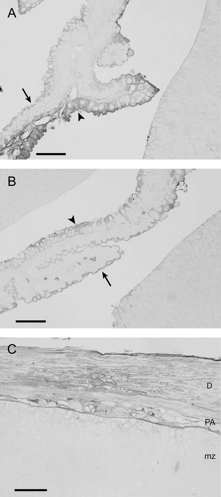
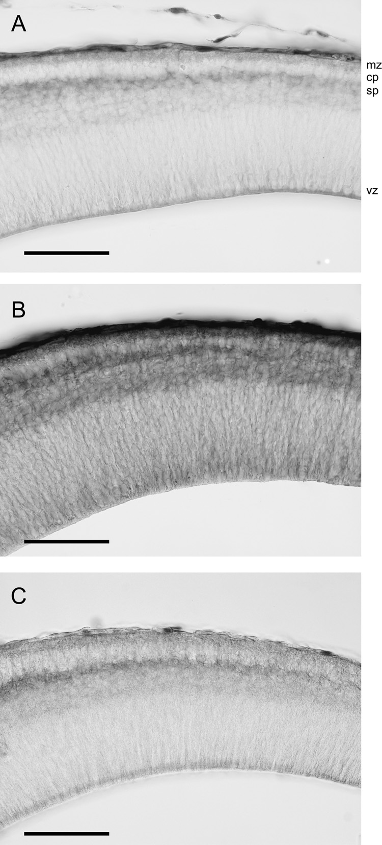
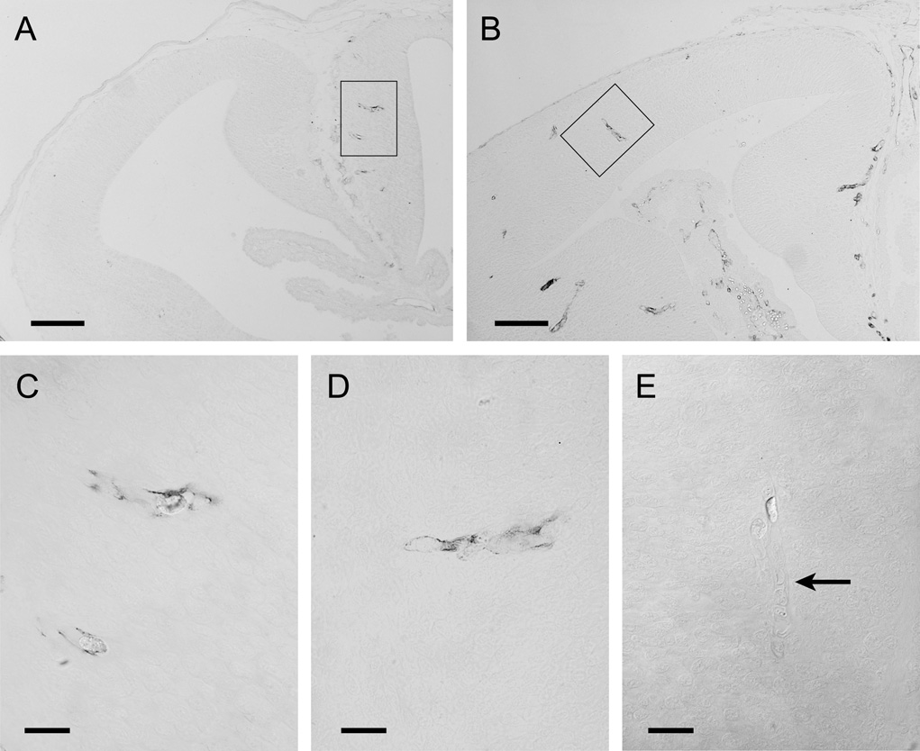
We have evaluated a small water-soluble molecule, biotin ethylenediamine (BED, 286 Da), as a permeability tracer across the blood-brain barrier. This molecule was found to have suitable characteristics in that it is stable in plasma, has low plasma protein binding, and appears to behave in a similar manner across brain barriers as established by permeability markers such as sucrose. BED, together with a 3000-Da biotin-dextran (BDA3000), was used to investigate the effectiveness of tight junctions in cortical vessels during development and adulthood of a marsupial opossum (Monodelphis domestica). Marsupial species are born at an early stage of brain development when cortical vessels are just beginning to appear. The tracers were administered systemically to opossums at various ages and localized in brains with light and electron microscopy. In adults, the tight junctions restricted the movement of both tracers. In neonates, as soon as vessels grow into the neocortex, their tight junctions are functionally restrictive, a finding supported by the presence of claudin-5 in endothelial cells. However, both tracers are also found within brain extracellular space soon after intraperitoneal administration. The main route of entry for the tracers into immature neocortex appears to be via the cerebrospinal fluid over the outer (subarachnoid) and inner (ventricular) surfaces of the brain. These experiments demonstrate that the previously described higher permeability of barriers to small molecules in the developing brain does not seem to be due to leakiness of cerebral endothelial tight junctions, but to a route of entry probably via the choroid plexuses and cerebrospinal fluid.


Figures

Figure 1
Figure 1
Three-hour CSF/plasma concentration ratios for inulin, sucrose, L-glucose, BED (biotin ethylenediamine) and fluorescent 3000Da biotin-dextran (D3308) in P16 opossums plotted against their estimated diffusion coefficients (D32) at 32°C (body temperature of opossum). Estimations of D32 were made from the Einstein-Stokes radii. Values are means±SEM; some error bars are obscured by symbols. Data for L-glucose, inulin and D3308 are from Ek et al. (2001). From this plot it can be seen that the ratios for all molecules are proportional to their D32 suggesting that their blood to CSF transfer route is by passive diffusion, and not subject to any inward transport mechanism.
Figure 2
Figure 2
Electron micrographs of the localisation of BED (biotin ethylenediamine) in blood vessels deep inside the cortex of a 2 month old opossum 10min after an intravenous injection. Similar staining is found after an intravenous injection of biotin-dextran (BDA3000). A) Low power micrograph showing two paired vessels with abundant reaction product within the lumen. No reaction product is visible in the surrounding tissue. Pairs of arteries and veins are characteristic of the vascular pattern in marsupial brains (Wislocki and Campbell, 1937). B) High power micrograph of an interendothelial cleft showing that the tight junctions in the young adult restrict the passage of BED through the cleft (arrowhead). Scale bars are 4µm in A, 300nm in B.
Figure 3
Figure 3
Light micrographs showing the localisation of BED in neocortex of opossums at P2 (A) and P5 (B–F) 20–25min after an intraperitoneal injection. A–C are vibratome sections (50µm) and D–F semithin sections (0.5µm). A) Note that at P2 there are no vessels present in the neocortex except for occasional one in the most lateral more developed parts (*). B) At P5 a few vessels are present within the neocortex. Note also that the staining of BED is most abundant in outer and inner surfaces of the cortex and in the choroid plexus (ch). C) High power micrograph showing vessels (paired vessels are characteristic of marsupial brains) growing into the neocortex. Note that the staining for BED is most visible within the vessels, in the marginal and subplate zones, similar to the staining for BDA3000 (see 4A). D) Section of the whole neocortex counterstained with toluidine blue. This section shows that in the cortex the reaction product is most visible within the marginal and the subplate zones (shown in high power in E). Note also how few vessels are visible in the neocortex at this age (one pair in lower box and shown at high power in F). E) Higher power view of upper box in D. Note reaction product in marginal zone (mz) and subplate zone (sp) indicated by *. F) Higher power magnification of two paired blood vessels from lower box in D showing that at the light microscopic level it appears that only the lumen of the vessels is stained and no reaction product is visible in the extracellular space surrounding the vessels. Scale bars are 100µm in A&B, 50µm in C, 100µm in D, 50µm in E and 20µm in F. Abbreviations: LV – lateral ventricle, mz – marginal zone, cp – cortical plate, sp - subplate, vz – ventricular zone.
Figure 4
Figure 4
The localisation of BDA3000 (biotin-dextran) in neocortex of opossums at P5 (A,B), P2 (C–E) and P12 (F) 20–25min (except A which is 40min) after an intraperitoneal injection. A and B are light micrographs of vibratome sections and C–F electron micrographs. A) Staining is seen within blood vessels and towards the inner and outer surfaces of the cortex. B) At high magnification it can be seen that two different pial vessels meet and grow into the brain together. Staining can only be seen inside the blood vessels and none in the surrounding tissue. C) Cross section of a blood vessel which shows that the reaction product is abundant inside the lumen of the vessel, but is not visible in the immediately surrounding tissue. The arrowheads point to the intercellular clefts of the endothelial cell. D) One of the interendothelial clefts from C (boxed in C) is shown at higher magnification. Arrowhead points to site of the tight junction. E) Although vesicles that contain the reaction product (arrowhead) are present in the endothelial cells, these are not common. F) A similar cleft to D at an older age (P12). Note in D and F that the reaction product is only present at the most luminal end of the cleft demonstrating that the tight junctions (arrowheads) between the endothelial cell restrict the movement of the tracer from blood into neural tissue. Scale bars are 200µm in A, 25µm in B, 2µm in C, 200nm in D&E and 400nm in F.
Figure 5
Figure 5
Electron micrographs showing the distribution of BED in the neocortex of a P5 opossum 25min after an intraperitoneal injection. A) A blood vessel that is penetrating into the neocortex from the pial plexus. Note that reaction product is visible in the perivascular (Virchow-Robin) space (arrow), inside the lumen of vessels and in the marginal zone. Some processes in the marginal zone are labelled (arrowhead). B) A pair of blood vessels in the intermediate zone. Note the reaction product within the vessel lumen. Some of the vessels within this area have reaction product in the perivascular space which is presumably an extension of the Virchow-Robin space seen in A. C) A high power micrograph of the pial surface and underlying marginal zone. Note that extracellular staining is apparent in between the cells (arrow) and some of the processes in the marginal zone (arrowhead). D) Micrograph of the intermediate zone showing that the reaction product is visible in the extracellular space (arrowhead). This micrograph has been chosen to illustrate the extracellular staining; however, in many regions of the cortex the staining is not as prominent. Scale bars are 5µm in A, 2µm in B, and 1µm in C&D.
Figure 6
Figure 6
Localisation of BED in choroid plexus at P2 (A), P5 (B) and in the meninges at P5 (C) 15min after an ip injection (1µm sections). A&B) Many epithelial cells of the choroid plexus contain the tracer (arrowheads). Note also the staining of the brush border of the epithelial cells (arrows) indicating the presence of the tracer within CSF. C) Apart from the tracer visible inside pial/arachnoid vessels, staining is also visible in the dura, subarachnoid space and extracellular space of the marginal zone. Note that there are no blood vessels present within the dura at this stage of development. Scale bars are 50µm in A&B and 25µm in C. Abbreviations: D – Dura mater, PA – pia-arachnoid, mz – marginal zone.
Figure 7
Figure 7
Penetration of tracers through the neocortex at P5 after submersion of brains in solutions containing BED or BDA3000. A) After 5min submersion of the brain BED has penetrated into the ventricular zone presumably from the lateral ventricle and into the subplate zone from the outer surface of the brain. B) After 25min submersion in BED solution the tracer penetrates the whole neocortex. C) After 25min submersion BDA3000 has a similar distribution in the neocortex as BED after 5min (see A). Scale bar is 100µm for all micrographs. Abbreviations: mz – marginal zone, cp – cortical plate, sb – subplate, vz – ventricular zone.
Figure 8
Figure 8
Localisation of claudin-5 at P0 (A&C) and P5 (B&D), and negative control of claudin-5 staining at P5 (E) in opossum brains. A) Although no vessels are present in the neocortex at P0, vessels in older parts of the brain are already immunoreactive for claudin-5 (box). B) At P5, vessels in the neocortex are also immunoreactive for claudin-5. Note that staining in the choroid plexus is only within the stroma and not between the epithelial cells where the tight junctions of the blood-CSF barrier are located. C) Two claudin-5 positive blood vessels shown at high power (box in A). D) One cortical vessel positive for claudin-5 shown at high power (box in B). E) Micrograph viewed with Nomarski optics showing that after absorption of anti claudin-5 antibody in adult lung homogenate, which is known to contain claudin-5, no staining is visible in brain blood vessels (arrow). Scale bars are 100µm in A&B and 20µm in C–E.

Similar articles

Cited by

References

    1. Bass NH, Lundborg P. Postnatal development of bulk flow in the cerebrospinal fluid system of the albino rat: clearance of carboxyl-(14C)inulin after intrathecal infusion. Brain Res. 1973;52:323–332. - PubMed
    1. Bauer HC, Bauer H, Lametschwandtner A, Amberger A, Ruiz P, Steiner M. Neovascularization and the appearance of morphological characteristics of the blood-brain barrier in the embryonic mouse central nervous system. Brain Res Dev Brain Res. 1993;75:269–278. - PubMed
    1. Bauer H, Sonnleitner U, Lametschwandtner A, Steiner M, Adam H, Bauer HC. Ontogenic expression of the erythroid-type glucose transporter (Glut 1) in the telencephalon of the mouse: correlation to the tightening of the blood-brain barrier. Brain Res Dev Brain Res. 86:317–325. - PubMed
    1. Brightman MW, Reese TS. Junctions between intimately apposed cell membranes in the vertebrate brain. J Cell Biol. 1969;40:648–677. - PMC - PubMed
    1. Claude P. Morphological factors influencing transepithelial permeability: a model for the resistance of the zonula occludens. J Membr Biol. 1978;3:219–232. - PubMed

Publication types

MeSH terms