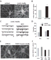
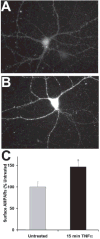
TNFalpha-induced AMPA-receptor trafficking in CNS neurons; relevance to excitotoxicity?
- PMID: 16520832
- PMCID: PMC1389713
- DOI: 10.1017/S1740925X05000608
TNFalpha-induced AMPA-receptor trafficking in CNS neurons; relevance to excitotoxicity?
Abstract
Injury and disease in the CNS increases the amount of tumor necrosis factor alpha (TNFalpha) that neurons are exposed to. This cytokine is central to the inflammatory response that occurs after injury and during prolonged CNS disease, and contributes to the process of neuronal cell death. Previous studies have addressed how long-term apoptotic-signaling pathways that are initiated by TNFalpha might influence these processes, but the effects of inflammation on neurons and synaptic function in the timescale of minutes after exposure are largely unexplored. Our published studies examining the effect of TNFalpha on trafficking of AMPA-type glutamate receptors (AMPARs) in hippocampal neurons demonstrate that glial-derived TNFalpha causes a rapid (<15 minute) increase in the number of neuronal, surface-localized, synaptic AMPARs leading to an increase in synaptic strength. This indicates that TNFalpha-signal transduction acts to facilitate increased surface localization of AMPARs from internal postsynaptic stores. Importantly, an excess of surface localized AMPARs might predispose the neuron to glutamate-mediated excitotoxicity and excessive intracellular calcium concentrations, leading to cell death. This suggests a new mechanism for excitotoxic TNFalpha-induced neuronal death that is initiated minutes after neurons are exposed to the products of the inflammatory response. Here we review the importance of AMPAR trafficking in normal neuronal function and how abnormalities that are mediated by glial-derived cytokines such as TNFalpha can be central in causing neuronal disorders. We have further investigated the effects of TNFalpha on different neuronal cell types and present new data from cortical and hippocampal neurons in culture. Finally, we have expanded our investigation of the temporal profile of the action of this cytokine relevant to neuronal damage. We conclude that TNFalpha-mediated effects on AMPAR trafficking are common in diverse neuronal cell types and very rapid in their onset. The abnormal AMPAR trafficking elicited by TNFalpha might present a novel target to aid the development of new neuroprotective drugs.
Figures


Similar articles
-
Cell death after spinal cord injury is exacerbated by rapid TNF alpha-induced trafficking of GluR2-lacking AMPARs to the plasma membrane.J Neurosci. 2008 Oct 29;28(44):11391-400. doi: 10.1523/JNEUROSCI.3708-08.2008. J Neurosci. 2008. PMID: 18971481 Free PMC article.
-
Rapid tumor necrosis factor alpha-induced exocytosis of glutamate receptor 2-lacking AMPA receptors to extrasynaptic plasma membrane potentiates excitotoxicity.J Neurosci. 2008 Feb 27;28(9):2119-30. doi: 10.1523/JNEUROSCI.5159-07.2008. J Neurosci. 2008. PMID: 18305246 Free PMC article.
-
Cannabinoid receptor activation reduces TNFalpha-induced surface localization of AMPAR-type glutamate receptors and excitotoxicity.Neuropharmacology. 2010 Feb;58(2):551-8. doi: 10.1016/j.neuropharm.2009.07.035. Epub 2009 Aug 4. Neuropharmacology. 2010. PMID: 19654014 Free PMC article.
-
Glutamate receptor antibodies in neurological diseases: anti-AMPA-GluR3 antibodies, anti-NMDA-NR1 antibodies, anti-NMDA-NR2A/B antibodies, anti-mGluR1 antibodies or anti-mGluR5 antibodies are present in subpopulations of patients with either: epilepsy, encephalitis, cerebellar ataxia, systemic lupus erythematosus (SLE) and neuropsychiatric SLE, Sjogren's syndrome, schizophrenia, mania or stroke. These autoimmune anti-glutamate receptor antibodies can bind neurons in few brain regions, activate glutamate receptors, decrease glutamate receptor's expression, impair glutamate-induced signaling and function, activate blood brain barrier endothelial cells, kill neurons, damage the brain, induce behavioral/psychiatric/cognitive abnormalities and ataxia in animal models, and can be removed or silenced in some patients by immunotherapy.J Neural Transm (Vienna). 2014 Aug;121(8):1029-75. doi: 10.1007/s00702-014-1193-3. Epub 2014 Aug 1. J Neural Transm (Vienna). 2014. PMID: 25081016 Review.
-
The AMPAR subunit GluR2: still front and center-stage.Brain Res. 2000 Dec 15;886(1-2):190-207. doi: 10.1016/s0006-8993(00)02951-6. Brain Res. 2000. PMID: 11119696 Review.
Cited by
-
A potential role for microglia in stress- and drug-induced plasticity in the nucleus accumbens: A mechanism for stress-induced vulnerability to substance use disorder.Neurosci Biobehav Rev. 2019 Dec;107:360-369. doi: 10.1016/j.neubiorev.2019.09.007. Epub 2019 Sep 21. Neurosci Biobehav Rev. 2019. PMID: 31550452 Free PMC article. Review.
-
Altered presymptomatic AMPA and cannabinoid receptor trafficking in motor neurons of ALS model mice: implications for excitotoxicity.Eur J Neurosci. 2008 Feb;27(3):572-9. doi: 10.1111/j.1460-9568.2008.06041.x. Eur J Neurosci. 2008. PMID: 18279310 Free PMC article.
-
Genetic deletion of TNF receptor suppresses excitatory synaptic transmission via reducing AMPA receptor synaptic localization in cortical neurons.FASEB J. 2012 Jan;26(1):334-45. doi: 10.1096/fj.11-192716. Epub 2011 Oct 7. FASEB J. 2012. PMID: 21982949 Free PMC article.
-
Oligodendrocytes in central nervous system diseases: the effect of cytokine regulation.Neural Regen Res. 2024 Oct 1;19(10):2132-2143. doi: 10.4103/1673-5374.392854. Epub 2024 Jan 8. Neural Regen Res. 2024. PMID: 38488548 Free PMC article.
-
Role of Aβ in Alzheimer's-related synaptic dysfunction.Front Cell Dev Biol. 2022 Aug 26;10:964075. doi: 10.3389/fcell.2022.964075. eCollection 2022. Front Cell Dev Biol. 2022. PMID: 36092715 Free PMC article. Review.
References
-
- Allan SM. Varied actions of proinflammatory cytokines on excitotoxic cell death in the rat central nervous system. Journal of Neuroscience Research. 2002;67:428–434. - PubMed
-
- Allan SM, Rothwell NJ. Cytokines and acute neurodegeneration. Nature Reviews Neuroscience. 2001;2:734–744. - PubMed
-
- Barger SW, Basile AS. Activation of microglia by secreted amyloid precursor protein evokes release of glutamate by cystine exchange and attenuates synaptic function. Journal of Neurochemistry. 2001;76:846–854. - PubMed
-
- Beattie EC, Carroll RC, Yu X, Morishita W, Yasuda H, von Zastrow M, et al. Regulation of AMPA receptor endocytosis by a signaling mechanism shared with LTD. Nature Neuroscience. 2000;3:1291–1300. - PubMed
Grants and funding
LinkOut - more resources
Full Text Sources
