. 2006 Mar;26(6):2215-25.
doi: 10.1128/MCB.26.6.2215-2225.2006.

Inhibition of ADP/ATP exchange in receptor-interacting protein-mediated necrosis

Affiliations

Inhibition of ADP/ATP exchange in receptor-interacting protein-mediated necrosis

Vladislav Temkin et al. Mol Cell Biol. 2006 Mar.

Abstract

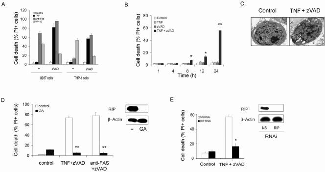
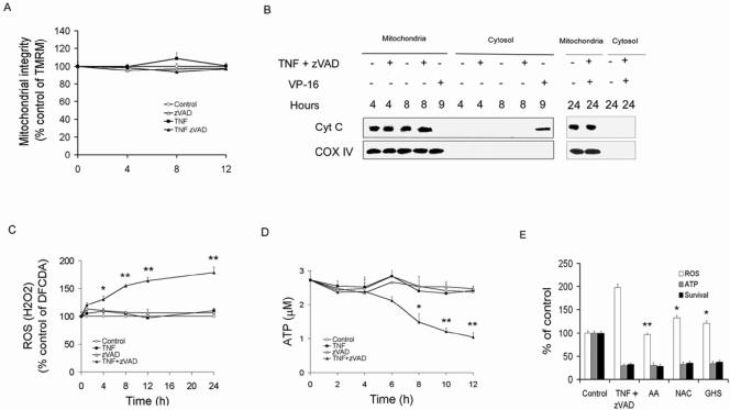
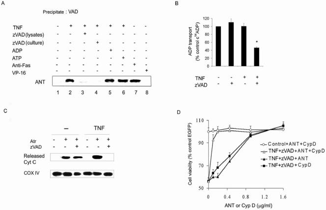
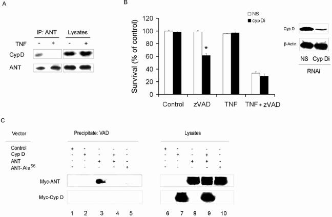
Receptor-interacting protein (RIP) has been implicated in the induction of death receptor-mediated, nonapoptotic cell death. However, the mechanisms remain to be elucidated. Here we show that tumor necrosis factor alpha induced RIP-dependent inhibition of adenine nucleotide translocase (ANT)-conducted transport of ADP into mitochondria, which resulted in reduced ATP and necrotic cell death. The inhibition of ADP/ATP exchange coincided with the loss of interaction between ANT and cyclophilin D and the inability of ANT to adopt the cytosolic conformational state, which prevented cytochrome c release. Neither overexpression of Bcl-xL nor inhibition of reactive oxygen species prevented necrosis. In contrast, the ectopic expression of ANT or cyclophilin D was effective at preventing cell death. These observations demonstrate a novel mechanism initiated through death receptor ligation and mediated by RIP that results in the suppression of ANT activity and necrosis.


Figures

FIG. 1.
FIG. 1.
RIP is required for necrotic cell death. (A to C) The induction of necrotic cell death. (A) Cell death of U937 and THP-1 cells. Cells were cultured for 24 h with TNF-α (10 ng/ml), anti-Fas (1 μg/ml), VP-16 (40 μM), or control medium in the presence or absence of zVAD (100 μM). Loss of viability was determined by a PI exclusion test. (B) Time course for TNF-α/zVAD-induced death of THP-1 cells. TNF-α was employed at 10 ng/ml and zVAD at 100 μM. (C) TNF-α/zVAD induces necrosis. U937 cells cultured in control medium (left panel) or TNF-α/zVAD (right panel) were examined by electron microscopy to identify the features of necrosis: membrane rupture, vacuolization of organelles, intact nucleus. Scale bar, 1 μm. (D and E) TNF-α/zVAD-induced cell death is prevented by the reduction of RIP. (D) Reduction of RIP with GA (0.5 μg/ml) protects against TNF-α/zVAD- or anti-Fas-induced cell death. (E) RIP suppression protects macrophages form TNF-α/zVAD-induced cell death. In vitro-differentiated human macrophages were transfected with NS or RIP RNAi for 96 h. Cell lysates were examined by immunoblot analysis for RIP (right panels). The cells were treated with TNF-α/zVAD and cell death defined by inability to exclude PI. The values in panels A, B, D, and E are the means ± 1 SE of three or four experiments, each performed in duplicate. **, P < 0.001; *, P < 0.05 (compared to medium alone).
FIG. 2.
FIG. 2.
Role of mitochondria in TNF-α/zVAD-induced necrosis. (A, B) Neither mitochondrial outer membrane permeabilization nor loss of ΔΨm is involved. (A) THP-1 cells were treated with zVAD, TNF-α, or control medium, and ΔΨm was determined by TMRM retention. (B) Cytochrome c is not released from mitochondria by TNF-α/zVAD. Mitochondrial and cytosolic fractions were isolated from THP-1 cells treated with TNF-α/zVAD or VP-16 for the times indicated. The mitochondrial fractions were assessed for cytochrome c and COX IV by immunoblot analysis. (C to E) Increased ROS production and ATP depletion in necrosis. (C) ROS/H2O2 induction by ΤNF-α/zVAD. THP-1 cells were treated as described for Fig. 1 and examined for ROS/H2O2 employing DCFDA as described in Materials and Methods. (D) ΤNF-α/zVAD results in ATP depletion. THP-1 cells were treated as described for panel A, and ATP levels were determined as described in Materials and Methods. (E) Antioxidants do not prevent TNF-α/zVAD-induced cell death or ATP depletion. THP-1 cells were pretreated with ascorbic acid (AA), glutathione (GSH), or N-acetyl-cysteine (NAC) prior to addition of TNF-α/zVAD or control medium. ROS and ATP were measured as described for panels A and B, and survival was determined according to the ability to exclude PI. The cells and supernatants were harvested 24 h after addition of TNF-α/zVAD. The results presented in panels A and C to E are the means ± 1 SE of three or four experiments, each performed in duplicate. The results in panel B are representative of three independent experiments. **, P < 0.001; *, P < 0.05 (compared to the TNF-α/zVAD-treated control).
FIG. 3.
FIG. 3.
Role of ANT in necrotic cell death. (A) TNF-α and Fas ligation promote binding of VAD to ANT. THP-1 cells were treated with TNF-α, anti-Fas antibody, VP-16, or zVAD for 8 h and harvested, and the lysates were incubated with zVAD, ADP, or ATP prior to biotinylated-VAD. The streptavidin-agarose precipitates were examined by immunoblot analysis for ANT. (B) TNF-α/zVAD inhibits ANT activity. Mitochondrial fractions from TNF-α-treated or control-treated (8 h) THP-1 cells were employed to determine the effect of zVAD on C14ADP transport into mitochondria as described in Materials and Methods. (C) TNF-α/zVAD inhibits cytochrome c release. Mitochondrial fractions of TNF-α-treated or control-treated (8 h) THP-1 cells were incubated with zVAD or control medium prior to adding atractyloside (2 mM) for 40 min. Following centrifugation at 10,000 × g, supernatants were analyzed for released cytochrome c and the pellets were analyzed for COX IV. (D) ANT and cyclophilin D protect against TNF-α/zVAD-induced death. RAW264.7 cells were transfected with control or cyclophilin D- or ANT- plus EGFP-expressing vectors at the indicated concentrations. After 24 h, cells were treated with TNF-α/zVAD or control medium for an additional 24 h and viability was assessed according to EGFP-positive cells results. The data in panels B and D are the means ± 1 SE of three orfour experiments performed in duplicate. The data presented in panels A and C are representative of three or four independent experiments. *,P < 0.05 versus control.
FIG. 4.
FIG. 4.
RIP is required for inhibition of ANT activity. (A) The reduction of RIP prevents the association of VAD with ANT. Macrophages were transfected with nonspecific RNAi (NS) or with RNAi for RIP (each for 96 h; left panel), or THP-1 cells were incubated with control medium or GA for 14 h (0.5 μg/ml, right panel). The cells were then treated with TNF-α or anti-Fas for 8 h, and then cell lysates were obtained and precipitated with biotinylated-VAD, as described for Fig. 5B. The precipitates were probed with antibody to ANT. IB, immunoblot. (B) The reduction of RIP by GA prevents the suppression of ANT activity induced by TNF-α/zVAD. THP-1 cells were incubated with GA (0.5 μg/ml) or control medium for 14 h and then treated with TNF-α for 8 h. The fractions containing the mitochondria were then isolated and treated with zVAD or DMSO (control) for 15 min. ADP transport activity was determined as described in Fig. 3B. (C) The reduction of RIP abrogates the inhibition of cytochrome c release by TNF-α/zVAD. THP-1 cells were treated with GA or control medium (14 h) and then TNF-α (8 h), prior to the isolation of the mitochondrial fractions, as described for panel B. The mitochondria were then incubated with zVAD for 15 min and then with atractyloside for 40 min. The supernatants and pellet were separated by centrifugation and examined for cytochrome c and COX IV, respectively. (D) RIP colocalizes with Mitotracker following treatment with TNF-α. (E) TNF-α-induced colocalization of RIP to the mitochondria was abrogated by reduction of RIP. Mitochondria were isolated from TNF-α-treated cells and analyzed for RIP by immunoblot analysis. The values in panel B represent the means ± 1 SE of three experiments performed in duplicate. The results presented in each panel are representative of three independent experiments. *, P < 0.05 versus zVAD alone.
FIG. 5.
FIG. 5.
Regulation of necrosis by cyclophilin D (CypD). (A) TNF-α suppresses ANT-cyclophilin D interaction. THP-1 cells were treated with TNF-α or control medium, and the cell lysates were used for immunoprecipitation (IP) with anti-ANT. The blots were probed for cyclophilin D and ANT. (B) Reduction of cyclophilin D facilitates zVAD-induced cell death. THP-1 cells were transfected with NS or cyclophilin D RNAi (Cyp Di) for 96 h. Some cells were harvested and examined for the expression of cyclophilin D by immunoblot analysis (right panel). The remaining cells were treated with zVAD and/or TNF-α as indicated for 24 h and were then examined by for survival (left panel) as determined by the ability to exclude PI. (C) Cyclophilin D reduces VAD/ANT interaction. 293Tcells were transfected with control, Myc-cyclophilin D (Myc-Cyp D)-, Myc-ANT-, or Myc-ANT-Ala56-expressing vectors, as described in Materials and Methods. After 24 h, the cells were harvested and the lysates incubated with biotinylated-VAD and then streptavidin-agarose. The precipitates were examined by immunoblot analysis for Myc. The values in panel B represent the means ± 1 SE of three experiments performed in duplicate. The results presented in each panel are representative of three independent experiments. *, P < 0.05 for cyclophilin D RNAi versus NS RNAi treatment.

Similar articles

Cited by

References

    1. Ahmad, M., S. M. Srinivasula, L. Wang, R. V. Talanian, G. Litwack, T. Fernandes-Alnemri, and E. S. Alnemri. 1997. CRADD, a novel human apoptotic adaptor molecule for caspase-2, and FasL/tumor necrosis factor receptor-interacting protein RIP. Cancer Res. 57:615-619. - PubMed
    1. Baines, C. P., R. A. Kaiser, N. H. Purcell, N. S. Blair, H. Osinska, M. A. Hambleton, E. W. Brunskill, M. R. Sayen, R. A. Gottlieb, G. W. Dorn, J. Robbins, and J. D. Molkentin. 2005. Loss of cyclophilin D reveals a critical role for mitochondrial permeability transition in cell death. Nature 434:658-662. - PubMed
    1. Belzacq, A. S., H. L. Vieira, F. Verrier, G. Vandecasteele, I. Cohen, M. C. Prevost, E. Larquet, F. Pariselli, P. X. Petit, A. Kahn, R. Rizzuto, C. Brenner, and G. Kroemer. 2003. Bcl-2 and Bax modulate adenine nucleotide translocase activity. Cancer Res. 63:541-546. - PubMed
    1. Bottero, V., F. Rossi, M. Samson, M. Mari, P. Hofman, and J. F. Peyron. 2001. Ikappa b-alpha, the NF-kappa B inhibitory subunit, interacts with ANT, the mitochondrial ATP/ADP translocator. J. Biol. Chem. 276:21317-21324. - PubMed
    1. Castedo, M., K. Ferri, T. Roumier, D. Metivier, N. Zamzami, and G. Kroemer. 2002. Quantitation of mitochondrial alterations associated with apoptosis. J. Immunol. Methods 265:39-47. - PubMed

Publication types

MeSH terms

Substances

LinkOut - more resources