. 2006 Mar;26(6):2187-201.
doi: 10.1128/MCB.26.6.2187-2201.2006.

A regulated nucleocytoplasmic shuttle contributes to Bright's function as a transcriptional activator of immunoglobulin genes

Affiliations

A regulated nucleocytoplasmic shuttle contributes to Bright's function as a transcriptional activator of immunoglobulin genes

Dongkyoon Kim et al. Mol Cell Biol. 2006 Mar.

Abstract

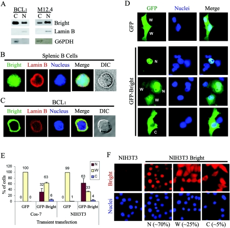
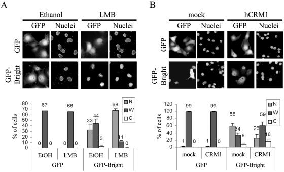
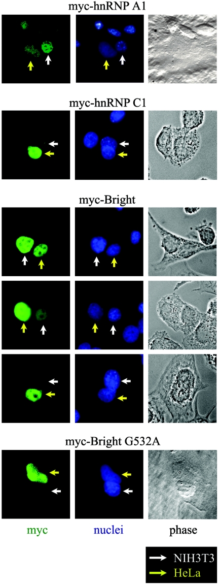
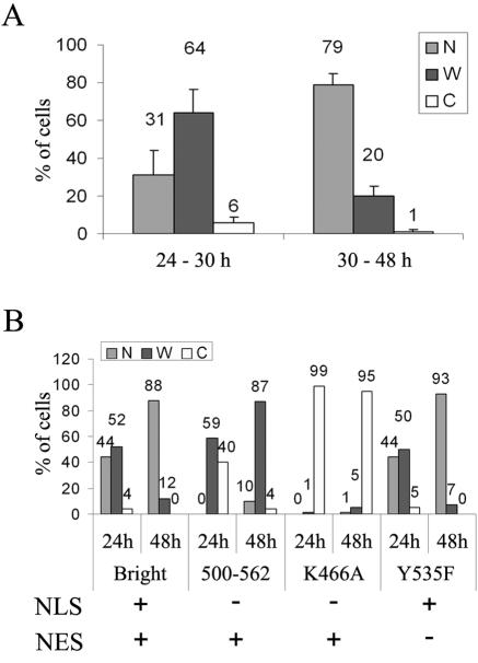
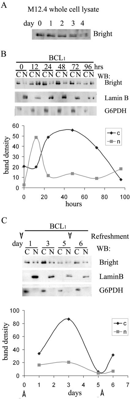
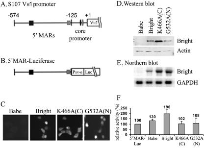
Bright/ARID3a has been implicated in mitogen- and growth factor-induced up-regulation of immunoglobulin heavy-chain (IgH) genes and in E2F1-dependent G1/S cell cycle progression. For IgH transactivation, Bright binds to nuclear matrix association regions upstream of certain variable region promoters and flanking the IgH intronic enhancer. While Bright protein was previously shown to reside within the nuclear matrix, we show here that a significant amount of Bright resides in the cytoplasm of normal and transformed B cells. Leptomycin B, chromosome region maintenance 1 (CRM1) overexpression, and heterokaryon experiments indicate that Bright actively shuttles between the nucleus and the cytoplasm in a CRM1-dependent manner. We mapped the functional nuclear localization signal to the N-terminal region of REKLES, a domain conserved within ARID3 paralogues. Residues within the C terminus of REKLES contain its nuclear export signal, whose regulation is primarily responsible for Bright shuttling. Growth factor depletion and cell synchronization experiments indicated that Bright shuttling during S phase of the cell cycle leads to an increase in its nuclear abundance. Finally, we show that shuttle-incompetent Bright point mutants, even if sequestered within the nucleus, are incapable of transactivating an IgH reporter gene. Therefore, regulation of Bright's cellular localization appears to be required for its function.


Figures

FIG. 1.
FIG. 1.
Nuclear and cytoplasmic localization of Bright. (A) Nuclear and cytoplasmic fractionation of Bright within B-cell lines BCL1 and M12.4. Five micrograms of nuclear or cytoplasmic proteins was analyzed by Western blotting with anti-Bright antiserum. The purity of the nuclear (lamin B) and cytoplasmic (G6PDH) fractions was confirmed by Western blotting of 20 μg of each extract with the appropriate antibodies. (B) Nucleocytoplasmic localization of Bright within mouse splenic B cells. Mouse CD43 splenic B cells were isolated from spleens using anti-CD43 antibody-conjugated magnetic beads. Costaining of the cells with anti-Bright (green, fluorescein isothiocyanate) and anti-lamin B (red, rhodamine) allowed discrimination between the nucleus and the relatively small volume of cytoplasm. DIC, differential interference contrast. (C) Cytoplasmic accumulation of Bright in a mature B-cell line, BCL1. Immunostaining with anti-Bright revealed various localization patterns, including a small portion of cells expressing Bright exclusively within their cytoplasm. (D) Cellular localization of GFP-Bright expressed ectopically in Cos-7 cells. The localization of GFP-Bright was determined at 24 h posttransfection of Cos-7 cells. W, dispersed across the whole cell; N, preferential nuclear localization; and C, preferential cytoplasmic localization. (E) Quantitative analysis of GFP-Bright and GFP-only localization in Cos-7 and NIH 3T3 cells after transient transfection. (F) Cellular localization of Bright ectopically and stably expressed in NIH 3T3 cells. Retroviral transduced, Bright-expressing NIH 3T3 cells or NIH 3T3 cells alone were immunostained with anti-Bright antiserum (red, rhodamine) and DAPI (blue). Population sizes are shown with the representative localization patterns. The data in panels E and F were quantified as described in Materials and Methods.
FIG. 2.
FIG. 2.
Bright is exported from the nucleus to the cytoplasm by CRM1. (A) LMB treatment produces enhanced nuclear accumulation of GFP-Bright. (Upper panel) Cos-7 cells were transiently transfected with GFP-Bright and after 12 h were treated with 10 ng/ml of LMB and incubated for another 12 h. The solvent used to dissolve LMB, ethanol (EtOH), was used for mock treatment. (Lower panel) Quantitation of increased nuclear localization of Bright after LMB treatment. (B) CRM1 enhanced the nuclear export of GFP-Bright. (Upper panel) Cos-7 cells were transiently cotransfected with GFP-Bright and CRM1, and GFP-Bright localization was observed at 24 h posttransfection. Cotransfection of Bright with pCR3.1 empty vector served as mock treatment. (Lower panel) Quantitation of increased cytoplasmic localization of Bright produced by CRM1 overexpression.
FIG.3.
FIG.3.
NLS and NES activities of Bright reside within different regions of the REKLES domain. (A) Schematic of Bright domains (boxes) and truncation mutants. REKLES is divided into N-terminal (α) and C-terminal (β) subdomains. The cellular localization phenotypes (W, N, and C; described in previous legend and in Materials and Methods) of each mutant analyzed with or without (mock) the addition of 10 ng/ml of LMB are shown to the right of each construct tested. The first character represents the most frequently observed pattern. Boxes at the bottom represent the minimal regions mapped for NLS and NES activities. (B, upper panel) The REKLESα subdomain and residues that are required for the nuclear import of Bright. Bold enlarged letters denote residues whose mutation abolished nuclear import. Mutation of the K (small bold) within the REKLES motif (underlined) had no effect on nuclear import. (Lower panel) Schematic of point mutation constructs and phenotypes. The vertical lines denote the positions of mutations within the REKLESα subdomain. (C) Residues within the REKLESβ subdomain required for the nuclear export of Bright. Point mutations, their positions, and localization phenotypes are represented as in panel B. (D) Exemplary images of the cellular localization of GFP-Bright and selected mutants collected 24 h posttransfection of Cos-7 cells.
FIG. 4.
FIG. 4.
Bright shuttles between the nucleus and cytoplasm. HeLa cells were transiently transfected with myc-Bright wild type or the NES-deficient G532A mutant and then fused with NIH 3T3 cells. Bright proteins were detected by immunostaining the resulting heterokaryons with anti-myc monoclonal antibody. myc-hnRNP A1 was previously shown to be a shuttling protein, whereas myc-hnRNP C1 served as a nucleus-only control (44).
FIG. 5.
FIG. 5.
Reduction in NES activity can lead to enhanced nuclear accumulation of ectopically expressed Bright following prolonged culture. (A) Summary of localization phenotypes for wild-type GFP-Bright analyzed early (24 to 30 h) or late (30 to 48 h) posttransfection of Cos-7 cells. (B) Comparison of localization phenotypes in short and prolonged cultures of the wild type and localization mutants in transiently transfected Cos-7 cells. Functions retained (+) or lost (−) in each protein are indicated below the corresponding data.
FIG. 6.
FIG. 6.
Fluctuation in the nucleocytoplasmic ratio of Bright in BCL1 B cells is induced by serum deprivation. (A) Overall Bright expression level is gradually reduced following replenishment. M12.4 cells were grown for 4 days without replenishment, and whole-cell lysates were prepared at the indicated time points. Five micrograms of lysates was analyzed for Bright expression. Equivalent protein loading among lanes was confirmed by ink staining (not shown). (B) Alteration of nuclear/cytoplasmic ratio of Bright in BCL1 B cells. (Upper panel) Western analysis of subcellular fractionation of Bright and nuclear (lamin B) or cytoplasmic (G6PDH) controls. Samples were prepared at 12-h intervals after replenishment (time zero). (Lower panel) Quantitation (Scion Image) of the data indicates an early (12 to 24 h) export-import phase, followed by a prolonged reduction in cytoplasmic Bright over 96 h. (C) Recovery of cytoplasmic expression of Bright following replenishment of cells. BCL1 cultures were manipulated as in panel B and then at day 5 were replenished with medium. Fractionations (at the indicated time points) and quantitation were performed as described above. Arrowheads represent the time points of replenishment.
FIG. 7.
FIG. 7.
Cellular localization of Bright is altered during S phase. (A) GFP-Bright-expressing Cos-7 cells are in S phase between 12 to 24 h posttransfection. DNA content of transfected Cos-7 cells was analyzed at the indicated time points through propidium iodide staining and fluorescence-activated cell sorter analysis. (B) Nuclear/cytoplasmic ratio of Bright increases during S phase in BCL1 cells. (Upper panel) Cell cycle analysis after synchronization of BCL1 cells at G1/S with serum reduction and aphidicolin treatment. Four hours after release of the cell cycle block, a significant number of the cells are in S phase. (Middle panel) Bright fractionation at the indicated times after release of the cell cycle block. Five micrograms of nuclear or cytoplasmic proteins was analyzed by Western blotting. (Lower panel) Quantitation (Scion Image) of the data indicates an increase in the nuclear/cytoplasmic ratio of Bright at 4 h after removing aphidicolin, coinciding with the majority of cells being in early S phase.
FIG. 8.
FIG. 8.
Shuttling activity is required for Bright to transactivate an integrated IgH promoter-associated MAR reporter in NIH 3T3 cells. (A and B) Schematic of (A) the S107 VH1 5′ upstream region and (B) the 5′ MAR luciferase reporter construct. Squares represent MARs previously shown (21) to contain specific Bright binding sites (centered at −574 and −125 with respect to transcriptional start of VH1) required for promoter activation by Bright in B cells. Small black and gray ovals denote octB and octamer binding sites. (C) Cellular localization of Bright wild type and mutants stably expressed in NIH 3T3 cells bearing integrated 5′ MAR luciferase reporters. Cytoplasmic-restricted expression (C) of NLS point mutant K466A, nuclearly restricted expression (N) of NES point mutant G532A, and variable expression of the wild type were confirmed by anti-Bright immunostaining. (D and E) Wild-type and mutant Bright expression levels in 5′ MAR luciferase/NIH 3T3. For the Western blot shown in panel D, 5 μg of each whole-cell lysate was analyzed with anti-Bright, and 20 μg of each lysate was used for the antiactin loading control. For the Northern blot shown in panel E, total RNA was hybridized with Bright or GAPDH-specific probes. (F) Relative Bright transactivation in each 5′ MAR luciferase/NIH 3T3 cell line. Luciferase activities were measured from 10 μg of whole-cell extract prepared from Bright wild-type or mutant transductants. Babe denotes mock infection with the pBabe retroviral backbone (10). K466A is the cytoplasmically restricted (C) mutant, and G532A is the nuclearly restricted (N) mutant. Each column and vertical bar represent the mean and standard deviation from four independent cultures of retrovirally transduced cell lines, respectively. Bright, P < 0.05; others, P > 0.05.
FIG. 9.
FIG. 9.
Functional domains and hypothesized regulation of Bright nucleocytoplasmic shuttling. (A) Domains contributing to Bright localization. In addition to motifs within the REKLESα (NLS-required) and REKLESβ (NES-required) subdomains, data from mutants shown in Fig. 3 indicate that regions (lower boxes) partially overlapping the ARID and the acidic domain (upper boxes) affect cellular localization of Bright through inter- or intramolecular interactions (arrows). Protein Y represents an unknown factor(s) required to mediate nuclear export in lieu of a conventional NES motif within REKLESβ. (B) Hypothesized regulation of Bright localization during B-cell growth under limiting growth factor conditions (fasting). At the top is a reproduction of Bright subcellular fractionation in serum-deprived BCL1 cells. This fluctuation pattern may be controlled by two distinct mechanisms. First, nuclear export may be regulated. After replenishing cells with growth factors, overall Bright expression (denoted by the number of circles) increases, but the nuclear export activity remains low, resulting in a unidirectional (small arrows at 12 h) nuclear accumulation of Bright. Shortly thereafter, robust nuclear export activity (large arrows) results in a rapid (day 1, 2) accumulation of Bright in the cytoplasm. However, our data indicate that a pool of Bright may remain behind, by virtue of interaction with an unknown nuclear factor(s) (X) that mediates a second control mechanism, nuclear retention. This ensures that a minimal level of Bright must be retained in the nucleus irrespective of its overall expression level. The remaining Bright pool undergoes nucleocytoplasmic shuttling (bidirectional arrows), which persists throughout the course of the fasting/growth factor-depleting culture.

Similar articles

Cited by

References

    1. Alt, J. R., J. L. Cleveland, M. Hannink, and J. A. Diehl. 2000. Phosphorylation-dependent regulation of cyclin D1 nuclear export and cyclin D1-dependent cellular transformation. Genes Dev. 14:3102-3114. - PMC - PubMed
    1. Alt, J. R., A. B. Gladden, and J. A. Diehl. 2002. p21(Cip1) promotes cyclin D1 nuclear accumulation via direct inhibition of nuclear export. J. Biol. Chem. 277:8517-8523. - PubMed
    1. Avitahl, N., and K. A. Calame. 1996. 125 bp region of the Ig VH1 promoter is sufficient to confer lymphocyte-specific expression in transgenic mice. Int. Immunol. 8:1359-1366. - PubMed
    1. Benditt, J. O., C. Meyer, H. Fasold, F. C. Barnard, and N. Riedel. 1989. Interaction of a nuclear location signal with isolated nuclear envelopes and identification of signal-binding proteins by photoaffinity labeling. Proc. Natl. Acad. Sci. USA 86:9327-9331. - PMC - PubMed
    1. Blanc, V., S. Kennedy, and N. O. Davidson. 2003. A novel nuclear localization signal in the auxiliary domain of apobec-1 complementation factor regulates nucleocytoplasmic import and shuttling. J. Biol. Chem. 278:41198-41204. - PubMed

Publication types

MeSH terms