. 2006 Feb 15;26(7):2041-52.
doi: 10.1523/JNEUROSCI.4566-05.2006.

Functional dissection of Reelin signaling by site-directed disruption of Disabled-1 adaptor binding to apolipoprotein E receptor 2: distinct roles in development and synaptic plasticity

Affiliations

Functional dissection of Reelin signaling by site-directed disruption of Disabled-1 adaptor binding to apolipoprotein E receptor 2: distinct roles in development and synaptic plasticity

Uwe Beffert et al. J Neurosci. .

Abstract

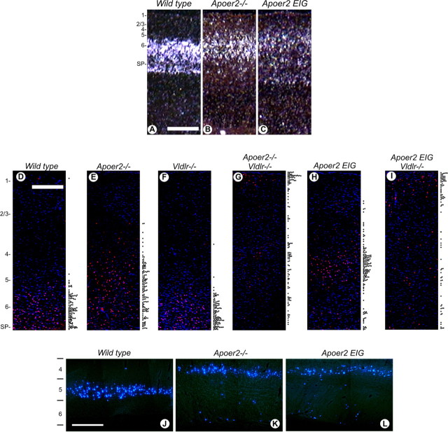
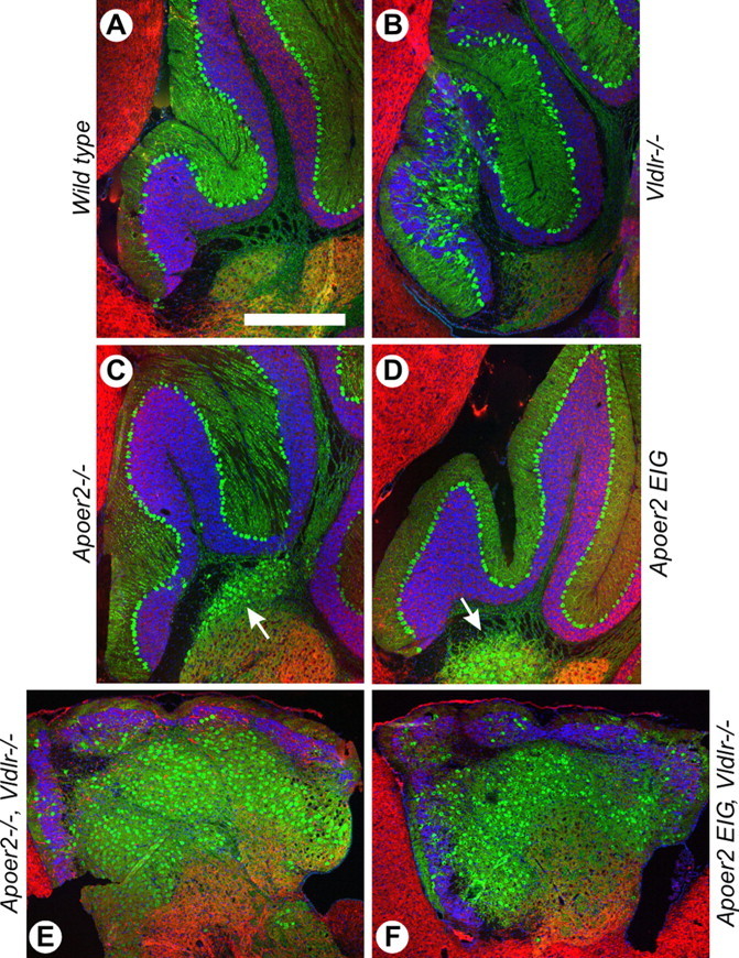
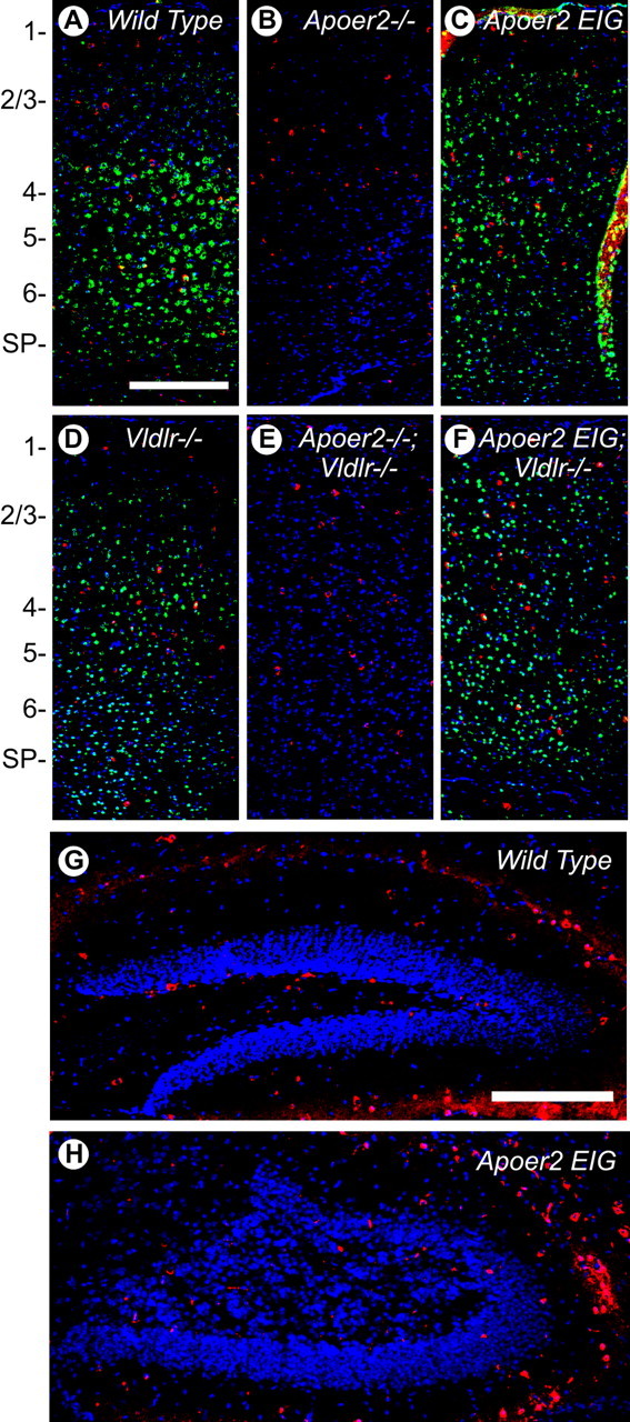
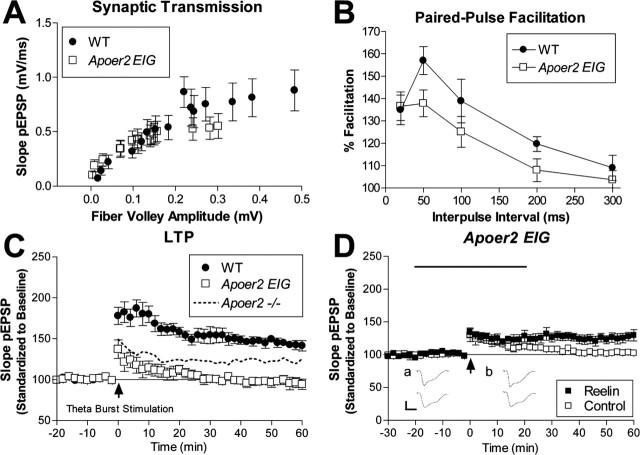
The Reelin signaling pathway controls neuronal positioning in human and mouse brain during development as well as modulation of long-term potentiation (LTP) and behavior in the adult. Reelin signals by binding to two transmembrane receptors, apolipoprotein E receptor 2 (Apoer2) and very-low-density lipoprotein receptor. After Reelin binds to the receptors, Disabled-1 (Dab1), an intracellular adaptor protein that binds to the cytoplasmic tails of the receptors, becomes phosphorylated on tyrosine residues, initiating a signaling cascade that includes activation of Src-family kinases and Akt. Here, we have created a line of mutant mice (Apoer2 EIG) in which the Apoer2 NFDNPVY motif has been altered to EIGNPVY to disrupt the Apoer2-Dab1 interaction to further study Reelin signaling in development and adult brain. Using primary neuronal cultures stimulated with recombinant Reelin, we find that normal Reelin signaling requires the wild-type NFDNPVY sequence and likely the interaction of Apoer2 with Dab1. Furthermore, examination of hippocampal, cortical, and cerebellar layering reveals that the NFDNPVY sequence of Apoer2 is indispensable for normal neuronal positioning during development of the brain. Adult Apoer2 EIG mice display severe abnormalities in LTP and behavior that are distinct from those observed for mice lacking Apoer2. In Apoer2 EIG slices, LTP degraded to baseline within 30 min, and this was prevented in the presence of Reelin. Together, these findings emphasize the complexity of Reelin signaling in the adult brain, which likely requires multiple adaptor protein interactions with the intracellular domain of Apoer2.


Figures

Figure 1.
Figure 1.
Generation and basic characterization of Apoer2 EIG mice. A, Schematic representation of mouse Apoer2 demonstrating the cytoplasmic location of the wild-type NFDNPVY sequence and the mutated sequence EIGNPVY introduced into Apoer2 EIG. The position of the alternatively spliced exon 19 is indicated. The diagram is not drawn to scale. B, PCR genotyping of Apoer2 EIG homozygous (lanes 1 and 4), heterozygous (lane 2), and wild-type (lane 3) mice. C, The Dab1 protein interacts with wild-type Apoer2 but not the Apoer2 EIG mutant receptor. GST fusion proteins containing the cytoplasmic domains of wild-type Vldlr (lane 3) and Apoer2 (lane 5), but not the mutated Apoer2 EIG (lane 4) or GST control (lane 2), bound Dab1 from transfected human embryonic kidney 293 cells. D, Reelin signaling in Apoer2 EIG embryos is disrupted. Cultured primary neurons (E16; 5 d in culture) from Vldlr−/− (lanes 1 and 2), Apoer2−/−;Vldlr−/− (lanes 3 and 4), and Apoer2 EIG;Vldlr−/− (lanes 5 and 6) mice were treated with control (lanes 1, 3, and 5) or Reelin-conditioned (lanes 2, 4, and 6) medium. Lysates were collected, run on SDS-PAGE, transferred to nitrocellulose, and immunoblotted using antibodies against the Apoer2 extracellular domain (α-ED) or C terminus (α-CT), phosphotyrosine to detect Dab1 phosphorylation (4G10), total Dab1, p-SFK, CDK5, serine phosphorylated Akt, total Akt, and serine phosphorylated GSK3β.
Figure 2.
Figure 2.
Structural differences in binding of Dab1 by native Apoer2 and the Apoer2 EIG mutant. A, Interaction between the Dab1 PTB domain and the native ApoER2 NFDNPVYRKT peptide sequence (from PDB coordinates 1NTV). Based on previous convention, Y is designated as the “0” position. The Dab1 PTB domain is shown as a molecular surface representation (gray), whereas the Apoer2 peptide is shown in ball-and-stick form (yellow; Corey–Pauling–Koltun). Amino acids in the peptide sequence are labeled in yellow. Residues of Dab1 that form hydrogen bonds with the N or D residues of the peptide are labeled in black, and the hydrogen bonds are represented by orange dashed lines. Residues of Dab1 that form hydrophobic interactions with the F residue of the peptide are labeled in blue. B, Modeled interaction of the Dab1 PTB domain and the Apoer2 EIG mutant EIGNPVYRKT sequence. The Dab1 PTB domain, its labeled residues, and the unmodified residues of the peptide are shown as in A. The mutated peptide residues and the corresponding labels are colored salmon.
Figure 3.
Figure 3.
Brain histopathology of neuronal positioning defects in Apoer2 mouse mutants in the presence and absence of Vldlr. A–R, Hematoxylin and eosin staining of sagittal brain sections illustrating neuronal positioning in P21 mouse cortex layers 1–3 (A–F), hippocampus (G–L), and cerebellum (M–R) in wild-type (A, G, M), Apoer2−/− (B, H, N), Apoer2 EIG (C, I, O), Vldlr−/− (D, J, P), Apoer2−/−; Vldlr−/− (E, K, Q), and Apoer2 EIG; Vldlr−/− (F, L, R) mice. Scale bars: (in A) cortex and cerebellum, 500 μm; (in G) hippocampus, 250 μm.
Figure 4.
Figure 4.
Molecular marker analyses of cortical layering in Apoer2 mouse mutants. In situ hybridization for TLE4 (A–C) in P1 mouse cortex of wild-type (A), Apoer2−/− (B), and Apoer2 EIG (C) mutant mice demonstrating misplaced layer 6 and subplate neurons is shown. Indirect immunofluorescence detection of Foxp2-labeled cells in wild-type (D), Apoer2−/− (E), Vldlr−/− (F), Apoer2−/−; Vldlr−/− (G), Apoer2 EIG (H), and Apoer2 EIG; Vldlr−/− (I) mutant cortex is shown at P21. The relative position of individual Foxp2-expressing neurons (red) within the neocortical layers is represented by the scatter plot to the right of each section (D–I). Nuclei (blue) are labeled with DAPI. J–L, Direct immunofluorescence detection of corticospinal neurons labeled with Fast Blue highlighting cortical layer 5 neuron displacement in P28 brain. The numbers 1–6 represent cortical layers. SP, Subplate. Scale bars, 500 μm.
Figure 5.
Figure 5.
Cerebellar neuronal positioning defects in Apoer2 mouse mutants. A–F, Immunohistochemistry for the calcium binding protein calbindin (green) and MAP2 (red) in P21 mouse cerebellum in wild-type (A), Vldlr−/− (B), Apoer2−/− (C), Apoer2 EIG (D), Apoer2−/−; Vldlr−/− (E), and Apoer2 EIG; Vldlr−/− (F) mice. Nuclei are stained blue with DAPI. The arrows in C and D indicate ectopic calbindin staining in the cerebellar medulla. Rostral is to the left, and dorsal to the top. Scale bar: (in A) A–F, 500 μm.
Figure 6.
Figure 6.
Abnormal neuronal positioning in the dentate gyrus. A–F, Immunohistochemistry for the calcium binding protein calbindin (green) and MAP2 (red) in P21 mouse dentate gyrus in wild-type (A), Vldlr−/− (B), Apoer2−/− (C), Apoer2−/−; Vldlr−/− (D), Apoer2 EIG (E), and Apoer2 EIG; Vldlr−/− (F) mice. Nuclei are stained blue with DAPI. Scale bar, 250 μm.
Figure 7.
Figure 7.
Reelin is expressed in adult cortex and hippocampus. A–F, Indirect immunofluorescence for Reelin (red) and Apoer2 (green) in wild-type (A), Apoer2−/− (B), Vldlr−/− (C), Apoer2 EIG (D), Apoer2−/−; Vldlr−/− (E), and Apoer2 EIG; Vldlr−/− (F) cortex from P21 mice. G, H, Representative sections from wild type (G) and Apoer2 EIG (H) showing indirect Reelin immunofluorescence (red) in the dentate region of the hippocampus. Nuclei are stained blue with DAPI. The numbers 1–6 represent cortical layers. SP, Subplate. Scale bars: A, 500 μm; G, 250 μm.
Figure 8.
Figure 8.
Electrophysiological defects in Apoer2 EIG mutants distinct from Apoer2−/−. A, Synaptic transmission is represented as the slope of the field EPSP versus the fiber volley amplitude at increasing stimulus intensities for wild type (•; n = 9) and Apoer2 EIG (□; n = 14). B, Short-term synaptic plasticity is evaluated by the amount of paired-pulse facilitation with interpulse intervals of 20, 50, 100, 200, and 300 ms in wild type (•; n = 9) and Apoer2 EIG (□; n = 9). C, LTP induced with theta-burst stimulation consisting of five trains of four pulses at 100 Hz with an interburst interval of 20 s. Apoer2 EIG mutants display reduced LTP (□; n = 6) compared with wild-type mice (•; n = 6). [For comparison, the dotted line represents LTP induced in Apoer2 −/− (Weeber et al., 2002).] D, Hippocampal slices perfused with Reelin (▪), in combination with theta-burst stimulation, caused a slight but significant increase of LTP compared with control medium (□) in Apoer2 EIG (▪, n = 9; □, n = 8). Insets, Representative pEPSP traces from Reelin perfusion experiments (mean ± SEM of 6 successive EPSPs) immediately before HFS (a) and at 60 min after tetanus (b) obtained from Reelin (top traces) or control (bottom traces) perfusion experiments.
Figure 9.
Figure 9.
Normal associative and spatial learning requires Dab1 interaction with Apoer2. Fear conditioning/associative learning. Two-trial fear conditioning to assess associative learning 1 and 24 h after two CS–unconditioned stimulus pairings. A, No significant difference in the conditioned response was observed during the cue test between wild-type (n = 19) and Apoer2 EIG (n = 12) mice at either time point. B, Assessment of freezing to the context revealed a conditioned response that was greater in wild type than in Apoer2 EIG (n = 12) at both times tested. Morris water maze task/spatial learning. C–F, Results of escape latency (C), distance to platform (D), wall hugging (thigmotaxis) (E), and swim speed (F) for wild type and Apoer2 EIG during the acquisition phase in the hidden platform task. G, H, Percentage of time spent in the target or opposite quadrant (G) or the number of platform crossings (H) during a probe trial performed on day 12 (mean ± SEM; number of n for training consistent with probe trails; *p < 0.05 compared with wild type).

Similar articles

Cited by

References

    1. Alcantara S, Ruiz M, D’Arcangelo G, Ezan F, de Lecea L, Curran T, Sotelo C, Soriano E (1998). Regional and cellular patterns of reelin mRNA expression in the forebrain of the developing and adult mouse. J Neurosci 18:7779–7799. - PMC - PubMed
    1. Andersen OM, Yeung CH, Vorum H, Wellner M, Andreassen TK, Erdmann B, Mueller EC, Herz J, Otto A, Cooper TG, Willnow TE (2003). Essential role of the apolipoprotein E receptor-2 in sperm development. J Biol Chem 278:23989–23995. - PubMed
    1. Arnaud L, Ballif BA, Forster E, Cooper JA (2003). Fyn tyrosine kinase is a critical regulator of disabled-1 during brain development. Curr Biol 13:9–17. - PubMed
    1. Assadi AH, Zhang G, Beffert U, McNeil RS, Renfro AL, Niu S, Quattrocchi CC, Antalffy BA, Sheldon M, Armstrong DD, Wynshaw-Boris A, Herz J, D’Arcangelo G, Clark GD (2003). Interaction of reelin signaling and Lis1 in brain development. Nat Genet 35:270–276. - PubMed
    1. Beffert U, Morfini G, Bock HH, Reyna H, Brady ST, Herz J (2002). Reelin-mediated signaling locally regulates protein kinase B/Akt and glycogen synthase kinase 3beta. J Biol Chem 277:49958–49964. - PubMed

Publication types

Substances

LinkOut - more resources