

Coherent corticomuscular oscillations originate from primary motor cortex: evidence from patients with early brain lesions
- PMID: 16475178
- PMCID: PMC6871432
- DOI: 10.1002/hbm.20220
Coherent corticomuscular oscillations originate from primary motor cortex: evidence from patients with early brain lesions
Abstract
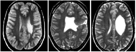
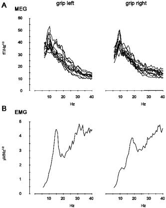
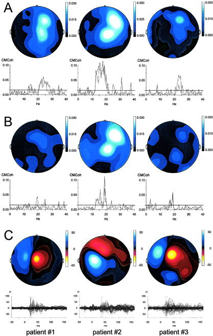
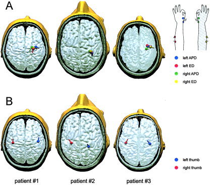
Coherent oscillations of neurons in the primary motor cortex (M1) have been shown to be involved in the corticospinal control of muscle activity. This interaction between M1 and muscle can be measured by the analysis of corticomuscular coherence in the beta-frequency range (beta-CMCoh; 14-30 Hz). Largely based on magnetoencephalographic (MEG) source-modeling data, it is widely assumed that beta-CMCoh reflects direct coupling between M1 and muscle. Deafferentation is capable of modulating beta-CMCoh, however, and therefore the influence of reafferent somatosensory signaling and corresponding neuronal activity in the somatosensory cortex (S1) has been unclear. We present transcranial magnetic stimulation (TMS) and MEG data from three adult patients suffering from congenital hemiparesis due to pre- and perinatally acquired lesions of the pyramidal tract. In these patients, interhemispheric reorganization had resulted in relocation of M1 to the contralesional hemisphere, ipsilateral to the paretic hand, whereas S1 had remained in the lesioned hemisphere. This topographic dichotomy allowed for an unequivocal topographic differentiation of M1 and S1 with MEG (which is not possible if M1 and S1 are directly adjacent within one hemisphere). In all patients, beta-CMCoh originated from the contralesional M1, in accordance with the TMS-evoked motor responses, and in contrast to the somatosensory evoked fields (SEFs) for which the sources (N20m) were localized in S1 of the lesioned hemisphere. These data provide direct evidence for the concept that beta-CMCoh reflects the motorcortical efferent drive from M1 to the spinal motoneuron pool and muscle. No evidence was found for a relevant contribution of neuronal activity in S1 to beta-CMCoh.
Figures
Similar articles
-
Cerebro-muscular and cerebro-cerebral coherence in patients with pre- and perinatally acquired unilateral brain lesions.Neuroimage. 2007 Oct 1;37(4):1301-14. doi: 10.1016/j.neuroimage.2007.05.053. Epub 2007 Jun 12. Neuroimage. 2007. PMID: 17669666
-
Two types of exercise-induced neuroplasticity in congenital hemiparesis: a transcranial magnetic stimulation, functional MRI, and magnetoencephalography study.Dev Med Child Neurol. 2013 Oct;55(10):941-51. doi: 10.1111/dmcn.12209. Epub 2013 Aug 13. Dev Med Child Neurol. 2013. PMID: 23937719
-
(Re-)organization of basal ganglia in congenital hemiparesis with ipsilateral cortico-spinal projections.Neuropediatrics. 2008 Oct;39(5):252-8. doi: 10.1055/s-0029-1202284. Epub 2009 Mar 17. Neuropediatrics. 2008. PMID: 19294597
-
Neuromagnetic integrated methods tracking human brain mechanisms of sensorimotor areas 'plastic' reorganisation.Brain Res Brain Res Rev. 2000 Sep;33(2-3):131-54. doi: 10.1016/s0169-328x(00)00090-5. Brain Res Brain Res Rev. 2000. PMID: 11011062 Review.
-
Integrated technology for evaluation of brain function and neural plasticity.Phys Med Rehabil Clin N Am. 2004 Feb;15(1):263-306. doi: 10.1016/s1047-9651(03)00124-4. Phys Med Rehabil Clin N Am. 2004. PMID: 15029909 Review.
Cited by
-
Developmental tuning and decay in senescence of oscillations linking the corticospinal system.J Neurosci. 2010 Mar 10;30(10):3663-74. doi: 10.1523/JNEUROSCI.5621-09.2010. J Neurosci. 2010. PMID: 20220000 Free PMC article.
-
Corticomuscular coherence between motor cortex, somatosensory areas and forearm muscles in the monkey.Front Syst Neurosci. 2010 Jul 30;4:38. doi: 10.3389/fnsys.2010.00038. eCollection 2010. Front Syst Neurosci. 2010. PMID: 20740079 Free PMC article.
-
Reorganization of functional and directed corticomuscular connectivity during precision grip from childhood to adulthood.Sci Rep. 2021 Nov 24;11(1):22870. doi: 10.1038/s41598-021-01903-1. Sci Rep. 2021. PMID: 34819532 Free PMC article.
-
Distinct sensorimotor feedback loops for dynamic and static control of primate precision grip.Commun Biol. 2020 Apr 2;3(1):156. doi: 10.1038/s42003-020-0861-0. Commun Biol. 2020. PMID: 32242085 Free PMC article.
-
Corticomuscular Coherence and Its Applications: A Review.Front Hum Neurosci. 2019 Mar 20;13:100. doi: 10.3389/fnhum.2019.00100. eCollection 2019. Front Hum Neurosci. 2019. PMID: 30949041 Free PMC article. Review.
References
-
- Braun C, Heinz U, Schweizer R, Wiech K, Birbaumer N, Topka H ( 2001): Dynamic organization of the somatosensory cortex induced by motor activity. Brain 124: 2259–2267. - PubMed
-
- Braun C, Schweizer R, Heinz U, Wiech K, Birbaumer N, Topka H ( 2003): Task‐specific plasticity of somatosensory cortex in patients with writer's cramp. Neuroimage 20: 1329–1338. - PubMed
-
- Brown P, Salenius S, Rothwell JC, Hari R ( 1998): Cortical correlate of the Piper rhythm in humans. J Neurophysiol 80: 2911–2917. - PubMed
-
- Carr LJ, Harrison LM, Evans AL, Stephens JA ( 1993): Patterns of central motor reorganization in hemiplegic cerebral palsy. Brain 116: 1223–1247. - PubMed
Publication types
MeSH terms
LinkOut - more resources
Full Text Sources
