. 2006 Apr;17(4):1768-78.
doi: 10.1091/mbc.e05-09-0839. Epub 2006 Feb 8.

Yeast nuclear envelope subdomains with distinct abilities to resist membrane expansion

Affiliations

Yeast nuclear envelope subdomains with distinct abilities to resist membrane expansion

Joseph L Campbell et al. Mol Biol Cell. 2006 Apr.

Abstract

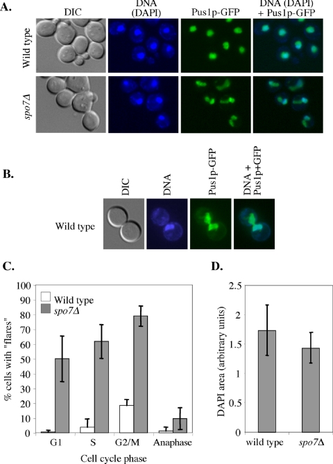
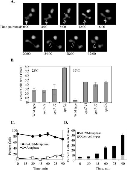
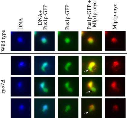
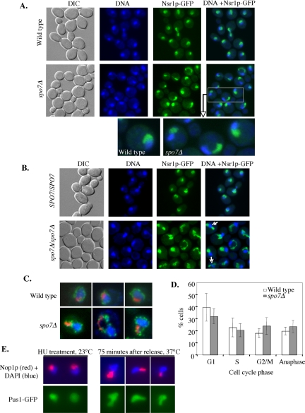
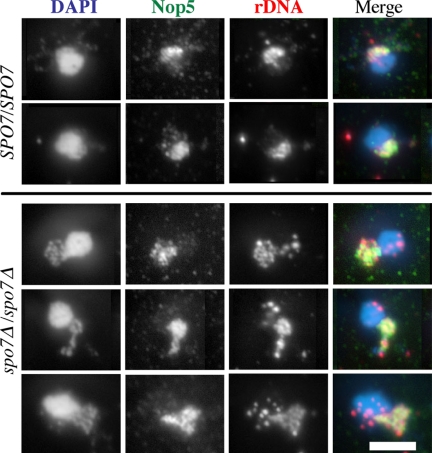
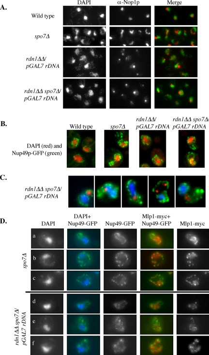
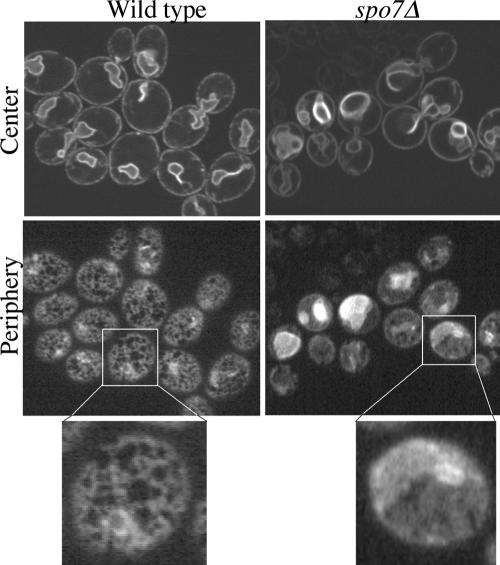
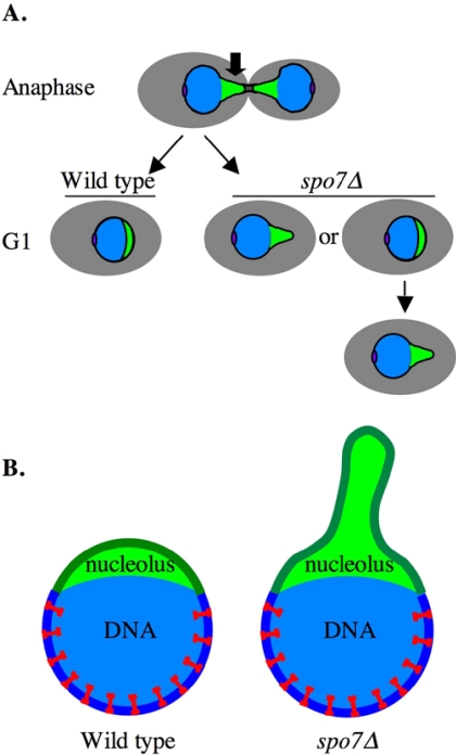
Little is known about what dictates the round shape of the yeast Saccharomyces cerevisiae nucleus. In spo7Delta mutants, the nucleus is misshapen, exhibiting a single protrusion. The Spo7 protein is part of a phosphatase complex that represses phospholipid biosynthesis. Here, we report that the nuclear protrusion of spo7Delta mutants colocalizes with the nucleolus, whereas the nuclear compartment containing the bulk of the DNA is unaffected. Using strains in which the nucleolus is not intimately associated with the nuclear envelope, we show that the single nuclear protrusion of spo7Delta mutants is not a result of nucleolar expansion, but rather a property of the nuclear membrane. We found that in spo7Delta mutants the peripheral endoplasmic reticulum (ER) membrane was also expanded. Because the nuclear membrane and the ER are contiguous, this finding indicates that in spo7Delta mutants all ER membranes, with the exception of the membrane surrounding the bulk of the DNA, undergo expansion. Our results suggest that the nuclear envelope has distinct domains that differ in their ability to resist membrane expansion in response to increased phospholipid biosynthesis. We further propose that in budding yeast there is a mechanism, or structure, that restricts nuclear membrane expansion around the bulk of the DNA.


Figures

Figure 1.
Figure 1.
The presence of flares in the spo7Δ mutant is cell cycle regulated. (A) Strains JCY619 (SPO7 pPUS1-GFP) and JCY617 (spo7Δ pPUS1-GFP) were grown to early log phase and imaged for GFP (nucleus, green) and DAPI (chromosomes, blue). “Flares” were defined as nuclear protrusions that do not contain DAPI staining material. (B) A typical flare of wild-type cells (JCY619). (C) Cell cycle distribution of flares. The data are shown as the percentage of cells with flares out of the total number of cells in a particular cell cycle phase. G1, unbudded cells with a single nucleus; S, small budded cells with a single nucleus; G2/M, large budded cells with a single nucleus; anaphase, large budded cells with two DNA masses. White columns, wild type; gray columns, spo7Δ. Strains are as described in A. (D) Measurements of the area occupied by DAPI staining material in wild-type and spo7Δ strains, described in A. Stacked images were taken at 2-μm intervals, and the DAPI area was measured at the focal plane with largest circumference. The number of nuclei counted was 24 and 20 for wild-type and spo7Δ cells, respectively.
Figure 2.
Figure 2.
Flare formation before and after anaphase. (A) Strain JCY617 (spo7Δ pPUS1-GFP) were embedded on 25% gelatin slabs prepared in YPD and visualized by confocal microscopy, taking a series of stacks every 4 min. A 32-min segment is shown. Note that at time 0, there are two nuclei in the field: the nucleus in the upper right corner remained in interphase for the duration of the experiment, whereas the nucleus in the lower left corner underwent nuclear division. The arrow points to the flare in the latter nucleus, and the arrowhead points to a remnant of the nuclear membrane that failed to be removed after nuclear division. The nucleus in the lower right corner at the 32:00-min time point also contains a flare, but because of the geometry of the plane of division it was not possible to determine whether this flare formed “de novo” or whether it was also a result of the failure to remove excess nuclear membrane after nuclear division. (B) Wild-type (JCY619), spo7-11 (JCY680), spo7-12 (JCY681), and spo7Δ (JCY617) strains, all containing the pPUS1-GFP plasmid, were grown at either 23 or 37°C for 16 h to mid-log phase and scored for the appearance of flares as described in Figure 1. (C) Cell cycle distribution of spo7-12 (JCY681) cells that were released from an S-phase arrest at 37°C at time 0; samples were taken at the indicated time points. The S/G2/metaphase cells represent large budded cells with a single nucleus, whereas anaphase cells are large budded with an elongated nucleus or two separate nuclei. A significant fraction of cells were in anaphase at 120 min after the release (our unpublished data). (D) For each time point shown in C, the percentage of cells with flares was scored as a function of cell cycle stage (for each time point, n > 50 cells were scored in each of three separate experiments). Shown is the overall percentage of cells that had flares as well as the types of cells with flares (S/G2/metaphase versus other cell types), at each time point. Similar results were obtained with the spo7-11 allele (our unpublished data).
Figure 3.
Figure 3.
The Mlp1 protein is excluded from the flare. Strains OCF2189 (SPO7 MLP1-MYC pPUS1-GFP) and JCY545 (spo7Δ MLP1-MYC pPUS1-GFP) were grown to early log phase and processed for immunofluorescence using antibodies against the Myc tag, while maintaining the fluorescence of Pus1-GFP. Mlp1p-Myc, red; DNA, blue; and Pus1-GFP, green. Three typical images of spo7Δ cells are shown.
Figure 4.
Figure 4.
The flare colocalizes with the nucleolus. (A) Strains OCF2276-1–9B (SPO7 NSR1-GFP) and OCF2276-1-12C (spo7Δ NSR1-GFP) were grown to early log phase, fixed, and imaged for DNA (blue) and Nsr1p-GFP (green). Enlarged images of a wild-type and several spo7Δ cells are also shown. Identical results were obtained with Utp10p, Nop6p, and Srp1p tagged with GFP (our unpublished data). (B) Diploid strains OCF2285 (SPO7/SPO7 NSR1-GFP/NSR1-GFP) and OCF2284 (spo7Δ/spo7Δ NSR1-GFP/NSR1-GFP) were grown to early log phase, fixed, and examined for DNA (blue) and Nsr1p-GFP (green) as described in text. Note the appearance of two nucleoli in some of the spo7Δ/spo7Δ cells (arrows). (C) Strains OCF1533-1A (SPO7) and JCY565 (spo7Δ), both containing a plasmid coding for the nuclear pore protein Nup49p fused to GFP (pNUP49-URA), were grown to early log phase and processed for immunofluorescence using antibodies against the nucleolar protein Nop1p, while maintaining the GFP fluorescence of Nup49-GFP. Images taken for DAPI (blue), Nup49-GFP (green), and Nop1p (red) were overlaid. Three typical examples are shown. (D) Cell cycle distribution of wild-type and spo7Δ cultures. Cells were grown to early log phase, fixed, treated with DAPI, and counted for cell cycle stage. Data are shown as the average percentage of cells in a particular cell cycle stage out of the total number of cells counted. (E) spo7-12 pPUS1-GFP (JCY 681) cells, used in the HU experiment described in Figure 2, C and D, were collected at the HU arrest point (23°C) and at 75 min after release from the HU arrest (37°C) and analyzed by indirect immunofluorescence using antibodies against Nop1p while maintaining the Pus1-GFP fluorescence. Typical examples are shown.
Figure 5.
Figure 5.
The rDNA of spo7Δ mutants extends throughout the flare. Diploid wild type (JCY720) and spo7Δ/spo7Δ (JCY721) cells were grown to early log phase, fixed, and treated as described by Fuchs and Loidl (2004). The DNA was detected by DAPI staining (blue in the overlay), the nucleolus was detected with anti-Nop5p antibodies (green in the overlay), and the rDNA was detected with an rDNA probe (red in the overlay). Note that in the spo7Δ/spo7Δ cells the rDNA extends throughout the abnormally shaped nucleolus. Bar, 5 μm.
Figure 6.
Figure 6.
The formation of the spo7Δ flare is independent of nucleolar expansion. (A) Strains OCF1533-1A (SPO7), JCY565 (spo7Δ), NOY891 (rdn1ΔΔ pGAL7 rDNA), and JCY831 (rdn1ΔΔ pGAL7 rDNA spo7Δ) were grown to early log phase and processed for immunofluorescence using antibodies against Nop1p. (B) The same strains as in A, also carrying pNup49-URA, were fixed and stained for DAPI. Images are overlays of DAPI (red) and Nup49-GFP (green) images. (C) Strain JCY831 (rdn1ΔΔ pGAL7 rDNA spo7Δ) carrying pNup49-GFP was grown to early log phase and processed for immunofluorescence using antibodies against Nop1p. Images are overlays of DAPI (blue) Nup49-GFP (green), and Nop1p (red) images. For comparison with a spo7Δstrain, see Figure 3B. Images are representative of the different types of flare/nucleolus localizations observed in this strain background. (D) Strains spo7Δ MLP1-myc pNUP49-GFP (JCY831 and JCY832) and rdn1ΔΔ pGAL7 rDNA spo7Δ MLP1-myc pNUP49-GFP (JCY994 and JCY995) were grown to early log phase, fixed, and processed for indirect immunofluorescence as described above. In the overlays, DAPI is in blue, Nup49-GFP is in green, and Mlp1-Myc is in red.
Figure 7.
Figure 7.
spo7Δ mutants exhibit proliferation of the peripheral ER. Wild-type (JCY622) and spo7Δ (JCY620) cells expressing the ER-associated protein Sec63p-GFP from a centromeric plasmid (pJK59) were grown to early log phase and examined for Sec63p-GFP distribution. Confocal sections corresponding to the cell center (top row) and cell periphery (bottom row) are shown.
Figure 8.
Figure 8.
Schematic model to explain the mechanism of flare formation. (A) The structural changes experienced by the nucleus and nucleolus of dividing cells. Cell body, gray; the bulk of the DNA mass, blue; nucleolus, green; and spindle pole body, purple. Note that in anaphase, the nucleolus trails behind the bulk of the DNA. The arrow indicates the region of the nuclear membrane that has to be removed after the nucleus divides. In spo7Δ mutants, a flare may form due to incomplete removal of this membrane region at the end of anaphase, or it can form some time after nuclear division. (B) Model. The expansion of the nuclear membrane around the bulk of the DNA is restricted by a tethering mechanism, indicated as red staples, that is excluded from the nucleolus.

Similar articles

Cited by

References

    1. Andrulis, E. D., Zappulla, D. C., Ansari, A., Perrod, S., Laiosa, C. V., Gartenberg, M. R., and Sternglanz, R. (2002). Esc1, a nuclear periphery protein required for Sir4-based plasmid anchoring and partitioning. Mol. Cell. Biol. 22, 8292–8301. - PMC - PubMed
    1. Belgareh, N., and Doye, V. (1997). Dynamics of nuclear pore distribution in nucleoporin mutant yeast cells. J. Cell Biol. 136, 747–759. - PMC - PubMed
    1. Bystricky, K., Laroche, T., van Houwe, G., Blaszczyk, M., and Gasser, S. M. (2005). Chromosome looping in yeast: telomere pairing and coordinated movement reflect anchoring efficiency and territorial organization. J. Cell Biol. 168, 375–387. - PMC - PubMed
    1. Dundr, M., and Misteli, T. (2001). Functional architecture in the cell nucleus. Biochem. J. 356, 297–310. - PMC - PubMed
    1. Fabre, E., and Hurt, E. (1997). Yeast genetics to dissect the nuclear pore complex and nucleocytoplasmic trafficking. Annu. Rev. Genet. 31, 277–313. - PubMed

Publication types

MeSH terms

LinkOut - more resources