

Cellular identification of water gustatory receptor neurons and their central projection pattern in Drosophila
- PMID: 16415164
- PMCID: PMC1347963
- DOI: 10.1073/pnas.0502376103
Cellular identification of water gustatory receptor neurons and their central projection pattern in Drosophila
Abstract
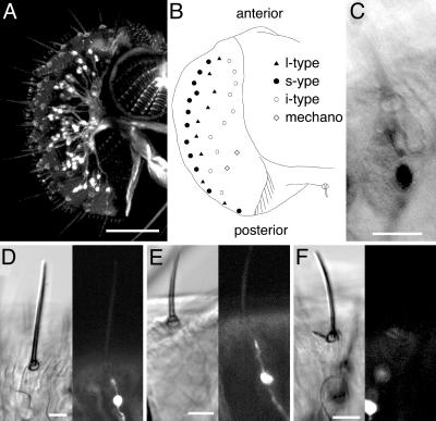
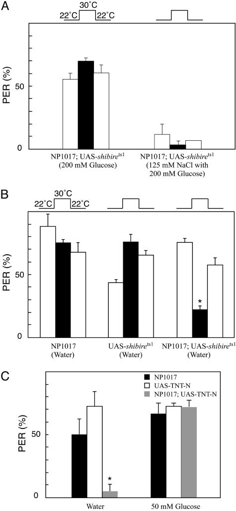
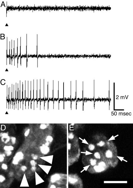
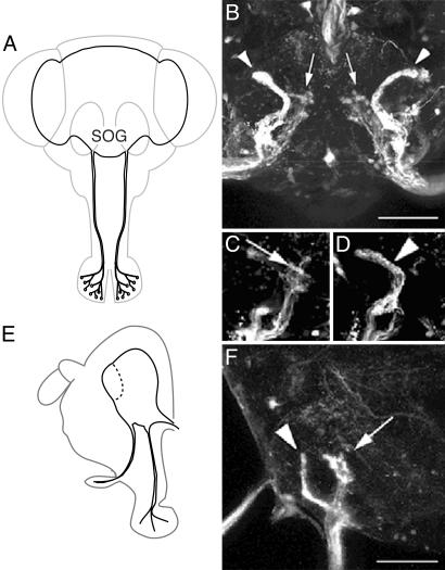
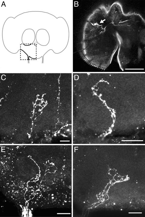
Water perception is important for insects, because they are particularly vulnerable to water loss because their body size is small. In Drosophila, gustatory receptor neurons are located at the base of the taste sensilla on the labellum, tarsi, and wing margins. One of the gustatory receptor neurons in typical sensilla is known to respond to water. To reveal the neural mechanisms of water perception in Drosophila, it is necessary to identify water receptor neurons and their projection patterns. We used a Gal4 enhancer trap strain in which GAL4 is expressed in a single gustatory receptor neuron in each sensillum on the labellum. We investigated the function of these neurons by expressing the upstream activating sequence transgenes, shibire(ts1), tetanus toxin light chain, or diphtheria toxin A chain. Results from the proboscis extension reflex test and electrophysiological recordings indicated that the GAL4-expressing neurons respond to water. We show here that the water receptor neurons project to a specific region in the subesophageal ganglion, thus revealing the water taste sensory map in Drosophila.
Figures
Similar articles
-
Spatially restricted expression of candidate taste receptors in the Drosophila gustatory system.Curr Biol. 2001 Jun 5;11(11):822-35. doi: 10.1016/s0960-9822(01)00258-5. Curr Biol. 2001. PMID: 11516643
-
Gustatory organs of Drosophila melanogaster: fine structure and expression of the putative odorant-binding protein PBPRP2.Cell Tissue Res. 2001 Jun;304(3):423-37. doi: 10.1007/s004410100388. Cell Tissue Res. 2001. PMID: 11456419
-
The molecular and cellular basis of taste coding in the legs of Drosophila.J Neurosci. 2014 May 21;34(21):7148-64. doi: 10.1523/JNEUROSCI.0649-14.2014. J Neurosci. 2014. PMID: 24849350 Free PMC article.
-
Molecular neurophysiology of taste in Drosophila.Cell Mol Life Sci. 2004 Jan;61(1):10-8. doi: 10.1007/s00018-003-3182-9. Cell Mol Life Sci. 2004. PMID: 14704850 Free PMC article. Review.
-
The organization of the chemosensory system in Drosophila melanogaster: a review.Cell Tissue Res. 1994 Jan;275(1):3-26. doi: 10.1007/BF00305372. Cell Tissue Res. 1994. PMID: 8118845 Review.
Cited by
-
The matricellular protein Drosophila Cellular Communication Network Factor is required for synaptic transmission and female fertility.Genetics. 2023 Mar 2;223(3):iyac190. doi: 10.1093/genetics/iyac190. Genetics. 2023. PMID: 36602539 Free PMC article.
-
A gustatory second-order neuron that connects sucrose-sensitive primary neurons and a distinct region of the gnathal ganglion in the Drosophila brain.J Neurogenet. 2015;29(2-3):144-55. doi: 10.3109/01677063.2015.1054993. Epub 2015 Aug 27. J Neurogenet. 2015. PMID: 26004543 Free PMC article.
-
Drosophila sensory receptors-a set of molecular Swiss Army Knives.Genetics. 2021 Mar 3;217(1):1-34. doi: 10.1093/genetics/iyaa011. Genetics. 2021. PMID: 33683373 Free PMC article. Review.
-
Common sense about taste: from mammals to insects.Cell. 2009 Oct 16;139(2):234-44. doi: 10.1016/j.cell.2009.10.001. Cell. 2009. PMID: 19837029 Free PMC article. Review.
-
Decaleside: a new class of natural insecticide targeting tarsal gustatory sites.Naturwissenschaften. 2012 Oct;99(10):843-52. doi: 10.1007/s00114-012-0966-5. Epub 2012 Sep 6. Naturwissenschaften. 2012. PMID: 22955371
References
-
- Falk, R., Bleiseravivi, N. & Atidia, J. (1976) J. Morphol. 150, 327–341. - PubMed
-
- Stocker, R. F. & Schorderet, M. (1981) Cell Tissue Res. 216, 513–523. - PubMed
-
- Fujishiro, N., Kijima, H. & Morita, H. (1984) J. Insect Physiol. 30, 317–325.
-
- Hiroi, M., Marion-Poll, F. & Tanimura, T. (2002) Zool. Sci. 19, 1009–1018. - PubMed
Publication types
MeSH terms
Substances
LinkOut - more resources
Full Text Sources
Molecular Biology Databases
