Neuronal or glial expression of human apolipoprotein e4 affects parenchymal and vascular amyloid pathology differentially in different brain regions of double- and triple-transgenic mice
- PMID: 16400027
- PMCID: PMC1592662
- DOI: 10.2353/ajpath.2006.050752
Neuronal or glial expression of human apolipoprotein e4 affects parenchymal and vascular amyloid pathology differentially in different brain regions of double- and triple-transgenic mice
Abstract
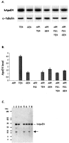
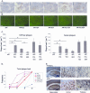
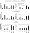
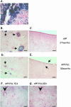
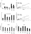
Apolipoprotein E4 (ApoE4) is associated with Alzheimer's disease by unknown mechanisms. We generated six transgenic mice strains expressing human ApoE4 in combination with mutant amyloid precursor protein (APP) and mutant presenilin-1 (PS1) in single-, double-, or triple-transgenic combinations. Diffuse, but not dense, amyloid plaque-load in subiculum and cortex was increased by neuronal but not glial ApoE4 in old (15 months) double-transgenic mice, whereas both diffuse and dense plaques formed in thalamus in both genotypes. Neuronal and glial ApoE4 promoted cerebral amyloid angiopathy as extensively as mutant PS1 but with pronounced regional differences: cortical angiopathy was induced by neuronal ApoE4 while thalamic angiopathy was again independent of ApoE4 source. Angiopathy correlated more strongly with soluble Abeta40 and Abeta42 levels in cortex than in thalamus throughout the six genotypes. Neither neuronal nor glial ApoE4 affected APP proteolytic processing, as opposed to mutant PS1. Neuronal ApoE4 increased soluble amyloid levels more than glial ApoE4, but the Abeta42/40 ratios were similar, although significantly higher than in single APP transgenic mice. We conclude that although the cellular origin of ApoE4 differentially affects regional amyloid pathology, ApoE4 acts on the disposition of amyloid peptides downstream from their excision from APP but without induction of tauopathy.
Figures


Similar articles
-
Prominent cerebral amyloid angiopathy in transgenic mice overexpressing the london mutant of human APP in neurons.Am J Pathol. 2000 Oct;157(4):1283-98. doi: 10.1016/S0002-9440(10)64644-5. Am J Pathol. 2000. PMID: 11021833 Free PMC article.
-
Lack of tau proteins rescues neuronal cell death and decreases amyloidogenic processing of APP in APP/PS1 mice.Am J Pathol. 2012 Dec;181(6):1928-40. doi: 10.1016/j.ajpath.2012.08.012. Epub 2012 Sep 28. Am J Pathol. 2012. PMID: 23026200
-
Human apolipoprotein E2 promotes parenchymal amyloid deposition and neuronal loss in vasculotropic mutant amyloid-β protein Tg-SwDI mice.J Alzheimers Dis. 2012;31(2):359-69. doi: 10.3233/JAD-2012-120421. J Alzheimers Dis. 2012. PMID: 22635103
-
Modeling Alzheimer's disease in transgenic mice: effect of age and of presenilin1 on amyloid biochemistry and pathology in APP/London mice.Exp Gerontol. 2000 Sep;35(6-7):831-41. doi: 10.1016/s0531-5565(00)00149-2. Exp Gerontol. 2000. PMID: 11053674 Review.
-
Cerebral amyloid angiopathy and dementia.Panminerva Med. 2004 Dec;46(4):253-64. Panminerva Med. 2004. PMID: 15876981 Review.
Cited by
-
Deletion of tumor necrosis factor death receptor inhibits amyloid beta generation and prevents learning and memory deficits in Alzheimer's mice.J Cell Biol. 2007 Aug 27;178(5):829-41. doi: 10.1083/jcb.200705042. J Cell Biol. 2007. PMID: 17724122 Free PMC article.
-
Chronic administration of anti-stroke herbal medicine TongLuoJiuNao reduces amyloidogenic processing of amyloid precursor protein in a mouse model of Alzheimer's disease.PLoS One. 2013;8(3):e58181. doi: 10.1371/journal.pone.0058181. Epub 2013 Mar 5. PLoS One. 2013. PMID: 23472157 Free PMC article.
-
Alzheimer's disease pathology in APOE transgenic mouse models: The Who, What, When, Where, Why, and How.Neurobiol Dis. 2020 Jun;139:104811. doi: 10.1016/j.nbd.2020.104811. Epub 2020 Feb 20. Neurobiol Dis. 2020. PMID: 32087290 Free PMC article. Review.
-
Rodent A beta modulates the solubility and distribution of amyloid deposits in transgenic mice.J Biol Chem. 2007 Aug 3;282(31):22707-20. doi: 10.1074/jbc.M611050200. Epub 2007 Jun 7. J Biol Chem. 2007. PMID: 17556372 Free PMC article.
-
Abeta40 inhibits amyloid deposition in vivo.J Neurosci. 2007 Jan 17;27(3):627-33. doi: 10.1523/JNEUROSCI.4849-06.2007. J Neurosci. 2007. PMID: 17234594 Free PMC article.
References
-
- Glenner GG, Wong CW. Alzheimer’s disease: initial report of the purification and characterization of a novel cerebrovascular amyloid protein. Biochem Biophys Res Commun. 1984;120:885–890. - PubMed
-
- Dewachter I, Van Leuven F. Secretases as targets for the treatment of Alzheimer’s disease: the prospects. Lancet Neurol. 2002;1:409–416. - PubMed
-
- Selkoe DJ. Defining molecular targets to prevent Alzheimer disease. Arch Neurol. 2005;2:192–195. - PubMed
-
- Corder EH, Saunders AM, Strittmatter WJ, Schmechel DE, Gaskell PC, Small GW, Roses AD, Haines JL, Pericak-Vance MA. Gene dose of apolipoprotein E type 4 allele and the risk of Alzheimer’s disease in late onset families. Science. 1993;261:921–923. - PubMed
-
- Bales KR, Dodart JC, DeMattos RB, Holtzman DM, Paul SM. Apolipoprotein E, amyloid, and Alzheimer disease. Mol Interv. 2002;6:363–375. - PubMed
Publication types
MeSH terms
Substances
LinkOut - more resources
Full Text Sources
Molecular Biology Databases

