. 2006 Jan 1;20(1):87-100.
doi: 10.1101/gad.357006.

Translational resistance of late alphavirus mRNA to eIF2alpha phosphorylation: a strategy to overcome the antiviral effect of protein kinase PKR

Affiliations

Translational resistance of late alphavirus mRNA to eIF2alpha phosphorylation: a strategy to overcome the antiviral effect of protein kinase PKR

Iván Ventoso et al. Genes Dev. .

Abstract

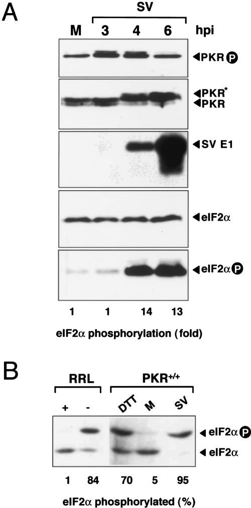
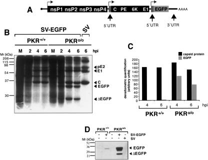
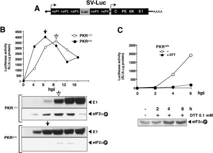
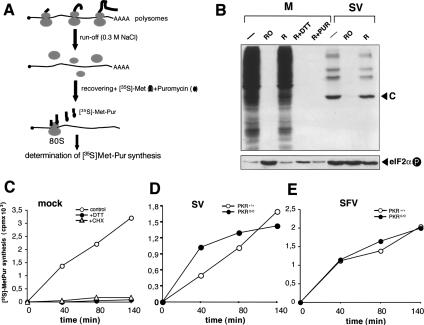
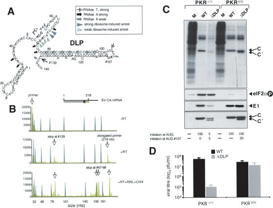
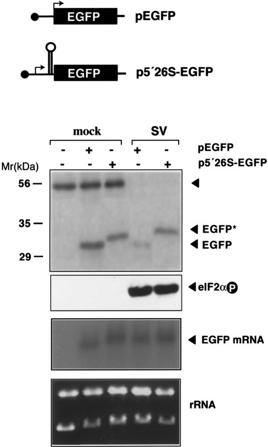
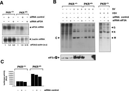
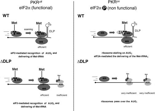
The double-stranded RNA-dependent protein kinase (PKR) is one of the four mammalian kinases that phosphorylates the translation initiation factor 2alpha in response to virus infection. This kinase is induced by interferon and activated by double-stranded RNA (dsRNA). Phosphorylation of eukaryotic initiation factor 2alpha (eIF2alpha) blocks translation initiation of both cellular and viral mRNA, inhibiting virus replication. To counteract this effect, most viruses express inhibitors that prevent PKR activation in infected cells. Here we report that PKR is highly activated following infection with alphaviruses Sindbis (SV) and Semliki Forest virus (SFV), leading to the almost complete phosphorylation of eIF2alpha. Notably, subgenomic SV 26S mRNA is translated efficiently in the presence of phosphorylated eIF2alpha. This modification of eIF2 does not restrict viral replication; SV 26S mRNA initiates translation with canonical methionine in the presence of high levels of phosphorylated eIF2alpha. Genetic and biochemical data showed a highly stable RNA hairpin loop located downstream of the AUG initiator codon that is necessary to provide translational resistance to eIF2alpha phosphorylation. This structure can stall the ribosomes on the correct site to initiate translation of SV 26S mRNA, thus bypassing the requirement for a functional eIF2. Our findings show the existence of an alternative way to locate the ribosomes on the initiation codon of mRNA that is exploited by a family of viruses to counteract the antiviral effect of PKR.


Figures

Figure 1.
Figure 1.
PKR activation and eIF2α phosphorylation in SV-infected cells. (A) 3T3 cells were infected with SV at an MOI of 25 PFU/cell. At the indicated times, cell extracts were made and analyzed by Western blot using the indicated antibodies. Bands corresponding to viral structural proteins E1, PKR, phospho-PKR, eIF2α, and phopho-eIF2α are shown. (Lower panel) The ratio of phosphorylated versus total eIF2α was estimated by densitometry of corresponding bands. (B) IEF analysis of eIF2α phosphorylation. 3T3 cells were SV-infected (4 h) or treated with 1 mM DTT (1 h), then analyzed by IEF (see Materials and Methods). Mock-infected cells (M) were included, as well as RRL-treated (+) or untreated (–), with hemine and EDTA as negative and positive controls of eIF2α phosphorylation, respectively. Phosphorylated and unphosphorylated eIF2α forms were quantified by densitometry.
Figure 2.
Figure 2.
SV 26S mRNA is specifically translated in PKR+/+ cells. (A) Scheme of recombinant SV expressing the EGFP gene under a second subgenomic promoter. Note that EGFP mRNA also contains the natural 5′ and 3′ UTRs present in 26S mRNA. Arrows indicate transcription initiation sites. (B) PKR+/+ and PKR0/0 cells were infected with SV-EGFP virus and metabolically labeled at indicated times. Bands corresponding to EGFP and a truncated form of the protein (ΔEGFP) are marked (see text for explanation). (C) Comparative analysis of EGFP versus SV C protein levels synthesized in PKR+/+ and PKR0/0 cells. Protein bands from the film shown in B were quantified by densitometry and plotted in arbitrary units. (D) Western blot analysis of EGFP accumulated in PKR+/+ and PKR0/0 cells. The blot was probed with a monoclonal anti-EGFP antibody (Clontech).
Figure 3.
Figure 3.
Premature phosphorylation of eIF2α blocks translation of genomic SV RNA. (A) Scheme of recombinant SV expressing luciferase from the genomic RNA. (B) Timing of eIF2α phosphorylation and translation of genomic and subgenomic 26S mRNA in PKR+/+ and PKR0/0 cells. Cells were infected with Toto-Luc1101 virus (MOI: 10 PFU/cell), and extracts were prepared at the times indicated. Luciferase activity was measured and used to quantitate translation from genomic SV RNA. Arrows indicate the time at which genomic RNA stopped translating. Translation from subgenomic mRNA was measured by Western blot of viral glycoprotein E1. (C) Effect of premature eIF2α phosphorylation on SV genomic RNA translation. PKR0/0 cells were infected with TotoLuc1101 and treated with 0.1 mM DTT from 0 hpi. At the times indicated, luciferase activity and the phosphorylation status of eIF2α were measured in extracts.
Figure 4.
Figure 4.
SV and SFV 26S mRNA initiate translation with Met in the presence of phosphorylated eIF2α.(A) Schematic overview of in vivo Met-Pur ([35S]Met-Pur) synthesis assay (see Materials and Methods for details). (B) Effect of different treatments on protein synthesis and eIF2α phosphorylation in mock- and SV-infected cells (MOI: 25 PFU/cell). (–) Untreated cells; (RO) cells treated with hypertonic medium (polysome run-off) for 40 min; (R) translation recovered in normal medium for 1 h after polysome run-off; (R + DTT) translation recovered in presence of 0.5 mM DTT; (R + Pur) translation recovered in presence of 50 μg/mL puromycin. In SV-infected cells, polysome run-off was initiated at 3.5 hpi followed by 1 h of recovery in normal medium. (Upper panel) SDS-PAGE followed by autoradiography of [35S]-labeled proteins. (Lower panel) Western blot for phosphorylated eIF2α. (C) [35S]Met-Pur synthesis in uninfected cells recovered in normal medium for 40, 80, and 140 min after polysome run-off (control). (+DTT) Recovery in 0.5 mM DTT; (CHX) recovery in 50 μg/mL CHX instead of puromycin. [35S]Met/Pur synthesis in PKR+/+ and PKR0/0 cells infected with SV (D) or SFV (E). All experiments were performed in parallel. (M) Mock-infected cells.
Figure 5.
Figure 5.
Analysis of DLP in SV 26S mRNA. Effect of disruption on viral translation and virus replication in PKR+/+ and PKR0/0 cells. (A) Structural analysis of a DLP of 26S mRNA. The MFold program secondary structure prediction for the first 145 nt of SV 26S mRNA is shown. The initiation codon (bold) is marked with a starting arrow; data from enzymatic probing are indicated (arrows). In-frame AUG at nucleotides 71 and 107 are shown in bold. The RT elongation arrest position at nucleotide 139 is marked. Toeprints generated by ribosome attachment to mRNA are indicated (arrowheads). The nucleotides replaced in ΔDLP virus are circled in gray. (B) Fluorochrome-based toeprinting analysis of 80S bound to SV CA mRNA. Negative control of reaction without RT addition (upper panel), primer extensions generated by RT addition (middle panel), and results after programming translation in RRL with SV CA mRNA in the presence of CHX, followed by RT addition (lower panel). Numbers indicate the position from the 5′ end at which primer extension was arrested (green lines). DNA weight markers are in gray. (C) Analysis of protein synthesis in PKR+/+ and PKR0/0 cells infected with wild-type or ΔDLP viruses (MOI: 25 PFU/cell). (Upper panels) Autoradiography of cell extracts metabolically labeled with [35S]Met/Cys at 5 hpi. (Middle panels) Western blot analysis of extracts using SV E1, SV capsid, and phospho eIF2α antibody, respectively. The initiation from AUGi and AUG#107 was quantified by densitometry of capsid bands and expressed in arbitrary units. (D) Replication of wild-type and ΔDLP mutant viruses in PKR+/+ and PKR0/0 cells. Cells were infected with the indicated viruses (MOI: 0.1 PFU/cell) and viral yields at 24 hpi were titrated on PKR0/0 cells.
Figure 6.
Figure 6.
Translation resistance to eIF2α phosphorylation promoted by the 5′ extreme of SV 26S mRNA. Diagram of EGFP constructs. p5′26S-EGFP contains the first 140 nt of SV before the EGFP-coding sequence. Arrows indicate translation initiation sites. BHK-21 cells were transfected with 2 μg of the indicated plasmids using JetPEI (Poly-Plus Transfection) and infected (SV) or not (mock) 48 h later with SV (MOI: 25 PFU/cell). (Upper panel) At 5 hpi, cells were labeled with [35S]Met/Cys (1 h) and immunoprecipitated with anti-EFGP antibodies. The autoradiogram of labeled products is shown. The protein band that cross-precipitated with anti-EGFP antibodies probably corresponds to actin and serves as an internal control. Western blot analysis of eIF2α phosphorylation and Northern blot analysis of EGFP mRNA levels are also shown (middle panel), as well as ethidium bromide staining of total RNA loaded in each sample (bottom panel). For Northern blot analysis, the membrane was probed with a 32P-labeled DNA fragment corresponding to the first 600 nt of the EGFP gene.
Figure 7.
Figure 7.
Silencing of eIF2A expression inhibits translation of SV 26S mRNA in PKR+/+, but not in PKR0/0 cells. The indicated cell type was transfected with eIF2A-specific or control siRNAs as described in Materials and Methods. (A) Fifty hours later, poly(A)+ mRNAs were isolated from cells and subjected to Northern blot analysis using a murine eIF2A probe. The ∼2-kb eIF2A transcript is indicated. The blot was also probed with a β-actin probe as a loading control. The level of eIF2A mRNA was quantified by densitometry and corrected by the amount of β-actin mRNA detected in each sample. The amount of eIF2A mRNA with respect to β-actin mRNA found in PKR+/+ and PKR0/0 cells was arbitrarily assigned as 1 a.u., arbitrary units. (B) Effect of eIF2A silencing on SV 26S mRNA translation. Cells transfected with the indicated siRNA were infected with SV or VSV at an MOI of 25 PFU/cell. (Upper panel) Six hours later, cells were pulsed with [35S]Met-Cys for 30 min and analyzed by SDS-PAGE followed by autoradiography. (Lower panel) Analysis of eIF2α phosphorylation in these samples is also shown. (C) Silencing of eIF2A does not affect translation of genomic SV mRNA. Transfected cells were infected with recombinant SV-Luc, and the luciferase activity of cell extracts was assayed 6 hpi.
Figure 8.
Figure 8.
A model for translational resistance to eIF2α phosphorylation of SV 26S mRNA. Proposed mechanism for translation initiation of SV 26S mRNA. When eIF2 is available (PKR0/0 cells), 26S mRNA can initiate by canonical eIF2-mediated recognition of AUGi. In this situation, ΔDLP can efficiently initiate translation and replicate. Under conditions in which eIF2 activity is absent or greatly limited (PKR+/+ cells), ribosomes could still position on AUGi by the stalling effect of DLP. Then, eIF2A could transfer the Met-tRNAi to the initiation complex. For virus lacking DLP, ribosomes cannot position on AUGi in the absence of eIF2 and continue scanning. Low-frequency initiation at AUG 107 in ΔDLP SV is shown in light gray. The efficient and inefficient translation shown is based on data in Figure 5. Met-tRNAi and eIF2 are in dark blue and yellow, respectively. eIF2A is marked in light blue. For simplicity, the rest of the eIFs were omitted.

Similar articles

Cited by

References

    1. Adams S.L., Safer, B., Anderson, W.F., and Merrick, W.C. 1975. Eukaryotic initiation complex formation. Evidence for two distinct pathways. J. Biol. Chem. 250: 9083–9089. - PubMed
    1. Anthony D.D. and Merrick, W.C. 1992. Analysis of 40 S and 80 S complexes with mRNA as measured by sucrose density gradients and primer extension inhibition. J. Biol. Chem. 267: 1554–1562. - PubMed
    1. Balachandran S. and Barber, G.N. 2004. Defective translational control facilitates vesicular stomatitis virus oncolysis. Cancer Cell 5: 51–65. - PubMed
    1. Balachandran S., Roberts, P.C., Brown, L.E., Truong, H., Pattnaik, A.K., Archer, D.R., and Barber, G.N. 2000. Essential role for the dsRNA-dependent protein kinase PKR in innate immunity to viral infection. Immunity 13: 129–141. - PubMed
    1. Bergmann M., Garcia-Sastre, A., Carnero, E., Pehamberger, H., Wolff, K., Palese, P., and Muster, T. 2000. Influenza virus NS1 protein counteracts PKR-mediated inhibition of replication. J. Virol. 74: 6203–6206. - PMC - PubMed

Publication types

MeSH terms

LinkOut - more resources