Review
. 2006 Jan;4(1):23-35.
doi: 10.1038/nrmicro1323.

Hendra and Nipah viruses: different and dangerous

Affiliations
Review

Hendra and Nipah viruses: different and dangerous

Bryan T Eaton et al. Nat Rev Microbiol. 2006 Jan.

Abstract

Hendra virus and Nipah virus are highly pathogenic paramyxoviruses that have recently emerged from flying foxes to cause serious disease outbreaks in humans and livestock in Australia, Malaysia, Singapore and Bangladesh. Their unique genetic constitution, high virulence and wide host range set them apart from other paramyxoviruses. These features led to their classification into the new genus Henipavirus within the family Paramyxoviridae and to their designation as Biosafety Level 4 pathogens. This review provides an overview of henipaviruses and the types of infection they cause, and describes how studies on the structure and function of henipavirus proteins expressed from cloned genes have provided insights into the unique biological properties of these emerging human pathogens.


Conflict of interest statement

The authors declare no competing financial interests.

Figures
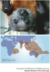
Figure 1
Figure 1. Flying foxes, their distribution and the locations of disease outbreaks caused by Hendra virus and Nipah virus.
a | Pteropus poliocephalus is an Australian flying fox and member of the family Pteropodidae, one of 18 bat families in the order Chiroptera. There are four Pteropus species in Australia. b | Sixty-five Pteropus species are distributed from Madagascar through the Indian subcontinent to south-eastern Asia and Australia and as far east as the Cook Islands. Some Pteropus species are among the largest of all bats, weighing as much as 1.2 kg and displaying a wing span of up to 1.7 m. Pteropus species are unique because they lack the complex neural and behavioural mechanisms required for echolocation that characterize the vast majority of bat species. Instead, they have large eyes and they navigate visually, feeding mainly on fruit and flowers, which they locate by smell. The sites of disease outbreaks caused by henipaviruses are indicated. Map modified with permission from Ref. © (2002) University of New South Wales Press.
Figure 2
Figure 2. Structure of henipaviruses and their genomes.
a | A schematic representation of henipavirus structure. Henipaviruses, like other paramyxoviruses, contain a linear ribonucleoprotein (RNP) core consisting of a single-stranded genomic RNA molecule of negative polarity to which nucleocapsid proteins (N) are tightly bound in a ratio of one N for every six nucleotides,. The RNP also contains smaller numbers of the phosphoprotein (P) and the large (L) polymerase protein, both of which are required to transcribe genomic RNA into mRNA and anti-genome RNA. The RNP core is surrounded by an envelope from which two spikes protrude; one is the receptor-binding glycoprotein (G) and the other the fusion (F) protein. The G and F proteins are arranged as homotetramers and homotrimers, respectively. The matrix protein (M) which underlies the viral envelope is important in determining virion architecture and is released from the RNP core on its entry into cells. b | Electron micrograph of Hendra virus (HeV). The ultrastructural characteristics of HeV and Nipah virus have been reviewed. c | The henipavirus genome. The negative-sense genomic RNA is presented in the 3′ to 5′ orientation. The open reading frames indicated by the yellow arrows encode the nucleocapsid protein (N), phosphoprotein (P), matrix protein (M), fusion protein (F), glycoprotein or attachment protein (G) and large protein (L) or RNA polymerase, in the order 3′-N-P-M-F-G-L-5′. The vertical lines represent gene start and stop signals. Note the long untranslated 3′ regions in all genes except the L gene. All genes except the P gene are monocistronic. The P gene of henipaviruses encodes not only the P protein, but also V, C and W proteins (Box 3). Genomic RNA in RNPs is transcribed by the viral polymerase which associates with the RNP at the 3′ terminus and sequentially generates discrete mRNAs from each of the viral genes. The mRNAs are not produced in equimolar amounts and there is a transcription gradient from the N to the L gene, with significant attenuation at the M–F and G–L gene junctions of HeV, a pattern of attenuation more closely resembling that observed in Sendai virus than in measles virus,,.
Figure 3
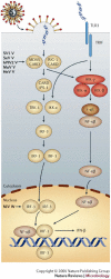
Figure 3. Interferon (IFN) induction or double-stranded (ds)RNA signalling.
The innate immune system depends on the ability of cells to detect the presence of unique, pathogen-specific molecules. The molecule considered most likely to be seen as foreign by virus-infected cells and activate the innate immune system is dsRNA, generated as a result of virus infection. Several cellular sensors detect the dsRNA signal and respond by activating pre-existing transcription factors such as IFN-regulatory factor 3 (IRF-3) and the general transcription factor nuclear factor (NF)-κB,,,,. Activated IRF-3 and NF-κB are redistributed to the nucleus, where they cooperate with other transcriptional activators to induce transcription of the interferon (IFN)-α/β genes. In one pathway, the sensor is an intracellular RNA helicase encoded by the retinoic inducible gene-1 (RIG1) or the melanoma differentiation-associated gene 5 (MDA5),. RIG-1 and MDA5 proteins are DExD/H-box RNA helicases that unwind dsRNA by virtue of their intrinsic ATPase activity. They also contain caspase-recruitment domains (CARD). The binding of dsRNA to the helicase has been hypothesized to result in the activation of the ATPase leading to conformational changes in CARD. An activated CARD acts as an interface between signalling molecules, and the CARD of RIG-1 and MDA5 has been shown to interact with the CARD-like domain of a protein called IFN-β promoter stimulator 1 (IPS-1) to transmit a signal downstream, resulting in the phosphorylation of IRF-3 by the kinases TANK-binding kinase 1 (TBK-1) and IκB kinase ε (IKK-ε),. Activated IRF-3 dimerizes and is translocated to the nucleus. Intracellular dsRNA signalling through RNA helicases also activates NF-κB. The inhibitor of NF-κB, I-κB, is phosphorylated by an activated member of the IKK complex, and I-κB is destroyed in proteasomes. NF-κB is therefore released and translocated to the nucleus. A second IFN-induction pathway, likely to be activated after the helicase-dependant pathway, uses Toll-like receptor 3 (TLR3), which probably detects dsRNA released from virus-infected cells. Signalling through TLR3 is mediated by an intracellular adaptor protein called TRIF, which signals two protein-kinase complexes, TBK-1–IKK-ε and IKK-α–IKK-β, and leads to the activation of both IRF-3 and NF-κB. The sites where henipavirus and other paramyxoviruses are known to interfere with dsRNA signalling and the viral proteins responsible are indicated (see text). HeV, Hendra virus; hPIV2, human parainfluenza virus 2; MuV, mumps virus; NiV, Nipah virus; SeV, Sendai virus; SV5, simian parainfluenza virus 5.
Figure 4
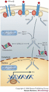
Figure 4. Interferon (IFN) signalling.
Type I IFN (IFN-β and a subset of IFN-α) induced as a result of virus infection activates IFN-inducible genes using the Jak-STAT pathway, a signalling pathway shared by many cytokines and growth factors that use members of the Janus tyrosine kinase family (TYK2 and JAK1) and a family of proteins called signal transducers and activators of transcription (STAT),,,. Type I IFN binds to two heterologous receptor subunits (IFNAR1 and IFNAR2) on the cell surface, and their dimerization leads to the activation of TYK2 and JAK1 tyrosine kinases bound to IFNAR1 and IFNAR2, respectively. TYK2 and JAK1 cross-activate each other and phosphorylate STATs. STAT1 and STAT2 form heterodimers and translocate to the nucleus where, in association with a DNA-binding protein p48 in a complex called IFN-stimulated gene factor 3 (ISGF3), they activate the transcription of genes containing IFN-stimulated response elements within their promoters. Antiviral activity and the additional, profound effects that IFNs have on cellular physiology are mediated by hundreds of IFN-induced proteins. The best characterized antiviral IFN-inducible gene products include dsRNA-dependent protein kinase (PKR), 2′,5′-oligoadenylate synthetases (2-5A) and RNase L and Mx proteins, which inhibit virus replication in various ways. Paramyxoviruses block IFN signalling by targeting specific components of the Jak-STAT pathway (see text).
None
Timeline | Emergence of henipaviruses Download Timeline PDF

Similar articles

Cited by

References

    1. Sulkin SE, Allen R. Virus Infections in Bats. 1974. - PubMed
    1. Li W, et al. Bats are natural reservoirs of SARS-like coronaviruses. Science. 2005;310:676–679. - PubMed
    1. Lau SK, et al. Severe acute respiratory syndrome coronavirus-like virus in Chinese horseshoe bats. Proc. Natl Acad. Sci. USA. 2005;102:14040–14045. - PMC - PubMed
    1. Hall L, Richards G. Flying Foxes: Fruit and Blossom Bats of Australia. 2000.
    1. Philbey AW, et al. An apparently new virus (family Paramyxoviridae) infectious for pigs, humans, and fruit bats. Emerg. Infect. Dis. 1998;4:269–271. - PMC - PubMed

Publication types