. 2006 Jan 11;25(1):174-83.
doi: 10.1038/sj.emboj.7600905. Epub 2005 Dec 8.

The Nfs1 interacting protein Isd11 has an essential role in Fe/S cluster biogenesis in mitochondria

Affiliations

The Nfs1 interacting protein Isd11 has an essential role in Fe/S cluster biogenesis in mitochondria

Alexander C Adam et al. EMBO J. .

Abstract

Formation of iron/sulfur (Fe/S) clusters, protein translocation and protein folding are essential processes in the mitochondria of Saccharomyces cerevisiae. In a systematic approach to characterize essential proteins involved in these processes, we identified a novel essential protein of the mitochondrial matrix, which is highly conserved from yeast to human and which we termed Isd11. Depletion of Isd11 caused a strong reduction in the levels of the Fe/S proteins aconitase and the Rieske protein, and a massive decrease in the enzymatic activities of aconitase and succinate dehydrogenase. Incorporation of iron into the Fe/S protein Leu1 and formation of the Fe/S cluster containing holoform of the mitochondrial ferredoxin Yah1 were inhibited in the absence of Isd11. This strongly suggests that Isd11 is required for the assembly of Fe/S proteins. We show that Isd11 forms a stable complex with Nfs1, the cysteine desulfurase of the mitochondrial machinery for Fe/S cluster assembly. In the absence of Isd11, Nfs1 is prone to aggregation. We propose that Isd11 acts together with Nfs1 in an early step in the biogenesis of Fe/S proteins.


Figures

Figure 1
Figure 1
Isd11 is a protein of the mitochondrial matrix. (A) Sequence alignment of Isd11 proteins of various species. The alignment was generated using ClustalX (1.8). Amino-acid residues identical in at least four of the aligned proteins are shown in black and similar residues are shown in gray: S. cerevisiae (Sc), S. pombe (Sp), N. crassa (Nc), H. sapiens (Hs) and D. melanogaster (Dm). (B) Isd11 is located in the mitochondria. Yeast cells were subfractionated and equal amounts of protein were subjected to SDS–PAGE and immunodecoration with antibodies against Isd11, a cytosolic protein (Bmh2), a protein of the ER (Erp1) and a mitochondrial protein (Mge1). (C) Isd11 is located in the mitochondrial matrix. Isolated mitochondria, mitoplasts prepared by osmotic shock and Triton-solubilized mitochondria were treated with or without proteinase K (PK). Mitochondria were subjected to carbonate extraction and supernatant (S) and pellet (P) fractions were separated. Samples were analyzed by SDS–PAGE and immunodecoration with antibodies against the indicated proteins. (D) Isd11 is attached to the inner membrane. WT mitochondria were resuspended in buffer containing different concentrations of KCl and opened by sonication. Supernatant fractions (Sup) containing soluble proteins and the membrane fractions were separated by centrifugation. Proteins in the supernatant fraction were collected by TCA precipitation. Laemmli buffer was added to both fractions and subjected to SDS–PAGE and immunodecoration with antibodies against Isd11, the mitochondrial desulfurase Nfs1, a protein of the inner membrane (ADP/ATP carrier, Aac2) and a matrix protein (Mge1). (E) Isd11 is imported into isolated mitochondria in a membrane-potential-dependent manner. Reticulocyte lysate containing 35S-labelled Isd11 was incubated with mitochondria in the presence or absence of membrane potential ΔΨ. After import, mitochondria were splitted and treated with or without PK under isotonic or hypotonic swelling (Sw) conditions, as indicated. Samples were analyzed by SDS–PAGE and autoradiography. 50% Input, 50% of the amount of lysate used per lane.
Figure 2
Figure 2
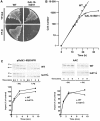
Downregulation of Isd11 in yeast cells affects cell growth, but not the capacity of the mitochondria to import preproteins. (A) Cells carrying the ISD11 gene under control of the GAL10 promoter and WT cells were grown for two rounds on plates containing rich medium with galactose (YP-Gal) or glucose (YP-D). (B) GAL10-ISD11 cells and WT cells were first grown on galactose and then incubated in glucose-containing medium for the indicated times. At time zero, the cell number was set to 1. (C) Isd11 is not required for the import of mitochondrial precursor proteins. The indicated radiolabelled precursor proteins of the TIM23 complex pathway (pSu9(1–69)DHFR) and of the TIM22 complex pathway (AAC) were imported into WT mitochondria and mitochondria depleted of Isd11 (Isd11↓) for different time periods. Mitochondria were reisolated, treated with or without PK and analyzed by SDS–PAGE and autoradiography. The mature forms of the proteins were quantified after PK treatment of mitochondria. Import into WT mitochondria at the longest time point was set to 100% (control). p, precursor form; m, mature form.
Figure 3
Figure 3
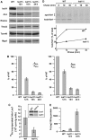
Downregulation of Isd11 affects the levels and activities of FeS cluster-containing proteins. (A) GAL10-ISD11 cells were grown at 30°C for 12, 18 and 24 h after shift to glucose-containing medium. Protein levels of mitochondria (100 μg) isolated from these cells and from WT cells were analyzed by SDS–PAGE and immunodecoration with antibodies against the indicated proteins. (B) The activities of SDH and aconitase (Aco) were measured in mitochondria isolated from WT cells and cells downregulated of Isd11 (Isd11↓) for the indicated time periods. As a standard, the non-FeS cluster-containing protein MDH was measured. The ratio of the activities of aconitase and MDH or SDH and MDH was calculated and expressed as percent of the ratio in WT cells. (C) The in vivo assembly of the Fe/S cluster of the cytoplasmic Leu1 protein was inhibited in the absence of Isd11. GAL-Isd11 cells were depleted of Isd11 by growth for 18 h in glucose-containing medium. These cells and WT cells were incubated with 55Fe for 2 h. Cell lysates were prepared and an immunoprecipitation with antibodies against Leu1 was performed. Incorporated 55Fe into Leu1 was determined by liquid scintillation counting. The inset shows the immunostaining of Leu1 and Isd11 present in these cell lysates. (D) The formation of holoYah1 was dependent on Isd11. The 35S-labelled precursor protein of Yah1 was imported into the mitochondria isolated of WT and Isd11↓ cells for 10 min at 25°C. The import reactions were stopped by the addition of valinomycin and incubated further for the indicated time periods (‘chase'). Aliquots were taken and the nonimported precursor proteins were removed by treatment with PK. The samples were analyzed by native gel electrophoresis and autoradiography. At the indicated time points, the amounts of apo- and holoYah1 were quantified and their ratios were expressed as percent holoYah1 of the sum of both forms (total). (E) The iron content of 100 μg of mitochondria isolated from WT and Isd11 downregulated cells was determined.
Figure 4
Figure 4
Isd11 is present in a complex with Nfs1. (A) Coisolation of Nfs1 with His-tagged Isd11. Isolated mitochondria of WT cells or cells expressing Isd11 with a C-terminal heptahistidinyl tag (Isd11-7his) were solubilized with DDM and incubated with NiNTA beads. Bound material was eluted and analyzed by SDS–PAGE and Coomassie staining. Nfs1 and Isd11 were identified by mass spectrometry. (B) Copurification of Nfs1 with Isd11. Mitochondria of a WT strain and a strain carrying a triple HA-tagged Isd11 were solubilized with digitonin. Following clarifying spin, the supernatants were incubated with antibodies against the HA tag coupled to protein A-Sepharose beads. The beads were harvested, washed and bound proteins were eluted with Laemmli buffer. Samples were analyzed by SDS–PAGE and immunodecoration with antibodies against Nfs1 and Isd11. In all, 10% of total and supernatant were loaded. f, Fragment of Nfs1. (C) Co-immunoprecipitation of Isd11 and Nfs1. WT mitochondria were solubilized with DDM. The co-immunoprecipitation was performed and analyzed as in (B), using antibodies against either Nfs1 or Isd11. (D) Isd11 and Nfs1 cofractionate on a gel-filtration column. Mitochondrial membranes were opened in the presence of 250 mM KCl by sonication. After a clarifying spin, the supernatant was subjected to a Superose-12 gel-filtration column. Fractions were analyzed by SDS–PAGE and immunodecoration with antibodies against Isd11 and Nfs1.
Figure 5
Figure 5
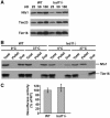
Nfs1 in Isd11-depleted mitochondria has desulfurase activity, but is prone to aggregation. (A) Protein levels of Nfs1 in mitochondria (25, 50, 100 μg) isolated from cells depleted of Isd11 for 18 h (Isd11↓) and from WT cells were analyzed by SDS–PAGE and immunodecoration. (B) Aggregation of Nfs1 in cells lacking Isd11. Mitochondria isolated from WT and Isd11-depleted cells were incubated for 20 min at 0 or 37°C. Mitochondria were lysed with 0.5% Triton X-100 and aggregated material was pelleted by centrifugation. Total and the supernatant fractions (Sup) were precipitated with TCA. Samples were analyzed by SDS–PAGE and immunodecoration. (C) Nfs1 has desulfurase activity in the absence of Isd11. Mitochondria (100 μg) from WT and Isd11↓ cells were lysed with 0.2% DDM and then incubated for 30 min at 30°C in buffer containing 5 mM DTT and 4 mM cysteine. Sulfide produced was converted to methylene blue, whose absorption was measured at 667 nm. Background activity was determined by incubation of the mitochondrial extracts in the absence of cysteine and subtracted. The desulfurase activity was expressed as percent of activity present in WT mitochondria.

Similar articles

Cited by

References

    1. Babcock M, de Silva D, Oaks R, Davis-Kaplan S, Jiralerspong S, Montermini L, Pandolfo M, Kaplan J (1997) Regulation of mitochondrial iron accumulation by Yfh1p, a putative homolog of frataxin. Science 276: 1709–1712 - PubMed
    1. Bateman A, Coin L, Durbin R, Finn RD, Hollich V, Griffiths-Jones S, Khanna A, Marshall M, Moxon S, Sonnhammer EL, Studholme DJ, Yeats C, Eddy SR (2004) The Pfam protein families database. Nucleic Acids Res 32 (database issue): D138–D141 - PMC - PubMed
    1. Cheng MY, Hartl FU, Martin J, Pollock RA, Kalousek F, Neupert W, Hallberg EM, Hallberg RL, Horwich AL (1989) Mitochondrial heat-shock protein hsp60 is essential for assembly of proteins imported into yeast mitochondria. Nature 337: 620–625 - PubMed
    1. Craig EA, Marszalek J (2002) A specialized mitochondrial molecular chaperone system: a role in formation of Fe/S centers. Cell Mol Life Sci 59: 1658–1665 - PMC - PubMed
    1. Craig EA, Voisine C, Schilke B (1999) Mitochondrial iron metabolism in the yeast Saccharomyces cerevisiae. Biol Chem 380: 1167–1173 - PubMed

Publication types

MeSH terms