. 2005 Dec 5;171(5):811-21.
doi: 10.1083/jcb.200506006.

Dendritic BC1 RNA in translational control mechanisms

Affiliations

Dendritic BC1 RNA in translational control mechanisms

Huidong Wang et al. J Cell Biol. .

Erratum in

  • J Cell Biol. 2006 Feb 13;172(4):635

Abstract

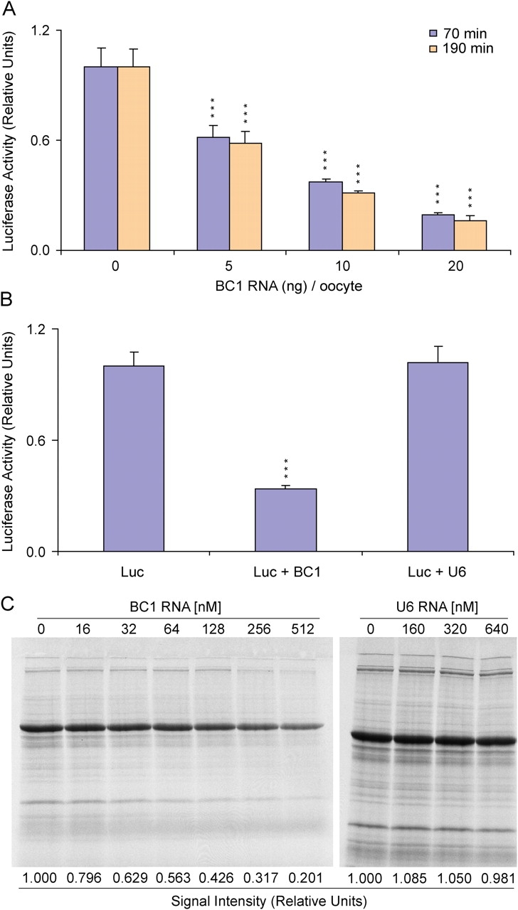
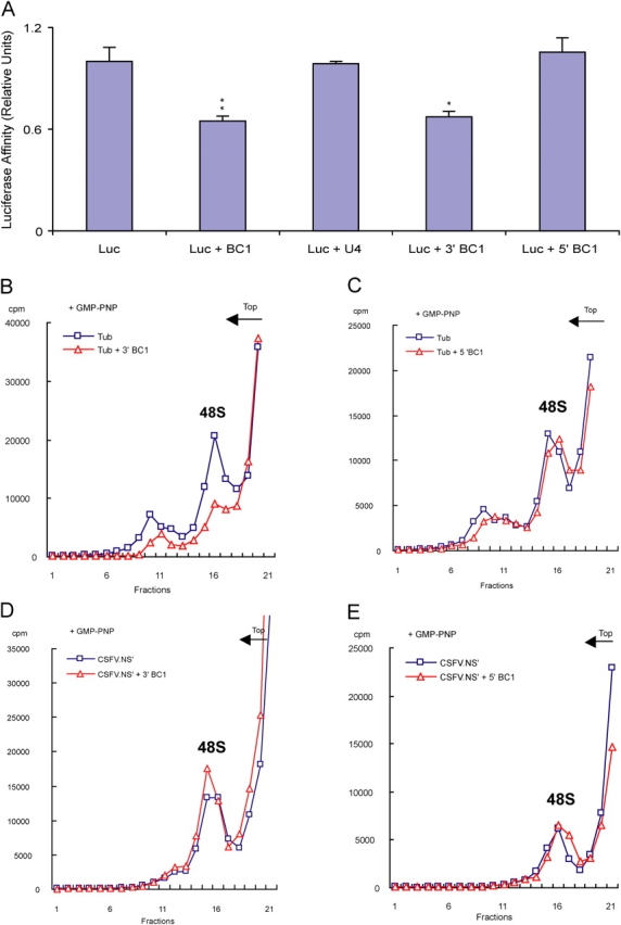
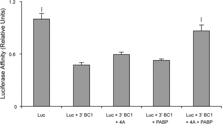
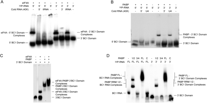
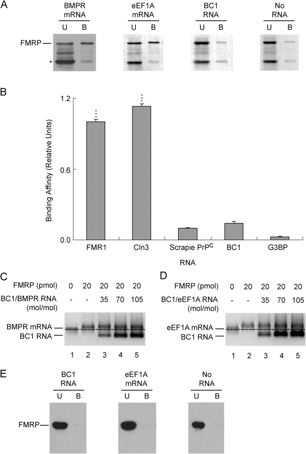
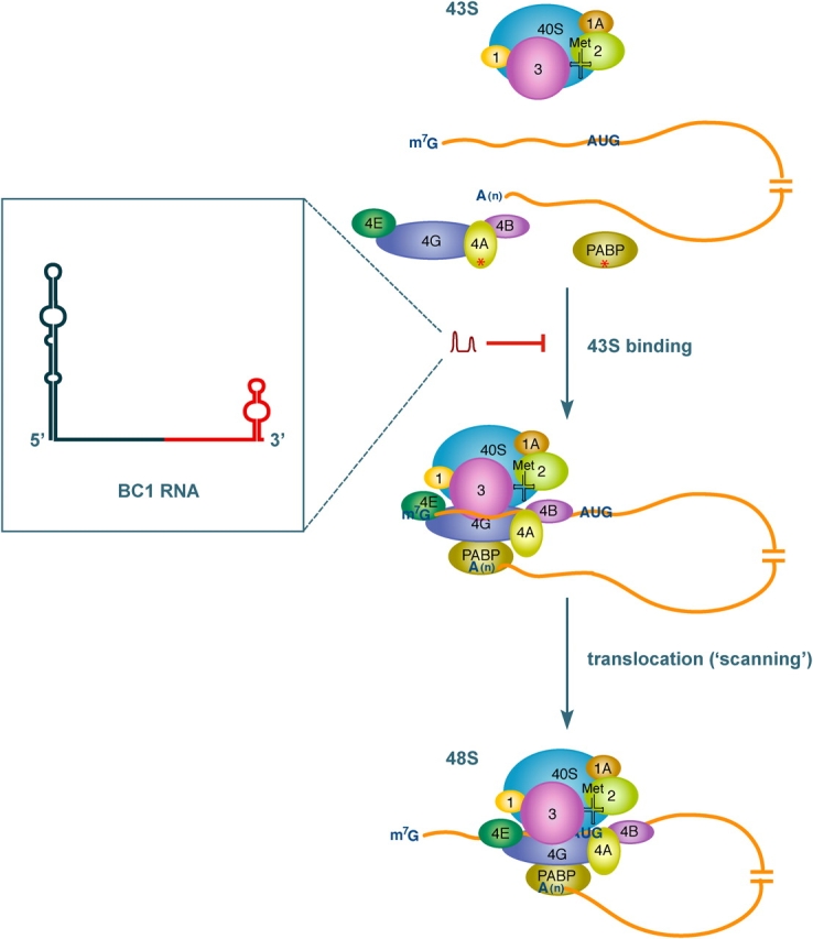
Translational control at the synapse is thought to be a key determinant of neuronal plasticity. How is such control implemented? We report that small untranslated BC1 RNA is a specific effector of translational control both in vitro and in vivo. BC1 RNA, expressed in neurons and germ cells, inhibits a rate-limiting step in the assembly of translation initiation complexes. A translational repression element is contained within the unique 3' domain of BC1 RNA. Interactions of this domain with eukaryotic initiation factor 4A and poly(A) binding protein mediate repression, indicating that the 3' BC1 domain targets a functional interaction between these factors. In contrast, interactions of BC1 RNA with the fragile X mental retardation protein could not be documented. Thus, BC1 RNA modulates translation-dependent processes in neurons and germs cells by directly interacting with translation initiation factors.


Figures

Figure 1.
Figure 1.
BC1 RNA represses translation in X. laevis oocytes. (A) 10 ng of capped and polyadenylated luciferase mRNA was coinjected into stage VI oocytes with increasing amounts of BC1 RNA (10 ng BC1 RNA corresponds to ∼200 nM intracellular). The average luminescence signal in the absence of BC1 RNA was assigned a relative luciferase activity value of 1. Quantitative analysis of four experiments revealed a significant reduction of luciferase activity in the presence of BC1 RNA (one-way analysis of variance [ANOVA], P < 0.001; Scheffe's multiple comparison post hoc analysis [comparison with 0 nM BC1 RNA control]: ***, P < 0.001). (B) In control experiments, 400 nM BC1 or U6 RNA was coinjected with luciferase mRNA (Luc). Luminescence was measured after 55 min. Quantitative analysis of four experiments revealed a significant decrease of luciferase activity in the presence of BC1 RNA but not in the presence of U6 RNA (one-way ANOVA, P < 0.001; Scheffe's multiple comparison post hoc analysis [comparison with Luc control]: ***, P < 0.001). (C) For comparison, translational repression by BC1 RNA is shown in RRL. Incorporation of 35S-methionine into endogenous proteins was examined in untreated RRL and visualized by SDS-PAGE as described previously (Wang et al., 2002). U6 RNA was not repression competent. Quantification is shown for the major bands.
Figure 2.
Figure 2.
The 3′ BC1 domain is repression competent. (A) Luciferase mRNA (Luc) was coinjected with 100 nM of small RNAs into stage VI oocytes and incubated for 55 min. Results from four experiments were quantified by luminescence (one-way ANOVA, P < 0.001; Scheffe's multiple comparison post hoc analysis [comparison with Luc control]: **, P < 0.01; *, P < 0.05). (B and C) 48S complex formation was analyzed in the presence of the 3′ (B) or the 5′ (C) BC1 domain (500 nM), using capped polyadenylated α-tubulin mRNA as a programming mRNA. (D and E) Analogously, 48S complex formation was probed in the presence of the 3′ (D) or the 5′ (E) BC1 domain, using uncapped polyadenylated CSFV.NS' mRNA as a programming mRNA.
Figure 3.
Figure 3.
Translation in X. laevis oocytes, repressed by the 3′ BC1 domain, is restored by joint replenishment with eIF4A and PABP. Luciferase mRNA (Luc) was coinjected into stage VI oocytes with ∼400 nM 3′ BC1 domain, 100 nM eIF4A, and/or 100 nM PABP. Luminescence was measured after 1 h. Only replenishment with eIF4A and PABP in combination restored 3′ BC1–repressed translation to a level that was statistically indistinguishable from unrepressed translation. Quantitative analysis of four experiments is shown (one-way ANOVA, Scheffe's multiple comparison post hoc analysis, ***, P < 0.001). The observed differences among 3′ BC1, 3′ BC1 + eIF4A, and 3′ BC1 + PABP were not statistically significant.
Figure 4.
Figure 4.
The 3′ BC1 domain binds specifically to eIF4A and PABP. EMSA experiments were performed with 32P-labeled 5′ or 3′ BC1 domains. (A) BC1 domains were incubated with eIF4A in the absence or presence of unlabeled competitor RNAs. Note that the 5′ BC1 domain produced a shift with eIF4A that was abolished by unlabeled tRNAs. (B) BC1 domains were incubated with an NH2-terminal segment of PABP (aa 1–182) in the absence or presence of unlabeled competitor RNAs. (C) When used in combination, eIF4A and PABP produced a more substantial mobility shift with the 3′ BC1 domain than did either factor alone, indicating that both factors can bind simultaneously. (D) A PABP segment containing RRM 1/2 but not a PABP segment containing RRM 3/4 or a PABP segment containing the COOH-terminal domain produced mobility shifts with BC1 RNA (left half of gel) and with the 3′ BC1 domain (right half of the gel). Incubation was performed in the presence of 5 mg/ml heparin, followed by RNase T1 digestion (Muddashetty et al., 2002). Omission of heparin/T1 resulted in a weak shift of BC1 RNA by PABP (RRM 3/4), suggesting binding competence of lower affinity and/or specificity of this domain (not depicted).
Figure 5.
Figure 5.
BC1 RNA does not bind specifically to FMRP. (A) Affinity capture reactions were performed between 35S-labeled FMRP and 1 μg of biotinylated BMPR mRNA, 1 μg of biotinylated eEF1A mRNA, and 1 μg of biotinylated BC1 RNA. (Note that on a molar basis, this amount represents ∼10 times more BC1 RNA than eEF1A mRNA and 15 times more BC1 RNA than BMPR mRNA.) Full-length FMRP is indicated; asterisk marks incomplete or proteolytic products that were generated in the in vitro translation reaction. Incubation in the absence of RNA was performed as a control for nonspecific binding of FMRP to the resin. (B) A solution RNA binding assay was performed to quantify interactions of FMRP with target RNAs. In these experiments, FMRP was biotinylated and RNAs were 32P labeled. Binding of FMRP to its own mRNA (FMR1 mRNA) served as a positive control; Scrapie PrPc mRNA was used as a negative control. Quantitative analysis of three experiments revealed that binding of Scrapie PrPc mRNA to FMRP was significantly different from binding of FMR1 and Cln3 mRNA but not from binding of BC1 RNA or partial G3BP mRNA (one-way ANOVA, P < 0.001; Scheffe's multiple comparison post hoc analysis [comparison with Scrapie PrPc mRNA, negative control]: ***, P < 0.001). (C) AGESA FMRP competition assay between BMPR mRNA and BC1 RNA. Specific FMRP–BMPR mRNA complexes were formed (lane 2, compare with unbound BMPR mRNA in lane 1, indicated by dashed line) and remained resistant to titration with BC1 RNA (lanes 3–5). (D) Analogous AGESA FMRP competition assay between eEF1A mRNA and BC1 RNA. FMRP–eEF1A mRNA complexes were resistant to titration with BC1 RNA. (E) Affinity capture assays with FMRP1-280 and biotinylated eEF1A mRNA or biotinylated BC1 RNA. (The amount of BC1 RNA used was again 10 times higher than that of eEF1A mRNA.) FMRP1-280 was visualized by Western blotting.
Figure 6.
Figure 6.
An interaction of the 3′ BC1 domain with eIF4A and PABP is responsible for BC1-mediated repression of translation initiation. As a consequence of this interaction, formation of the 48S initiation complex is inhibited. BC1 RNA, shown here targeting recruitment, may in addition interact with one or both factors during translocation. BC1 targets (eIF4A and PABP) are indicated by red asterisks. The 3′ BC1 domain is shown in red; the central A22 domain is directly 5′ adjacent to the 3′ domain. For clarity, some factors that are part of the complex (e.g., eIF5) have been omitted from this sketch. (Reprinted with permission from the Annual Review of Cell and Developmental Biology, Vol. 21, 2005, by Annual Reviews, www.annualreviews.org)

Similar articles

Cited by

References

    1. Adinolfi, S., C. Bagni, G. Musco, T. Gibson, L. Mazzarella, and A. Pastore. 1999. Dissecting FMR1, the protein responsible for fragile X syndrome, in its structural and functional domains. RNA. 5:1248–1258. - PMC - PubMed
    1. Antar, L.N., and G.J. Bassell. 2003. Sunrise at the synapse: the FMRP mRNP shaping the synaptic interface. Neuron. 37:555–558. - PubMed
    1. Audet, R.G., J. Goodchild, and J.D. Richter. 1987. Eukaryotic initiation factor 4A stimulates translation in microinjected Xenopus oocytes. Dev. Biol. 121:58–68. - PubMed
    1. Baer, B.W., and R.D. Kornberg. 1980. Repeating structure of cytoplasmic poly(A)-ribonucleoprotein. Proc. Natl. Acad. Sci. USA. 77:1890–1892. - PMC - PubMed
    1. Barciszewski, J., and V.A. Erdmann. 2003. Noncoding RNAs: Molecular Biology and Molecular Medicine. Landes Bioscience, Georgetown, TX. 292 pp.

Publication types

MeSH terms