. 2005 Dec;25(24):10684-94.
doi: 10.1128/MCB.25.24.10684-10694.2005.

PGC-1alpha coactivates PDK4 gene expression via the orphan nuclear receptor ERRalpha: a mechanism for transcriptional control of muscle glucose metabolism

Affiliations

PGC-1alpha coactivates PDK4 gene expression via the orphan nuclear receptor ERRalpha: a mechanism for transcriptional control of muscle glucose metabolism

Adam R Wende et al. Mol Cell Biol. 2005 Dec.

Abstract

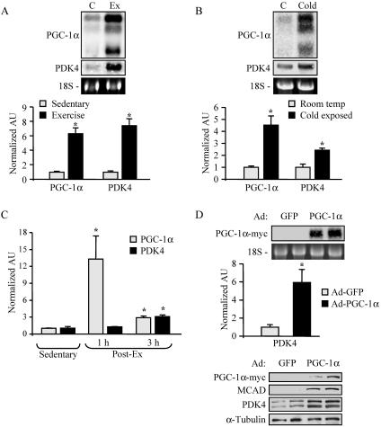
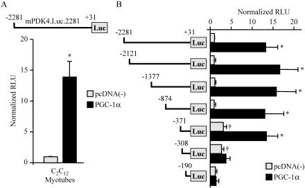
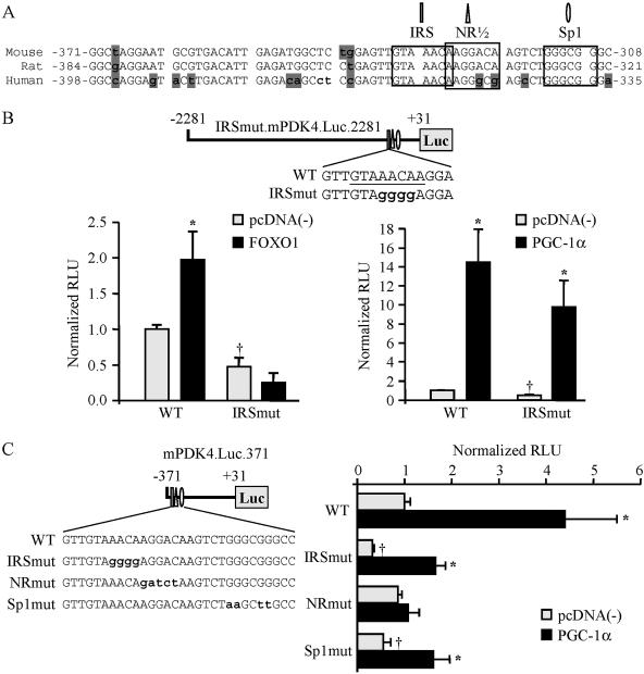
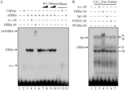
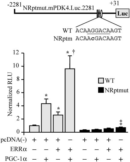
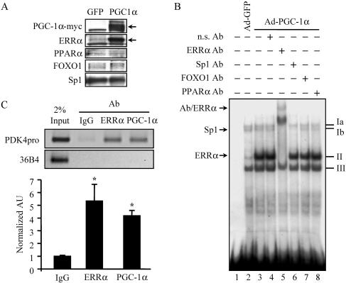
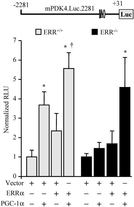
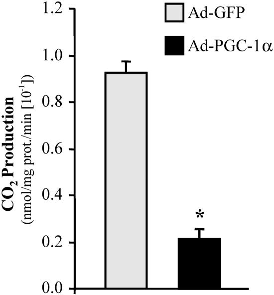
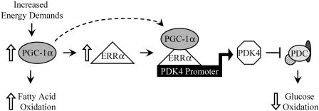
The transcriptional coactivator PGC-1alpha is a key regulator of energy metabolism, yet little is known about its role in control of substrate selection. We found that physiological stimuli known to induce PGC-1alpha expression in skeletal muscle coordinately upregulate the expression of pyruvate dehydrogenase kinase 4 (PDK4), a negative regulator of glucose oxidation. Forced expression of PGC-1alpha in C(2)C(12) myotubes induced PDK4 mRNA and protein expression. PGC-1alpha-mediated activation of PDK4 expression was shown to occur at the transcriptional level and was mapped to a putative nuclear receptor binding site. Gel shift assays demonstrated that the PGC-1alpha-responsive element bound the estrogen-related receptor alpha (ERRalpha), a recently identified component of the PGC-1alpha signaling pathway. In addition, PGC-1alpha was shown to activate ERRalpha expression. Chromatin immunoprecipitation assays confirmed that PGC-1alpha and ERRalpha occupied the mPDK4 promoter in C(2)C(12) myotubes. Additionally, transfection studies using ERRalpha-null primary fibroblasts demonstrated that ERRalpha is required for PGC-1alpha-mediated activation of the mPDK4 promoter. As predicted by the effects of PGC-1alpha on PDK4 gene transcription, overexpression of PGC-1alpha in C(2)C(12) myotubes decreased glucose oxidation rates. These results identify the PDK4 gene as a new PGC-1alpha/ERRalpha target and suggest a mechanism whereby PGC-1alpha exerts reciprocal inhibitory influences on glucose catabolism while increasing alternate mitochondrial oxidative pathways in skeletal muscle.


Figures

FIG. 1.
FIG. 1.
Activation of PDK4 gene expression by physiological stimuli and by overexpression of PGC-1α. (A) Northern blot studies performed with 15 μg of total RNA isolated from the gastrocnemii of animals following two bouts of exercise as described in Materials and Methods (Ex) or from sedentary controls (C) (n = 4). Blots were sequentially hybridized with probes specific for the genes encoding PGC-1α and PDK4. Representative autoradiographs are shown at the top. Phosphorimage-based quantification of Northern blot signal intensities is shown in the graph. Data represent mean arbitrary units (±SE) normalized to sedentary control values (taken as 1.0). (B) Results of Northern blot analysis performed as described above on gastrocnemius samples immediately following 6 h of cold exposure (Cold) and from room temperature controls (C) (n = 6). Graph represents mean arbitrary units (±SE) as determined by real-time PCR quantification corrected to the 36B4 transcript and normalized to the values for control littermates (taken as 1.0). (C) RNA analysis of PGC-1α and PDK4 gastrocnemius gene expression 1 h and 3 h after a single bout of exercise (n = 4). The graph represents mean arbitrary units (±SE) as determined by RT-PCR corrected to the 36B4 transcript and normalized to the value for sedentary controls (taken as 1.0). (D) RNA analyses and Western blot analysis were performed with samples isolated from Ad-GFP- or Ad-PGC-1α-infected C2C12 myotubes. (Top) Results of Northern blotting performed as described above. Data represent mean arbitrary units (±SE) as determined by RT-PCR corrected to the 36B4 transcript and normalized to value for control Ad-GFP-infected samples (taken as 1.0). (Bottom) Western blot analysis of whole-cell extracts (25 μg) prepared from C2C12 myotubes infected with adenovirus vectors expressing GFP or PGC-1α. Asterisks indicate significant differences (P < 0.05) from controls.
FIG. 2.
FIG. 2.
PGC-1α activates the mPDK4 gene promoter through a cis-acting element within the proximal region. (A) A schematic of the reporter construct mPDK4.Luc.2281, containing the −2281-to-+31 region of the mouse PDK4 gene promoter, is shown at the top. The graph contains results of cotransfection studies with mPDK4.Luc.2281 in the presence (PGC-1α) or absence [pcDNA(−)] of pcDNA-PGC-1α in C2C12 myotubes. The graph represents mean (±SE) relative light units (RLU) corrected for Renilla luciferase activity and normalized to the activity of mPDK4.Luc.2281 (taken as 1.0). (B) Transient transfections performed with a 5′ deletion series of mPDK4.Luc.2281. All values represent at least three independent transfections conducted in triplicate. Asterisks indicate significant differences compared to individual reporters cotransfected with empty vector (P < 0.05). Daggers indicate significant differences in activity compared to mPDK4.Luc.2281 cotransfected with empty vector (P < 0.05).
FIG. 3.
FIG. 3.
PGC-1α activates PDK4 gene transcription through a nuclear receptor binding site. (A) Species comparison of the nucleotide sequence (sense strand is shown) of the PGC-1α-activated region of the PDK4 promoter, demonstrating a high level of sequence identity between mouse, rat, and human genes. The numbers are relative to the transcription start site (+1). Shaded nucleotides indicate sequence that is not conserved. Computer analysis revealed numerous candidate response elements, including a nuclear receptor half-site (NR1/2), an Sp1 site, and the previously identified FOXO1 binding site (IRS) (9, 27), indicated by labels, boxes, and symbols. (B) (Top) Site-directed mutagenesis was used to abolish the FOXO1 response element. Mutated nucleotides are lowercased. (Bottom) The mPDK4.Luc.2281 (wild type [WT]) or IRSmut.mPDK4.Luc.2281 promoter reporter was used in cotransfections in C2C12 myotubes in the presence or absence of FOXO1 (left graph) or PGC-1α (right graph). (C) (Left) Schematic showing the mutations generated by site-directed mutagenesis of the minimal PGC-1α-responsive mPDK4.Luc.371 reporter in FOXO1 (IRSmut), NR (NRmut), or Sp1 (Sp1mut) candidate response elements (mutated nucleotides are lowercased). (Right) Cotransfection studies using WT or mutant mPDK4 promoter reporters were performed with C2C12 myotubes. Data represent mean (±SE) relative light units (RLU) corrected for rLuc and normalized to values for the WT reporter (taken as 1.0). All transient transfection studies represent at least three independent transfections conducted in triplicate. Asterisks indicate significant differences from individual reporters cotransfected with empty vector (P < 0.05). Daggers indicate significant differences from the wild-type reporter cotransfected with empty vector (P < 0.05).
FIG. 4.
FIG. 4.
The orphan nuclear receptor ERRα binds to the PGC-1α-responsive region of the mPDK4 promoter. (A) Representative autoradiograph of EMSA conducted with 32P-labeled probes of the 63-bp region of the mPDK4 promoter identified in Fig. 2B. Mutant probe corresponds to the same nucleotide substitutions for NRmut as in Fig. 3C. Probes were incubated with recombinant in vitro-translated ERRα (rERRα) or unprogrammed reticulocyte lysate (Unprog.) as indicated at the top. Antibody (Ab) supershift studies were performed by incubation with an anti-ERRα (lane 5) or nonspecific (n.s.) (lane 4) Ab. Competition analysis was performed by adding a molar excess of the unlabeled wild-type (lanes 7 and 8) or NRmut (lanes 9 and 10) probe in increasing amounts as indicated. For lanes 11 to 13, the 32P-labeled NRmut probe was used. n.s. bands represent protein complexes identified in unprogrammed reticulocyte lysate. (B) Autoradiograph of an EMSA using nuclear (Nuc.) extracts isolated from C2C12 myotubes incubated with the 32P-labeled wild-type probe. Four prominent complexes, labeled Ia, Ib, II, and III, were observed. Complexes were identified by antibody supershift analysis using antibodies specific for the transcription factors indicated at the top.
FIG. 5.
FIG. 5.
ERRα and PGC-1α regulate the mPDK4 promoter through the NR response element. (Top) Schematic showing the mutation (lowercase) of the NR response element in mPDK4.Luc.2281 (NRptmut.mPDK4.Luc.2281). (Bottom) Graph depicting the results of transient cotransfections in CV-1 cells performed with either the mPDK4.Luc.2281 reporter (wild type [WT]) or NRptmut.mPDK4.Luc.2281 (NRptmut). Asterisks indicate significant differences from WT mPDK4.Luc.2281 cotransfected with empty vector (P < 0.05). Dagger indicates a significant difference from mPDK4.Luc.2281 with ERRα (P < 0.05). Double dagger indicates a significant difference from the value obtained using NRptmut.mPDK4.Luc.2281 with empty vector (P < 0.05).
FIG. 6.
FIG. 6.
PGC-1α induces expression of the endogenous ERRα gene in skeletal muscle cells.(A) Representative autoradiograph of Western blot analysis performed on nuclear extracts isolated from C2C12 myotubes following infection with Ad-GFP or Ad-PGC-1α as indicated at the top. The antibodies used are given on the left. Arrows on the right indicate specific (PGC-1α and ERRα) bands. (B) EMSA study performed with nuclear extracts isolated from C2C12 myotubes following infection with Ad-GFP (lane 2) or Ad-PGC-1α (lanes 3 to 8) and the wild-type probe described in the legend to Fig. 4. Complexes II and Ib were identified by antibody (Ab) supershift analysis using Abs specific for ERRα and Sp1, respectively (lanes 5 and 6). n.s., nonspecific. (C) Chromatin immunoprecipitation assays performed on C2C12 myotubes following infection by Ad-PGC-1α. Formaldehyde cross-linked protein-DNA complexes were isolated. Sheared protein-DNA complexes were immunoprecipitated with a nonspecific antibody (IgG), anti-ERRα, or anti-PGC-1α. Isolated fragments were amplified by PCR to detect the enrichment of amplicons corresponding to a 190-bp region of the 36B4 gene (negative control) or a 164-bp region encompassing the NR response element of the mPDK4 gene promoter. (Top) Results of agarose gel analysis of a representative trial showing relative band intensities. Input represents 2% of the total chromatin used in the immunoprecipitation reactions. (Bottom) Graphs contain mean values as determined by SYBR green quantification of three independent chromatin isolations and immunoprecipitations (arbitrary units ± SE) normalized to the value for the IgG control (taken as 1.0). Asterisks indicate significant differences from the IgG control (P < 0.05).
FIG. 7.
FIG. 7.
ERRα is required for PGC-1α-mediated activation of PDK4 gene transcription. Primary fibroblasts isolated from wild-type or ERR−/− mice were cotransfected with mPDK4.Luc.2281 and an expression vector for PGC-1α or ERRα, or both. Asterisks indicate significant differences from wild-type mPDK4.Luc.2281 cotransfected with empty vector (P < 0.05). Dagger indicates a significant difference from mPDK4.Luc.2281 cotransfected with ERRα (P < 0.05).
FIG. 8.
FIG. 8.
PGC-1α decreases glucose oxidation in C2C12 myotubes. The graph represents mean (±SE) [U-14C]glucose oxidation rates in C2C12 myotubes infected with Ad-PGC-1α or Ad-GFP (control). Results are based on three independent experiments performed in triplicate. Asterisk indicates a significant difference from the GFP-infected control (P < 0.001).
FIG. 9.
FIG. 9.
Proposed mechanism for the regulation of muscle glucose metabolism by PGC-1α/ERRα. Increased energy demands related to physiological stress (e.g., exercise) result in the induction of muscle PGC-1α gene expression, which in turn induces ERRα gene expression. ERRα then binds to the PDK4 gene promoter, where PGC-1α directly coactivates ERRα to induce PDK4 gene expression. PDK4 has known roles in the negative regulation of the PDC, resulting in decreased glucose oxidation coincident with increased mitochondrial fatty acid oxidation driven by PGC-1α.

Similar articles

Cited by

References

    1. Arany, Z., H. He, J. Lin, K. Hoyer, C. Handschin, O. Toka, F. Ahmad, T. Matsui, S. Chin, P.-H. Wu, I. I. Rybkin, J. M. Shelton, M. Manieri, S. Cinti, F. J. Schoen, R. Bassel-Duby, A. Rosenzweig, J. S. Ingwall, and B. M. Spiegelman. 2005. Transcriptional coactivator PGC-1α controls the energy state and contractile function of cardiac muscle. Cell Metab. 1:259-271. - PubMed
    1. Baar, K., A. R. Wende, T. E. Jones, M. Marison, L. A. Nolte, M. Chen, D. P. Kelly, and J. O. Holloszy. 2002. Adaptations of skeletal muscle to exercise: rapid increase in the transcriptional coactivator PGC-1α. FASEB J. 16:1879-1886. - PubMed
    1. Brandt, J., F. Djouadi, and D. P. Kelly. 1998. Fatty acids activate transcription of the muscle carnitine palmitoyltransferase I gene in cardiac myocytes via the peroxisome proliferator-activated receptor α. J. Biol. Chem. 273:23786-23792. - PubMed
    1. Carter, M. E., T. Gulick, B. D. Raisher, T. Caira, J. A. Ladias, D. D. Moore, and D. P. Kelly. 1993. Hepatocyte nuclear factor-4 activates medium chain acyl-CoA dehydrogenase gene transcription by interacting with a complex regulatory element. J. Biol. Chem. 268:13805-13810. - PubMed
    1. Caruso, M., C. Miele, P. Formisano, G. Condorelli, G. Bifulco, A. Oliva, R. Auricchio, G. Riccardi, B. Capaaldo, and F. Beguinot. 1997. In skeletal muscle, glucose storage and oxidation are differentially impaired by the IR1152 mutant receptor. J. Biol. Chem. 272:7290-7297. - PubMed

Publication types

MeSH terms

LinkOut - more resources