. 2005 Dec;79(24):15246-57.
doi: 10.1128/JVI.79.24.15246-15257.2005.

Genetic analysis of influenza virus NS1 gene: a temperature-sensitive mutant shows defective formation of virus particles

Affiliations

Genetic analysis of influenza virus NS1 gene: a temperature-sensitive mutant shows defective formation of virus particles

Urtzi Garaigorta et al. J Virol. 2005 Dec.

Abstract

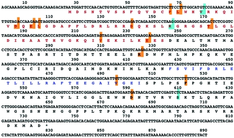
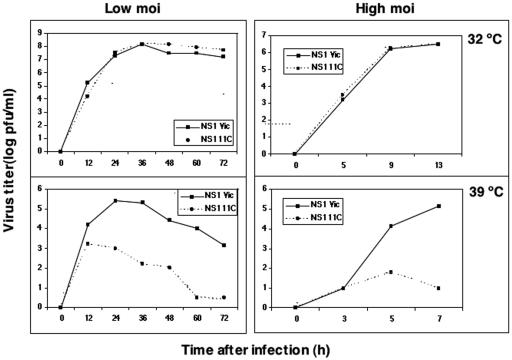
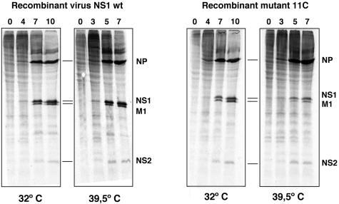
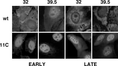
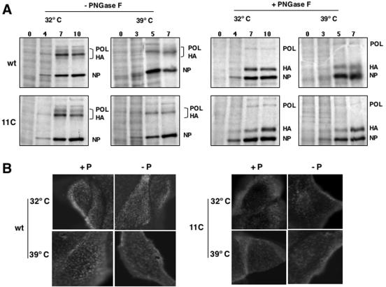
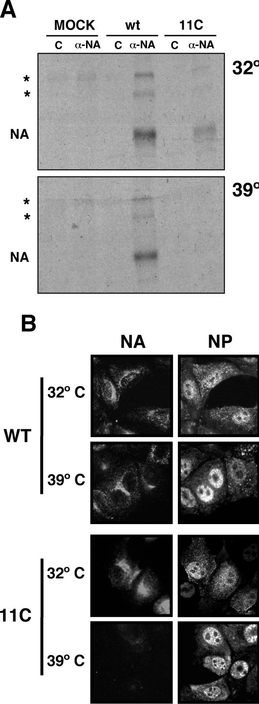
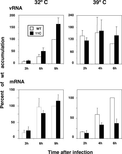
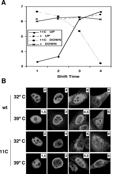
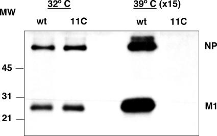
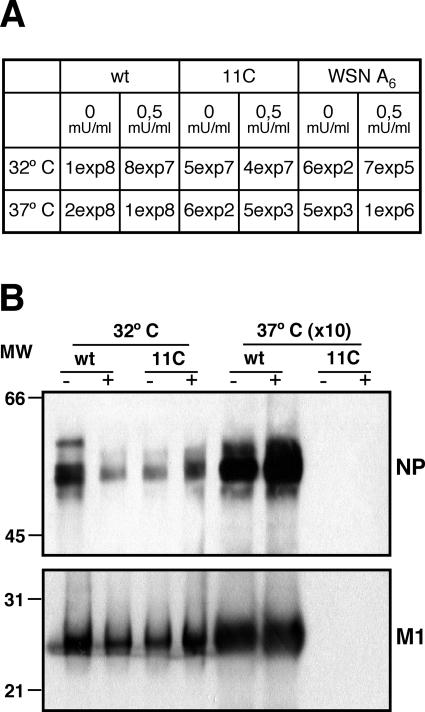
To perform a genetic analysis of the influenza A virus NS1 gene, a library of NS1 mutants was generated by PCR-mediated mutagenesis. A collection of mutant ribonucleic proteins containing the nonstructural genes was generated from the library that were rescued for an infectious virus mutant library by a novel RNP competition virus rescue procedure. Several temperature-sensitive (ts) mutant viruses were obtained by screening of the mutant library, and the sequences of their NS1 genes were determined. Most of the mutations identified led to amino acid exchanges and concentrated in the N-terminal region of the protein, but some of them occurred in the C-terminal region. Mutant 11C contained three mutations that led to amino acid exchanges, V18A, R44K, and S195P, all of which were required for the ts phenotype, and was characterized further. Several steps in the infection were slightly altered: (i) M1, M2, NS1, and neuraminidase (NA) accumulations were reduced and (ii) NS1 protein was retained in the nucleus in a temperature-independent manner, but these modifications could not justify the strong virus titer reduction at restrictive temperature. The most dramatic phenotype was the almost complete absence of virus particles in the culture medium, in spite of normal accumulation and nucleocytoplasmic export of virus RNPs. The function affected in the 11C mutant was required late in the infection, as documented by shift-up and shift-down experiments. The defect in virion production was not due to reduced NA expression, as virus yield could not be rescued by exogenous neuraminidase treatment. All together, the analysis of 11C mutant phenotype may indicate a role for NS1 protein in a late event in virus morphogenesis.


Figures

FIG. 1.
FIG. 1.
Rescue of virus genes by competition of RNPs. The virus yield from a mixed transfection with WSN recombinant NS RNP and total VIC virion RNPs was used to infect MDCK cells (REC. MIX). Total cell extract was analyzed by Western blotting with anti-NS1 serum. As controls, cells were infected with WSN virus or VIC virus or were mock infected (MOCK). The mobility of VIC NS1 protein is indicated by stars and that of WSN NS1 protein is denoted by diamonds. Numbers to the left indicate the mobility of molecular mass markers (in kilodaltons).
FIG. 2.
FIG. 2.
Amino acid replacements in temperature-sensitive mutants affected in NS1 protein. The NS RNA segments of influenza virus temperature-sensitive mutants isolated in this study were cloned and sequenced. The positions in which amino acid substitutions were detected in the NS1 open reading frame are boxed in light brown on the NS1 protein sequence. Those present in mutant 11C are boxed in light blue. The protein regions acting as RNA-binding domain (red) and as effector domain (blue) are indicated (37, 43, 57).
FIG. 3.
FIG. 3.
Replication of 11C virus is temperature sensitive. Cultures of MDCK cells were infected with mutant 11C or wt recombinant viruses at an MOI of 10−3 PFU/cell (left panels) or 10 PFU/cell (right panels) and incubated at either 32 or 39.5°C. At the times indicated, samples were obtained from the supernatant of each culture and assayed for virus infectivity by a plaque assay at permissive temperature.
FIG. 4.
FIG. 4.
Synthesis of viral proteins in 11C mutant-infected cells. Cultures of MDCK cells were infected at an MOI of 10 PFU/cell with either wt or 11C recombinant viruses. The cultures were incubated at 32 or 39.5°C and at the times after infection indicated in the figure they were labeled with [35S]met-cys as indicated in Materials and Methods. Total extracts of each culture were analyzed by polyacrylamide gel electrophoresis and autoradiography. The position of selected viral proteins is indicated to the right of each panel.
FIG. 5.
FIG. 5.
Viral protein accumulation in 11C mutant-infected cells. Cultures of MDCK cells were infected at an MOI of 10 PFU/cell with either wt virus or 11C mutant, and total cell extracts were prepared at the times (in hours) indicated in the figure. The accumulation of NP, NS1, M1, and M2 proteins was determined by Western blotting with specific antibodies, as described in Materials and Methods. The position of viral proteins is indicated to the right of the figure and the mobility of molecular mass markers (MW) is shown to the left.
FIG. 6.
FIG. 6.
Intracellular localization of 11C mutant NS1 protein. Cultures of MDCK cells were infected with either wt or 11C mutant virus and incubated at permissive or restrictive temperature. At 4 and 9 hpi (32°C) or 3 and 7 hpi (39.5°C) the cells were fixed and processed for immunofluorescence to visualize NS1 protein, as indicated in Materials and Methods.
FIG. 7.
FIG. 7.
Expression and localization of HA in wt and mutant virus-infected cells. Cultures of MDCK cells were infected with wt or mutant recombinant viruses at an MOI of 10 PFU/cell. (A) At the times postinfection indicated on the top of each lane, the cells were pulse labeled with [35S]met-cys. The cell extracts were treated with PNGase F or left untreated and were analyzed by polyacrylamide gel electrophoresis and autoradiography. The position of relevant virus protein bands is indicated to the right of the images. POL, polymerase; NP, nucleoprotein. (B) At 8 hpi (32°C) or 6 hpi (39.5°C) the cultures were fixed with paraformaldehyde. Some cultures were permeabilized with Triton X-100 (+P), and others were not (−P). The cultures were analyzed by immunofluorescence with monoclonal H179 specific for WSN HA. Mock-infected cells gave no fluorescence signal (data not shown).
FIG. 8.
FIG. 8.
Expression and localization of NA in wt and mutant virus-infected cells. Cultures of MDCK cells were infected with wt or mutant recombinant viruses at an MOI of 10 PFU/cell. (A) The cells were labeled continuously during the infection with [35S]met-cys. Cell extracts were prepared at 10 hpi (32°C) or 8 hpi (39.5°C) and immunoprecipitated with either the control or the M234/1G10 anti-NA monoclonal antibody. The protein immunoprecipitates were analyzed by polyacrylamide gel electrophoresis and autoradiography. The position of the NA band is indicated to the left of the images. The asterisks mark the positions of dimeric and tetrameric NA bands. (B) At 8 hpi (32°C) or 6 hpi (39.5°C) the cultures were fixed with paraformaldehyde. The cultures were analyzed by immunofluorescence with the M234/1G10 monoclonal, specific for NA, or a rat serum specific for NP. Mock-infected cells gave no fluorescence signal (data not shown).
FIG. 9.
FIG. 9.
Accumulation of NA-specific vRNA and mRNA. Cultures of MDCK cells were infected with wt or mutant recombinant viruses at an MOI of 10 PFU/cell and incubated at either 32 or 39.5°C. At the times indicated in the figure, total cell RNA was isolated and used for dot blot hybridization with positive- or negative-polarity riboprobes generated from NA cDNA. The RNA loading on the filters was standardized by hybridization with a 18S rRNA probe. The results presented are average values and standard deviations derived from at least three independent infection experiments. The accumulation of RNA obtained at the latest time after infection tested was defined as 100%.
FIG. 10.
FIG. 10.
Temperature-shift experiments. (A) Cultures of MDCK cells were infected with wt or mutant recombinant viruses at an MOI of 10 PFU/cell and incubated initially at either 32 or 39.5°C. At 2, 4, and 6 hpi (32.5°C) or 1.5, 3, and 4.5 hpi (39.5°C), cultures were transferred to the alternate temperature and incubation was continued till 8 hpi (32.5°C) or 6 hpi (39.5°C). The virus titer of each culture supernatant was determined by a plaque assay at permissive temperature. (B) At each transfer time, parallel cultures were fixed and processed for immunofluorescence using anti-NP antibodies.
FIG. 11.
FIG. 11.
Production of virus particles in wt and mutant virus-infected cells. Cultures of MDCK cells were infected with wt or mutant recombinant viruses at an MOI of 10 PFU/cell, and at 9 h postinfection the supernatant was collected. The virus particles were purified by centrifugation as indicated in Materials and Methods and analyzed by Western blotting with a mixture of anti-M1 and anti-NP antibodies. The position of the NP and M1 bands is indicated to the right, and the mobility of molecular weight markers in shown to the left. Material from 15 times more infected cells was loaded on the gel for samples obtained at restrictive temperature.
FIG. 12.
FIG. 12.
Rescue experiments using exogenous neuraminidase. Cultures of MDCK cells were infected with wt viruses, mutant recombinant viruses, or the WSN A6 mutant at an MOI of 10−3 PFU/cell, and the cultures were incubated at either permissive or restrictive temperature. Bacterial neuraminidase was added (+) or not (−) to the culture medium, and the supernatant was collected when cytopathic effect was apparent. (A) The virus titers were determined on MDCK cells at permissive temperature. (B) Virus particles were purified and analyzed by Western blotting as indicated in Materials and Methods and the legend to Fig. 11. The positions of the NP and M1 bands are indicated to the right, and the mobility of molecular weight markers in shown to the left. Material from 10 times more infected cells was loaded on the gel for samples obtained at restrictive temperature.

Similar articles

Cited by

References

    1. Aragón, T., S. de la Luna, I. Novoa, L. Carrasco, J. Ortín, and A. Nieto. 2000. Translation factor eIF4GI is a cellular target for NS1 protein, a translational activator of influenza virus. Mol. Cel. Biol. 20:6259-6268. - PMC - PubMed
    1. Bergmann, M., A. García-Sastre, E. Carnero, H. Pehamberger, K. Wolff, P. Palese, and T. Muster. 2000. Influenza virus NS1 protein counteracts PKR-mediated inhibition of replication. J. Virol. 74:6203-6206. - PMC - PubMed
    1. Blom, N., S. Gammeltoft, and S. Brunak. 1999. Sequence and structure-based prediction of eukaryotic protein phosphorylation sites. J. Mol. Biol. 294:1351-1362. - PubMed
    1. Briedis, D. J., G. Conti, E. A. Munn, and B. W. J. Mahy. 1981. Migration of influenza virus-specific polypeptides from cytoplasm to nucleus of infected cells. Virology 111:154-164. - PubMed
    1. Burgui, I., T. Aragón, J. Ortín, and A. Nieto. 2003. PABP1 and eIF4GI associate to influenza virus NS1 protein in viral mRNA translation initiation complexes. J. Gen. Virol. 84:3263-3274. - PubMed

Publication types

LinkOut - more resources