. 2005 Dec 1;175(11):7372-9.
doi: 10.4049/jimmunol.175.11.7372.

Programmed death-1 (PD-1):PD-ligand 1 interactions inhibit TCR-mediated positive selection of thymocytes

Affiliations

Programmed death-1 (PD-1):PD-ligand 1 interactions inhibit TCR-mediated positive selection of thymocytes

Mary E Keir et al. J Immunol. .

Abstract

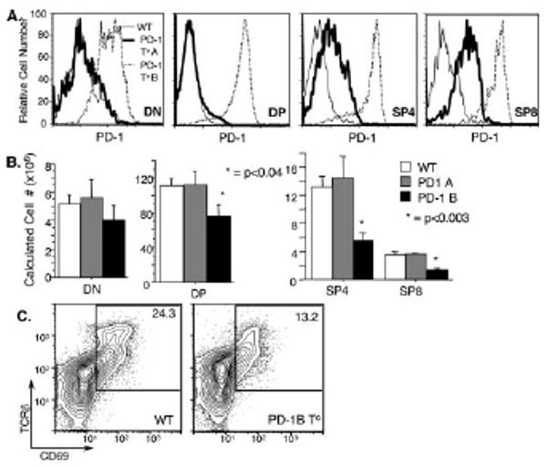
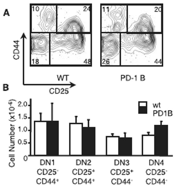
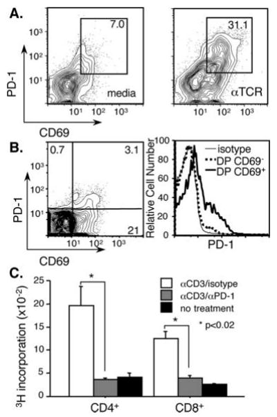
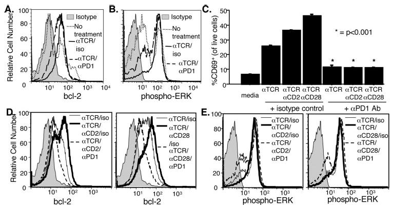
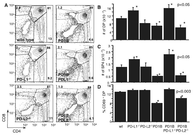
Positive selection during thymocyte development is driven by the affinity and avidity of the TCR for MHC-peptide complexes expressed in the thymus. In this study, we show that programmed death-1 (PD-1), a member of the B7/CD28 family of costimulatory receptors, inhibits TCR-mediated positive selection through PD-1 ligand 1 (PD-L1):PD-1 interactions. Transgenic mice that constitutively overexpress PD-1 on CD4+CD8+ thymocytes display defects in positive selection in vivo. Using an in vitro model system, we find that PD-1 is up-regulated following TCR engagement on CD4+CD8+ murine thymocytes. Coligation of TCR and PD-1 on CD4+CD8+ thymocytes with a novel PD-1 agonistic mAb inhibits the activation of ERK and up-regulation of bcl-2, both of which are downstream mediators essential for positive selection. Inhibitory signals through PD-1 can overcome the ability of positive costimulators, such as CD2 and CD28, to facilitate positive selection. Finally, defects in positive selection that result from PD-1 overexpression in thymocytes resolve upon elimination of PD-L1, but not PD-1 ligand 2, expression. PD-L1-deficient mice have increased numbers of CD4+CD8+ and CD4+ thymocytes, indicating that PD-L1 is involved in normal thymic selection. These data demonstrate that PD-1:PD-L1 interactions are critical to positive selection and play a role in shaping the T cell repertoire.


Conflict of interest statement

Disclosures: The authors have no financial conflict of interest.

Figures

FIGURE 1
FIGURE 1
PD-1B Tc mice have impaired thymic selection. PD-1 Tc mice constitutively overexpress PD-1 under the control of the CD2 promoter/enhancer. Two separate lines of PD-1 Tc mice have distinct PD-1 expression patterns, resulting in specific effects on thymocyte selection. A, PD-1 is expressed at the DN, DP, and SP stages of thymocyte development in PD-1 B Tc mice, while PD-1 A Tc mice express PD-1 at the SP stage of maturation. Thymocytes from the indicated mouse strains were isolated and stained for CD4, CD8, and PD-1. Live cells were gated on CD4CD8 (DN), CD4+CD8+ (DP), CD4+CD8 (SP4), or CD4CD8+ (SP8) cells, as indicated. B, PD-1 B Tc (n = 5) mice have fewer SP thymocytes than WT (n = 8) or PD-1 A Tc (n = 5) mice. Cell yields were calculated by multiplying the subset percentage by the total thymocyte yield. C, PD-1B Tc (n = 5) mice have reduced CD69 expression on DP thymocytes in comparison with WT (n = 4) littermates.
FIGURE 2
FIGURE 2
DN subsets are unchanged in PD-1 B Tc mice. Overexpression of PD-1 on DN thymocytes does not alter subset frequencies or cell numbers. A, CD3CD4CD8 thymocytes from PD-1 B Tc mice (n = 4) and WT (n = 3) littermates were analyzed for expression of CD25 and CD44. B, Thymocyte numbers for the WT and PD-1 B Tc mice were calculated by multiplying the absolute cell yield by the subset percentage. No significant difference was seen in DN thymocyte numbers.
FIGURE 3
FIGURE 3
PD-1 is expressed on DP thymocytes, and expression is up-regulated after TCR stimulation. A, PD-1 expression is enhanced on DP thymocytes from WT mice after TCR stimulation. DP thymocytes were isolated from WT mice and incubated overnight on uncoated plates (medium) or plates coated with anti-TCRβ Abs. After overnight incubation, thymocytes were washed and rested for an additional 18 h before analysis of CD4, CD8, PD-1, and CD69 expression. B, CD69+ DP WT thymocytes have increased PD-1 expression in comparison with CD69 DP thymocytes. Thymocytes were isolated from WT animals and stained for CD4, CD8, CD69, and PD-1. CD4+CD8+ thymocytes were analyzed for CD69 and PD-1 expression (left). Increased expression of PD-1 on CD69+ DP thymocytes is shown in a histogram (right). C, PD-1 agonistic mAb inhibits anti-CD3-induced proliferation of PD-1 Tc PD-L1−/− CD4+ or CD8+ peripheral T cells. Purified CD4+ or CD8+ peripheral T cells were stimulated with plate-bound anti-CD3 and anti-PD-1 mAbs, as indicated. At 24 h, proliferation was assessed by 3H incorporation. Data are representative of three experiments.
FIGURE 4
FIGURE 4
Cocross-linking of TCR and PD-1 inhibits TCR-induced CD69 up-regulation and CD4/CD8 coreceptor down-regulation without inducing apoptosis in thymocytes. Immature DP (>98% CD4+CD8+) thymocytes were isolated from WT mice and plated on untreated or Ab-coated plates, as indicated. After culture, thymocytes were stained for CD4, CD8, CD69, and TCRβ or annexin and analyzed by flow cytometry. A, Developing thymocytes up-regulate CD69 after culture on anti-TCR-coated plates, but coligation with anti-PD-1 and anti-TCR mAb inhibits CD69 up-regulation. B, Immature DP thymocytes down-regulate CD4 and CD8 after TCR stimulation. CD4 and CD8 down-regulation is inhibited by coligation with anti-PD-1 mAb treatment (50 μg/ml). C, Cross-linking of TCR and PD-1 does not increase apoptosis in immature thymocytes. Immature DP thymocytes were plated on Ab-coated plates, as indicated, and analyzed for apoptosis by flow cytometry (apoptotic cells were defined as annexin+7-aminoactinomycin D) after 4 or 20 h. There was no increase in annexin staining on anti-TCR-treated thymocytes cotreated with anti-PD-1 mAb, while thymocytes treated with anti-TCR and -CD28 mAbs showed a significant increase in apoptosis at 4 h. D, Cross-linking of TCR and PD-1 does not increase the accumulation of necrotic thymocytes. Thymocytes treated with anti-TCR and anti-CD28 mAbs showed significant increases in necrotic cells at 20 h. Immature DP thymocytes were treated as in C, and necrotic cells were defined as annexin+7-aminoactinomycin D+ cells. Data are representative of three independent experiments.
FIGURE 5
FIGURE 5
Cocross-linking of PD-1 and TCR inhibits up-regulation of bcl-2 and phosphorylation of ERK in developing thymocytes, even in the presence of costimulation. Downstream mediators of positive selection are impaired in developing thymocytes treated with PD-1 agonistic mAb. Immature DP thymocytes were isolated from WT mice and cultured as in Fig. 3. After 18 h, thymocytes were stained for developmental markers and intracellular proteins. A, Up-regulation of bcl-2 protein levels is inhibited in DP thymocytes treated with anti-TCR and anti-PD-1 mAb. Isotype control is shown as a shaded histogram. B, DP thymocytes that have undergone selection (DPdull cells, as in Fig. 4B) show a reduction in sustained phosphorylation of ERK after treatment with anti-TCR and anti-PD-1 mAb. C, CD69 up-regulation was increased when DP thymocytes were treated with TCR plus CD2 or CD28 cross-linking mAbs, but the increase was abrogated by coligation with anti-PD-1 mAb. D, CD2 or CD28 costimulation enhanced TCR-induced up-regulation of bcl-2. The increase in bcl-2 observed in thymocytes treated with TCR plus CD2 or CD28 cross-linking mAb was inhibited by the addition of PD-1 mAb. E, ERK phosphorylation is impaired in DPdull thymocytes treated with anti-PD-1 mAb plus anti-TCR and anti-CD2 or anti-CD28. Data are representative of three independent experiments.
FIGURE 6
FIGURE 6
PD-1:PD-L1 interactions inhibit positive selection in vivo. PD-L1−/−, PD-L2−/−, and WT mice were evaluated for changes in thymocyte subsets. PD-1 B Tc mice were backcrossed onto a PD-L1−/− or PD-L2−/− background, and thymocyte numbers were calculated. A, CD4/CD8 expression on live thymocytes from a representative animal from each group is shown. B, DP thymocyte numbers are increased in PD-L1−/− (n = 4) and PD-1 B Tc PD-L1−/− (n = 3) mice. DP thymocyte numbers are decreased in PD-1 B Tc (n = 8) and PD-1 B Tc PD-L2−/− (n = 6) mice. C, SP4 thymocytes are reduced in PD-1 B Tc and PD-1 B Tc PD-L2−/− mice, but not in PD-1 B Tc PD-L1−/− mice. No significant difference in SP4 thymocytes was observed between WT (n = 8) and PD-L2−/− (n = 6) mice, but the number of SP4 thymocytes in PD-L1−/− was significantly increased. D, CD69 expression on DP thymocytes is reduced in PD-1 B Tc and PD-1 B Tc PD-L2−/− mice, but not in PD-1 B Tc PD-L1−/− mice. There was no significant difference in CD69 expression observed between WT and PD-L1−/− or PD-L2−/− animals.
FIGURE 7
FIGURE 7
PD-1 overexpression decreases and PD-L1 loss increases cell numbers in an Ag-specific model of selection. PD-1 B Tc and PD-L1−/− animals were bred to the H-Y TCR tg+ strain and evaluated for positive selection (females) and negative selection (males). A, Female PD-1 B Tc mice (n = 3) have decreased numbers of SP8 thymocytes. Female PD-L1 −/− mice (n = 5) expressing the H-Y TCR tg have increased numbers of DN, DP, and SP8 thymocytes. PD-L1−/− H-Y TCR tg+ mice also have an increased number of peripheral CD8+ T cells. B, Male mice overexpressing PD-1 (n = 2) have no alteration in thymocyte subsets. PD-L1−/− H-Y TCR tg+ (n = 2) mice have increased numbers of DN and SP8 thymocytes, but substantially fewer peripheral CD8 T cells.

Similar articles

Cited by

References

    1. Starr TK, Jameson SC, Hogquist KA. Positive and negative selection of T cells. Annu Rev Immunol. 2003;21:139–176. - PubMed
    1. Greenwald RJ, Latchman YE, Sharpe AH. Negative co-receptors on lymphocytes. Curr Opin Immunol. 2002;14:391–396. - PubMed
    1. Ishida Y, Agata Y, Shibahara K, Honjo T. Induced expression of PD-1, a novel member of the immunoglobulin gene superfamily, upon programmed cell death. EMBO J. 1992;11:3887–3895. - PMC - PubMed
    1. Freeman GJ, Long AJ, Iwai Y, Bourque K, Chernova T, Nishimura H, Fitz LJ, Malenkovich N, Okazaki T, Byrne MC, et al. Engagement of the PD-1 immunoinhibitory receptor by a novel B7 family member leads to negative regulation of lymphocyte activation. J Exp Med. 2000;192:1027–1034. - PMC - PubMed
    1. Latchman Y, Wood CR, Chernova T, Chaudhary D, Borde M, Chernova I, Iwai Y, Long AJ, Brown JA, Nunes R, et al. PD-L2 is a second ligand for PD-1 and inhibits T cell activation. Nat Immunol. 2001;2:261–268. - PubMed

Publication types

MeSH terms