Comparative Study
. 2005 Nov 29;102(48):17366-71.
doi: 10.1073/pnas.0508885102. Epub 2005 Nov 21.

The DHHC protein Pfa3 affects vacuole-associated palmitoylation of the fusion factor Vac8

Affiliations
Comparative Study

The DHHC protein Pfa3 affects vacuole-associated palmitoylation of the fusion factor Vac8

Haitong Hou et al. Proc Natl Acad Sci U S A. .

Abstract

Vacuole biogenesis depends on specific targeting and retention of peripheral membrane proteins. At least three palmitoylated proteins are found exclusively on yeast vacuoles: the fusion factor Vac8, the kinase Yck3, and a novel adaptor protein implicated in microautophagy, Meh1. Here, we analyze the role that putative acyltransferases of the DHHC family play in their localization and function. We find that Pfa3/Ynl326c is required for efficient localization of Vac8 to vacuoles in vivo, while Yck3 or Meh1 localization is not impaired in any of the seven DHHC deletions. Vacuole-associated Vac8 appears to be palmitoylated in a pfa3 mutant, but this population is refractive to further palmitoylation on isolated vacuoles. Vacuole morphology and inheritance, which both depend on Vac8 palmitoylation, appear normal, although there is a reduction in vacuole fusion. Interestingly, Pfa3 is required for the vacuolar localization of not only an SH4 domain that is targeted by myristate/palmitate (as in Vac8) but also one that is targeted by a myristate/basic stretch (as in Src). Our data indicate that Pfa3 has an important but not exclusive function for Vac8 localization to the vacuole.


Figures
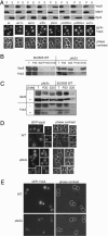
Fig. 1.
Fig. 1.
Analysis of the DHHC mutants for Vac8 and Yck3 localization. (A Upper) Subcellular fractionation. Cells from the respective deletion strains (BY4741, EUROSCARF) were lysed and separated by centrifugation (13,000 × g for 10 min at 4°C) into a pellet (P) and supernatant fraction (S) as described in Methods. Proteins were analyzed by SDS/PAGE and Western blotting with antibodies to Meh1, Vac8, and Rpl3, a ribosomal subunit. (A Lower) The indicated strains expressing GFP-Yck3 were analyzed by fluorescence microscopy. (B) Subcellular fractionation in BJ3505 cells. The lysate was centrifuged for 10 min at 20,000 × g to yield the P20 and S20 fractions. To get the P100 and S100 fractions, a similar amount of the S20 fraction was spun again (100,000 × g for 30 min at 4°C). Proteins in pellet and TCA-precipitated supernatant were analyzed by SDS/PAGE and Western blotting. (C) Mobility of Vac8 on gels. Subcellular fractionation was done as in B. Proteins were boiled in sample buffer with or without 2-mercaptoethanol and analyzed by SDS/PAGE and Western blotting with antibodies to Vac8 and Yck3. The top band in the Yck3 illustration corresponds to Vac8, which was detected before Yck3. (D and E) Localization of GFP-tagged Vac8 and Yck3 in BJ wild-type and pfa3Δ cells. Yeast strains expressing the indicated GFP variant were analyzed by fluorescence microscopy.
Fig. 2.
Fig. 2.
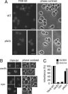
Vacuole morphology and inheritance. (A and B) Vacuoles from the indicated BJ wild-type, pfa3Δ, vac8Δ, and the Cys-mutant, which expresses a Vac8 mutant lacking the N-terminal cysteines (Cys– is Vac8-C4,5,7A) were stained with 50 μM FM4 – 64 and observed by fluorescence microscopy. (C) Vacuole inheritance was scored for the indicated strains as described in Methods.
Fig. 3.
Fig. 3.
Functional analysis of pfa3 deletion. (A) Vacuole fusion. Vacuoles from tester strains with the respective deletion were incubated at 26°C for 90 min and then analyzed for fusion (SD, n = 3). Fusion deficiency is stronger in the DKY background than in BJ (not shown). For details see Methods.(B) Sensitivity to inhibitors. Fusion was analyzed as in A in the presence or absence of the indicated antibodies. All antibodies were titrated into the reaction (not shown), and the strongest inhibition is shown. In general, antibody concentration was at ≈100 μg/ml. For Gdi1, the inhibitor was boiled to determine the buffer control. The control reaction was set to 100% for each combination. For additional information, see ref. . (C) Effect of Pal-CoA on fusion. Increasing Pal-CoA amounts were titrated into the fusion reaction, and fusion was determined as before. (D) Palmitoylation of Vac8 and Yck3 on vacuoles. Purified vacuoles (60 μg) from the respective strain were incubated in fusion reaction buffer with [3H]Pal-CoA for 10 min at 26°C. Vacuoles were then collected (12,000 × g for 10 min at 4°C), and proteins were analyzed by SDS/PAGE and fluorography. (E) Palmitoylation determined by the biotin-switch method. The indicated cells were lysed, free cysteines were quenched by N-ethylmaleimide, and the extract was subjected to the biotin-switch procedure by using hydroxylamine (HA) and biotin-BMCC as a crosslinker. Crosslinked proteins were captured on Neutravidin agarose (Pierce), eluted by boiling, and analyzed by SDS/PAGE. Western blots were decorated with antibodies to Vac8. Total (6%) refers to the faction of lysate removed before the Neutravidin pull-down. (F) Membrane association of Vac8. Vacuoles from the indicated strains (30 μg each) were diluted into 500 μl of 20 mM Hepes/KOH (pH 7.4), 1 mM PMSF, and the following conditions: 0.1 M Na2CO3 (carb.), 6 M urea, 1 M NaCl, or 1% Triton X-100, and incubated for 30 min on ice. Samples were then centrifuged (100,000 × g for 30 min at 4°C) and split into pellet (P) and supernatant (S). An aliquot corresponding to the membrane fraction used in each condition is shown for comparison (T). Proteins in pellet and TCA-precipitated supernatant were analyzed by SDS/PAGE and Western blotting. (G) Sizing of Pfa3. Vacuoles (200 μg) from BJ3505 carrying Pfa3-TAP were incubated for 10 min at 4°C in lysis buffer (20 mM Tris·HCl, pH 7.4/150 mM NaCl/0.5% Triton X-100/1× PIC), insoluble material was removed by centrifugation (20,000 × g for 10 min at 4°C), and the detergent extract was loaded onto a linear 10–30% glycerol gradient prepared in lysis buffer. The sample was then centrifuged (40,000 rpm in an SW40 rotor for 18 h at 4°C), 0.5-ml samples were collected from the top of the gradient, and TCA-precipitated proteins were analyzed by SDS/PAGE and Western blotting. The first 10 samples are shown. Vps41 (120 kDa) is the only protein that had a second peak at the bottom of the gradient (not shown); the SNARE Nyv1 (30 kDa) is found in a complex at 10S as part of the SNARE complex (26). (H) Physical associations. The detergent lysate from a BJ Pfa3-TAP strain expressing Vac8(1–18)-GFP was prepared as in G and incubated with IgG beads overnight at 4°C. Load (L) and flow-throw (FT) (1% each) were removed, and proteins were TCA-precipitated. Beads were washed two times in lysis buffer. Proteins were then eluted by boiling in sample buffer and analyzed as before. A BJ vac8Δ strain expressing Vac8-GFP (WT*) served as a negative control. The lower band in the Vac8 illustration is due to the Pfa3-TAP signal, which runs at a similar molecular weight.
Fig. 4.
Fig. 4.
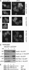
Localization of minimal targeting constructs in yeast. Vac8(1–18)-GFP (A), Src(1–16)-GFP (B), or HASPB.T6S(1–18)-GFP (HB.T6S) (C) were expressed in BY wild-type or all DHHC deletion strains and analyzed by fluorescence microscopy. Localization was similar in wild type and most DHHC deletion strains (AC Left). Only in pfa3Δ cells exclusive plasma membrane staining for Vac8(1–18) and Src(1–16) was visible (A and B Right). (D) Palmitoylation of the SH4(1–18)-GFP constructs. Palmitoylation of the indicated constructs was determined as described in Fig. 3E. Total (6%) corresponds to the fraction of lysate removed before the Neutravidin pull-down. Blots were decorated with antibodies to GFP. (E) Alignment of the N-terminal 15 aa of the Vac8, Src, and HASPB-T6S sequence.

Similar articles

Cited by

References

    1. Bonifacino, J. S. & Glick, B. S. (2004) Cell 116, 153–166. - PubMed
    1. Munro, S. (2005) Curr. Opin. Cell Biol. 17, 395–401. - PubMed
    1. Dietrich, L. E. & Ungermann, C. (2004) EMBO Rep. 5, 1053–1057. - PMC - PubMed
    1. Smotrys, J. E. & Linder, M. E. (2004) Annu. Rev. Biochem. 73, 559–587. - PubMed
    1. Magee, T. & Seabra, M. C. (2005) Curr. Opin. Cell Biol. 17, 190–196. - PubMed

Publication types

MeSH terms