

Expression of FGFR-2 and FGFR-3 in the normal human fetal orbit
- PMID: 16299148
- PMCID: PMC1772988
- DOI: 10.1136/bjo.2005.075978
Expression of FGFR-2 and FGFR-3 in the normal human fetal orbit
Abstract
Aims: To demonstrate the expression patterns of two fibroblast growth factor receptors (FGFR-2 and FGFR-3) in the normal human fetal orbit.
Methods: 6 microm orbital slide sections were prepared from 12 week old human fetal material obtained within established ethical guidelines. Radioactive in situ hybridisation techniques were used to demonstrate the expression patterns of FGFR-2 and FGFR-3 within these sections. Only one foetus had appropriate orbital sections taken.
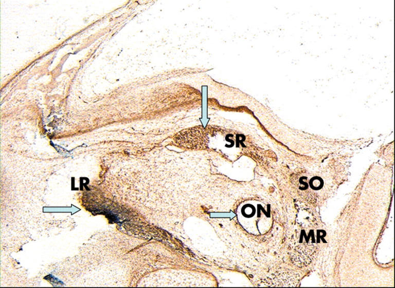
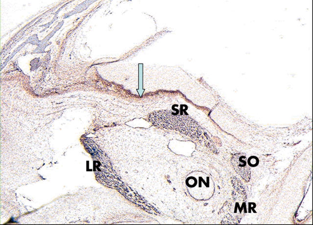
Results: FGFR-2 was expressed within the extraocular muscles (EOMs) and the optic nerve sheath and to a lesser degree within the orbital periosteal margins and the cranial sutures. FGFR-3 was expressed a lot within the periosteal margins and cranial sutures but not within either the EOMs or the optic nerve sheath.
Conclusions: FGFR-2 and FGFR-3 are differentially expressed within different orbital components. FGFR-2 gene mutations may be responsible for craniosynostotic syndromes such as Crouzon, Pfeiffer, and Apert, while those in the FGFR-3 gene may cause isolated unicoronal synostosis. EOMs may be histologically abnormal in cases of Apert, Pfeiffer, and Crouzon syndromes but not isolated unicoronal synostosis. The pattern of expression of FGFR-2 in the normal human fetal orbit may explain some of the EOM histological findings seen in some cases of Apert, Pfeiffer, and Crouzon syndromes.
Figures
Similar articles
-
Description of a new mutation and characterization of FGFR1, FGFR2, and FGFR3 mutations among Brazilian patients with syndromic craniosynostoses.Am J Med Genet. 1998 Jul 7;78(3):237-41. Am J Med Genet. 1998. PMID: 9677057
-
Increased calvaria cell differentiation and bone matrix formation induced by fibroblast growth factor receptor 2 mutations in Apert syndrome.J Clin Invest. 1998 Mar 15;101(6):1310-7. J Clin Invest. 1998. PMID: 9502772 Free PMC article.
-
Localisation and differential expression of the fibroblast growth factor receptor (FGFR) multigene family in normal and atherosclerotic human arteries.Cardiovasc Res. 1996 Sep;32(3):557-69. Cardiovasc Res. 1996. PMID: 8881516
-
[From gene to disease; craniosynostosis syndromes due to FGFR2-mutation].Ned Tijdschr Geneeskd. 2002 Jan 12;146(2):63-6. Ned Tijdschr Geneeskd. 2002. PMID: 11820058 Review. Dutch.
-
Ocular manifestations of Apert and Crouzon syndromes: qualitative and quantitative findings.J Craniofac Surg. 2010 Sep;21(5):1354-7. doi: 10.1097/SCS.0b013e3181ef2b53. J Craniofac Surg. 2010. PMID: 20856021 Review.
Cited by
-
Fibrous Band between Extraocular Muscles in Unilateral Coronal Synostosis.Korean J Ophthalmol. 2020 Feb;34(1):88-89. doi: 10.3341/kjo.2019.0072. Korean J Ophthalmol. 2020. PMID: 32037754 Free PMC article. No abstract available.
-
Clues from Crouzon: Insights into the potential role of growth factors in the pathogenesis of myelinated retinal nerve fibers.J Curr Ophthalmol. 2016 Aug 27;28(4):232-236. doi: 10.1016/j.joco.2016.07.008. eCollection 2016 Dec. J Curr Ophthalmol. 2016. PMID: 27830211 Free PMC article.
-
Use of neuroimaging measurements of optic nerve sheath diameter to assess intracranial pressure in craniosynostosis.Childs Nerv Syst. 2018 May;34(5):939-946. doi: 10.1007/s00381-018-3728-7. Epub 2018 Jan 29. Childs Nerv Syst. 2018. PMID: 29380112
-
Effect of Fibroblast Growth Factor 2 on Extraocular Muscle Structure and Function.Invest Ophthalmol Vis Sci. 2021 Jul 1;62(9):34. doi: 10.1167/iovs.62.9.34. Invest Ophthalmol Vis Sci. 2021. PMID: 34293078 Free PMC article.
-
Morphological Differences in the Inferior Oblique Muscles from Subjects with Over-elevation in Adduction.Invest Ophthalmol Vis Sci. 2020 Jun 3;61(6):33. doi: 10.1167/iovs.61.6.33. Invest Ophthalmol Vis Sci. 2020. PMID: 32539136 Free PMC article.
References
-
- Vajo Z, Francomano CA, Wilkin D. The molecular and genetic basis of fibroblast growth factor receptor 3 disorders: The achondroplasia family of skeletal dysplasias, Muenke craniosynostosis, and Crouzon syndrome with acanthosis nigricans. Endocrine Rev 2000;21:23–39. - PubMed
-
- De Moerlooze L, Dickson C. Skeletal disorders associated with fibroblast growth factor receptor mutations. Curr Opin Genet Develop 1997;7:378–85. - PubMed
-
- Captuo A, Lingua R. Abberrant muscle insertions in Crouzon’s disease. J Pediatr Ophthalmol Strabismus 1980;17:239–41. - PubMed
MeSH terms
Substances
LinkOut - more resources
Full Text Sources
Miscellaneous