. 2006 Jan;17(1):213-26.
doi: 10.1091/mbc.e05-06-0585. Epub 2005 Nov 2.

A genomewide screen for petite-negative yeast strains yields a new subunit of the i-AAA protease complex

Affiliations

A genomewide screen for petite-negative yeast strains yields a new subunit of the i-AAA protease complex

Cory D Dunn et al. Mol Biol Cell. 2006 Jan.

Abstract

Unlike many other organisms, the yeast Saccharomyces cerevisiae can tolerate the loss of mitochondrial DNA (mtDNA). Although a few proteins have been identified that are required for yeast cell viability without mtDNA, the mechanism of mtDNA-independent growth is not completely understood. To probe the relationship between the mitochondrial genome and cell viability, we conducted a microarray-based, genomewide screen for mitochondrial DNA-dependent yeast mutants. Among the several genes that we discovered is MGR1, which encodes a novel subunit of the i-AAA protease complex located in the mitochondrial inner membrane. mgr1Delta mutants retain some i-AAA protease activity, yet mitochondria lacking Mgr1p contain a misassembled i-AAA protease and are defective for turnover of mitochondrial inner membrane proteins. Our results highlight the importance of the i-AAA complex and proteolysis at the inner membrane in cells lacking mitochondrial DNA.


Figures
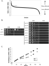
Figure 1.
Figure 1.
Yeast knockouts that require mtDNA for viability. (A) Distribution of YKOs after growth in EtBr-containing medium. In microarray analysis, the log2-converted ratio of the amount of hybridization to the probe from control cultures (–EtBr) and the probe from experimental cultures (+EtBr) for each YKO in our pool was determined. This ratio is plotted on the Y-axis. Thus, YKOs whose growth is most affected by EtBr are to the left, and those least affected are to the right. Strains above the dotted line were candidates for further testing. (B) Representative mutants from the genomewide screen. mgr1Δ (RJ2058), phb1Δ (RJ2083), opi1Δ (RJ2084), mgr2Δ (RJ2085), thr1Δ (RJ2086), thr4Δ (RJ2087), ira2Δ (RJ2088), pde2Δ (RJ2089), and yjr120wΔ (RJ2091) cells were grown 7 h in YEPD broth at 30°C without ethidium bromide (–EtBr) or for 22 h in YEPD containing 25 μg/ml EtBr (+EtBr). A 4-μl aliquot of cells at OD600 of 0.02, and serial 10-fold dilutions thereof, were spotted onto YEPD (–EtBr) or YEPD + EtBr (+EtBr) plates and grown at 30°C for 2 d. As controls, wild-type strain BY4742 and two known petite-negative mutants, atp2Δ (RJ1577) and yme1Δ (RJ2051) were also examined. (C) Growth of mgr1Δ cells is defective in EtBr medium. WT (BY4742) and mgr1Δ (RJ2058) strains were inoculated into YEPD medium and then transferred to YEPD with 25 μg/ml EtBr. At indicated times, the OD600 of each culture was determined and the number of doublings was calculated.
Figure 2.
Figure 2.
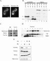
Mgr1p is a mitochondrial IM protein with its carboxyl-terminus facing the IMS. (A) Mgr1p is a mitochondrial protein. mgr1Δ strain RJ1688 with plasmid pM486, which expresses the Mgr1p-GFP fusion protein, was stained with 2 μM Mitofluor 589 (Molecular Probes) and examined by fluorescence microscopy. Bar, 2 μm. (B) Mgr1p is imported into isolated mitochondria. 35S-labeled Mgr1p or su9-DHFR were mixed with mitochondria from wild-type strain RJ1962 with (+Δψ) or without (–Δψ) an inner membrane potential. Some samples (+PK) were digested after the import reaction with 100 μg/ml proteinase K on ice for 20 min. After centrifugation, samples were analyzed by SDS-PAGE and phosphorimaging. The precursor (p) and mature (m) forms of su9-DHFR and Mgr1p are indicated. (C) Mgr1p is an integral membrane protein. Mitochondria from strain RJ1970, which expresses Mgr1p-myc, were resuspended in breaking buffer (BB; 0.6 M sorbitol, 20 mM HEPES-KOH, pH 7.4) or in 0.1 M sodium carbonate, pH 11. After centrifugation, equal amounts of the supernatant (S) or pellet (P) fractions were analyzed by Western blotting using antibodies to the myc epitope (Mgr1p-myc), Tim23p, an integral membrane protein, or Mas2p, a soluble matrix marker. (D) Mgr1p is located in the mitochondrial IM. Mgr1p-myc mitochondria from strain RJ1970 were sonicated and membrane vesicles separated on sucrose step gradients. Fractions from the gradients were analyzed by Western blotting with antibodies to the myc epitope (Mgr1p-myc), the IM protein F1β, or the OM protein, OM45. Fraction 1 represents the top of the gradient. (E) The carboxyl-terminus of Mgr1p-myc faces the IMS. Intact mitochondria from MGR1-MYC strain RJ1970, or mitochondria whose OM was disrupted by osmotic shock (+OS), were digested with 50 μg/ml PK for 30 min on ice and then analyzed by Western blotting using antibodies to the myc epitope (Mgr1p-myc), the matrix protein Mas2p, the OM protein Tom70p, or the IM protein Tim23p.
Figure 3.
Figure 3.
Mgr1p is a subunit of the i-AAA protease complex. (A) Yme1p coprecipitates with Mgr1p-myc. Mitochondria from MGR1-MYC strain RJ1970 were solubilized in buffer containing 1% digitonin, and the Mgr1p-myc protein was then precipitated using anti-myc antibodies coupled to agarose beads. Aliquots from the starting lysate (load) and the pellet fraction (IP) were analyzed by Western blotting with antibodies to the myc epitope (Mgr1p-myc), Phb1p, Phb2p, F1β, and Yme1p. As a control, mitochondrial lysates from wild-type mitochondria (MGR1) were analyzed in parallel. (B) Mgr1p-myc coprecipitates with Yme1p-HA. Mitochondria isolated from strain RJ1980, expressing both Mgr1p-myc and Yme1p-HA fusion proteins, were solubilized in digitonin-containing buffer, and the Yme1p-HA protein precipitated with agarose beads linked to anti-HA antibodies. As a control, mitochondria from YME1 MGR1-MYC strain RJ1970 were also analyzed. Aliquots from the mitochondrial lysate (load) and precipitated proteins (IP) were Western blotted using anti-HA (Yme1p-HA) or anti-myc (Mgr1p-myc) antibodies.
Figure 4.
Figure 4.
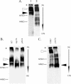
The i-AAA protease complex requires both Mgr1p and Yme1p. (A) Mgr1p comigrates with Yme1p during blue-native gel electrophoresis. Mitochondria isolated from strain RJ1980 were solubilized in digitonin-containing buffer, and protein complexes were separated on a 3–13% polyacrylamide blue-native gel. Western blotting was used to locate the (1) Yme1p-HA and (2) Mgr1p-myc proteins. The open and black arrows denote two apparent forms of the i-AAA complex. (B) The size of the Yme1p-containing complex decreases in the absence of Mgr1p. Mitochondria were isolated from WT (RJ1962), mgr1Δ (RJ1961), and phb1Δ (RJ2083) cells and then solubilized in digitonin buffer. Protein complexes were separated on a 3–8% polyacrylamide blue-native gel and analyzed by Western blotting with anti-Yme1p antibodies. The open and black arrows denote two apparent forms of the i-AAA complex. (C) The mobility of the main Mgr1p-containing complex changes in mitochondria lacking Yme1p. Mitochondria isolated from wild-type cells expressing Mgr1p-myc (RJ1970), or yme1Δ cells expressing Mgr1p-myc (RJ1972) were solubilized, run on a 3–13% polyacrylamide blue-native gel and analyzed by Western blotting with anti-myc antibodies. The arrows denote the two main forms of the Mgr1p-containing complex. In A, B, and C, Yme1p, Yme1p-HA, or Mgr1p-myc that has not entered the resolving gel is labeled with an asterisk.
Figure 5.
Figure 5.
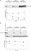
Mitochondria lacking Mgr1p are defective in turnover of IM proteins. (A) The Nde1p-HA fusion protein is more stable in mgr1Δ and yme1Δ cells. Wild-type strain RJ2077 (diamonds), mgr1Δ strain RJ2078 (squares), and yme1Δ strain RJ2079 (triangles) each expressing Nde1p-HA were grown to midlog phase at 30°C in YEP glycerol/ethanol medium. Cycloheximide was then added to 1 mg/ml and the cultures were shifted to 37°C. At the indicated times, cells were lysed, equal amounts of cellular protein were loaded in each lane, and the amount of Nde1p-HA was determined by Western blotting using antibodies to the HA-epitope (n = 3). (B) Turnover of the Yta10(161)-DHFRMUT fusion protein is reduced in mgr1Δ and yme1Δ mitochondria. The 35S-labeled Yta10(161)-DHFRMUT precursor was imported at 25°C for 10 min into mitochondria isolated from WT strain RJ1962 (diamonds), mgr1Δ strain RJ1961 (squares), and yme1Δ strain RJ1974 (triangles). Precursor not imported was digested with 100 μg/ml proteinase K on ice for 30 min. After the addition of 1 mM PMSF, samples were shifted to 25°C and incubated for the indicated times. Equal amounts of mitochondria were isolated by centrifugation at each time point, subjected to SDS-PAGE, and analyzed by phosphorimaging (n = 3). The processed forms of Yta10(161)-DHFRMUT (asterisk) were quantified.
Figure 6.
Figure 6.
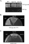
Several phenotypes of the yme1Δ mutant are not found in mgr1Δ cells. (A) Lack of Mgr1p does not increase mtDNA escape to the nucleus. trp1 strain PTY44, trp1 mgr1Δ strain RJ2001, and trp1 yme1Δ strain RJ2040, each containing the nuclear TRP1 marker in their mitochondria, were grown on YEP glycerol/ethanol medium for 4 d at 30°C. Strains were then replica-plated to SD medium lacking tryptophan and grown for a further 4 d at 30°C. (B) mgr1Δ mutants are not heat-sensitive for growth. WT (PTY44), mgr1Δ (RJ2001), and yme1Δ (RJ2040) strains were struck to YEP glycerol/ethanol medium and incubated at 37°C for 5 d. (C) mgr1Δ cells are not cold-sensitive. WT (BY4742), mgr1Δ (RJ2059), and yme1Δ (RJ2051) strains were struck to YEPD medium and incubated for 6 d at 18°C.

Similar articles

Cited by

References

    1. Adams, A., Gottschling, D., Kaiser, C., and Stearns, T. (1997). Methods in Yeast Genetics, Plainview, NY: Cold Spring Harbor Laboratory Press.
    1. Arnold, I., and Langer, T. (2002). Membrane protein degradation by AAA proteases in mitochondria. Biochim. Biophys. Acta 1592, 89–96. - PubMed
    1. Arnold, I., Pfeiffer, K., Neupert, W., Stuart, R. A., and Schagger, H. (1998). Yeast mitochondrial F1F0-ATP synthase exists as a dimer: identification of three dimer-specific subunits. EMBO J. 17, 7170–7178. - PMC - PubMed
    1. Attardi, G., and Schatz, G. (1988). Biogenesis of mitochondria. Annu. Rev. Cell Biol. 4, 289–333. - PubMed
    1. Augustin, S., Nolden, M., Muller, S., Hardt, O., Arnold, I., and Langer, T. (2005). Characterization of peptides released from mitochondria: evidence for constant proteolysis and peptide efflux. J. Biol. Chem. 280, 2691–2699. - PubMed

Publication types

MeSH terms