Transgene-induced CCWGG methylation does not alter CG methylation patterning in human kidney cells
- PMID: 16246913
- PMCID: PMC1266073
- DOI: 10.1093/nar/gki920
Transgene-induced CCWGG methylation does not alter CG methylation patterning in human kidney cells
Abstract
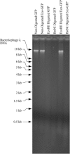
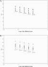
Several reports suggest that C(m)CWGG methylation tends not to co-exist with (m)CG methylation in human cells. We have asked whether or not methylation at CCWGG sites can influence CG methylation. DNA from cells expressing an M.EcoRII-GFP fusion was actively methylated at CCWGG sites. CG methylation as measured by R.HpaII/R.MspI ratios was unchanged in cells expressing the transgene. Cloned representatives of C(m)CWGG methylated DNA often contained, or were adjacent to an ALU repeat, suggesting that M.EcoRII-GFP actively methylated gene-rich R-band DNA. The transgenic methyltransferase applied C(m)CWGG methylation to a representative human promoter that was heavily methylated at CG dinucleotides (the SERPINB5 promoter) and to a representative promoter that was essentially unmethylated at CG dinucleotides (the APC promoter). In each case, the CG methylation pattern remained in its original state, unchanged by the presence of neighboring C(m)CWGG sites. Q-PCR measurements showed that RNA expression from the APC gene was not significantly altered by the presence of C(m)CWGG in its promoter. Kinetic studies suggested that an adjacent C(m)CWGG methylation site influences neither the maintenance nor the de novo methylation activities of purified human Dnmt1. We conclude that C(m)CWGG methylation does not exert a significant effect on CG methylation in human kidney cells.
Figures


Similar articles
-
Regulation of EcoRII methyltransferase: effect of mutations on gene expression and in vitro binding to the promoter region.Nucleic Acids Res. 1994 Dec 11;22(24):5347-53. doi: 10.1093/nar/22.24.5347. Nucleic Acids Res. 1994. PMID: 7816624 Free PMC article.
-
Cooperative activity of DNA methyltransferases for maintenance of symmetrical and non-symmetrical cytosine methylation in Arabidopsis thaliana.Plant J. 2008 Dec;56(5):814-23. doi: 10.1111/j.1365-313X.2008.03640.x. Epub 2008 Aug 27. Plant J. 2008. PMID: 18665914 Free PMC article.
-
Distribution of CWG and CCWGG in the human genome.Epigenetics. 2007 Sep;2(3):151-4. doi: 10.4161/epi.2.3.4748. Epub 2007 Jul 17. Epigenetics. 2007. PMID: 17965623
-
DNA methylation in plants.Curr Top Microbiol Immunol. 2006;301:67-122. doi: 10.1007/3-540-31390-7_4. Curr Top Microbiol Immunol. 2006. PMID: 16570846 Review.
-
Epigenetic DNA-(cytosine-5-carbon) modifications: 5-aza-2'-deoxycytidine and DNA-demethylation.Biochemistry (Mosc). 2009 Jun;74(6):613-9. doi: 10.1134/s0006297909060042. Biochemistry (Mosc). 2009. PMID: 19645665 Review.
Cited by
-
Global leukocyte DNA methylation is similar in African American and Caucasian women under conditions of controlled folate intake.Epigenetics. 2007 Jan-Mar;2(1):66-8. doi: 10.4161/epi.2.1.4066. Epub 2007 Feb 27. Epigenetics. 2007. PMID: 17965592 Free PMC article.
-
Human non-CG methylation: are human stem cells plant-like?Epigenetics. 2010 Oct 1;5(7):569-72. doi: 10.4161/epi.5.7.12702. Epub 2010 Oct 1. Epigenetics. 2010. PMID: 20647766 Free PMC article. Review.
-
Organomegaly and tumors in transgenic mice with targeted expression of HpaII methyltransferase in smooth muscle cells.Epigenetics. 2011 Mar;6(3):333-43. doi: 10.4161/epi.6.3.14089. Epub 2011 Mar 1. Epigenetics. 2011. PMID: 21107019 Free PMC article.
-
Recovery of bisulfite-converted genomic sequences in the methylation-sensitive QPCR.Nucleic Acids Res. 2007;35(9):2893-903. doi: 10.1093/nar/gkm055. Epub 2007 Apr 16. Nucleic Acids Res. 2007. PMID: 17439964 Free PMC article.
-
Duplex DNA from Sites of Helicase-Polymerase Uncoupling Links Non-B DNA Structure Formation to Replicative Stress.Cancer Genomics Proteomics. 2020 Mar-Apr;17(2):101-115. doi: 10.21873/cgp.20171. Cancer Genomics Proteomics. 2020. PMID: 32108033 Free PMC article.
References
-
- Smith S.S., Lingeman R.G., Kaplan B.E. Recognition of foldback DNA by the human DNA (cytosine-5-)-methyltransferase. Biochemistry. 1992;31:850–854. - PubMed
Publication types
MeSH terms
Substances
Grants and funding
LinkOut - more resources
Full Text Sources
Molecular Biology Databases
Miscellaneous

