. 2006 Jan;17(1):272-82.
doi: 10.1091/mbc.e05-08-0722. Epub 2005 Oct 19.

The Drosophila gamma-tubulin small complex subunit Dgrip84 is required for structural and functional integrity of the spindle apparatus

Affiliations

The Drosophila gamma-tubulin small complex subunit Dgrip84 is required for structural and functional integrity of the spindle apparatus

Nathalie Colombié et al. Mol Biol Cell. 2006 Jan.

Abstract

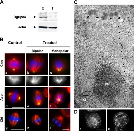
Gamma-tubulin, a protein critical for microtubule assembly, functions within multiprotein complexes. However, little is known about the respective role of gamma-tubulin partners in metazoans. For the first time in a multicellular organism, we have investigated the function of Dgrip84, the Drosophila orthologue of the Saccharomyces cerevisiae gamma-tubulin-associated protein Spc97p. Mutant analysis shows that Dgrip84 is essential for viability. Its depletion promotes a moderate increase in the mitotic index, correlated with the appearance of monopolar or unpolarized spindles, impairment of centrosome maturation, and increase of polyploid nuclei. This in vivo study is strengthened by an RNA interference approach in cultured S2 cells. Electron microscopy analysis suggests that monopolar spindles might result from a failure of centrosome separation and an unusual microtubule assembly pathway via centriolar triplets. Moreover, we point to an involvement of Dgrip84 in the spindle checkpoint regulation and in the maintenance of interphase microtubule dynamics. Dgrip84 also seems essential for male meiosis, ensuring spindle bipolarity and correct completion of cytokinesis. These data sustain that Dgrip84 is required in some aspects of microtubule dynamics and organization both in interphase and mitosis. The nature of a minimal gamma-tubulin complex necessary for proper microtubule organization in the metazoans is discussed.


Figures

Figure 1.
Figure 1.
Characterization of Dgrip84 mutants. (A) The three Dgrip84 mutations induce different patterns of lethality. One hundred first instars of each mutant genotype and wild-type (WT) were followed until adult stage. The percentage of live individuals— independently of the developmental stage— was determined. The PG66 and R20 alleles induce an early lethality, whereas the PG36 allele induces a belated lethality. (B) Dgrip84 protein is not detectable in the brain of the three mutants. Total protein extracts from L3 larval brains (∼10 brains for each genotype) were submitted to Western blot analyses with affinity-purified Dgrip84 antibodies (top). Actin was used as an internal loading control (bottom). (C) Dgrip84 protein is recruited at the centrosome during mitosis in wild-type larval brains but was not detectable in larval Dgrip84 mutant neuroblasts. L3 larval brains were probed with affinity-purified antibodies against Dgrip84 protein (green). Wild-type neuroblasts showed Dgrip84 protein recruitment at the centrosome throughout mitosis, whereas Dgrip84 is not detectable at the poles in R20 mutant neuroblasts at any mitotic stage. In all figures except Figure 6, the spindle is shown in red (anti-α-tubulin antibodies) and chromosomes in blue (DAPI staining). Bars, 5 μm.
Figure 2.
Figure 2.
Mitotic phenotypes in Dgrip84 mutants. Observations were made on L3 larval neuroblasts. (A) The mitotic index is increased in the three mutants. The percentage of mitotic figures was recorded on five brains for each genotype. Confidence intervals (in parentheses) are calculated for a probability of 95%. (B) Mutant neuroblasts accumulate in prometaphases/metaphases. For each genotype, the different mitotic stages were recorded in >250 cells undergoing mitosis (graph). The inset shows two typical control and mutant prometaphase figures, exhibiting BubR1 (in green) localized at the kinetochores. (C) Abnormal chromosomal plates are observed in R20 mutant neuroblasts. L3 larval brains were squashed and stained with DAPI. a, Wild-type metaphase plate showing eight (4N) normally condensed chromosomes; b, mutant cell showing eight overcondensed chromosomes; c, aneuploid mutant cell with overcondensed chromosomes; and d, hyperploid mutant cell showing ∼32 chromosomes (16N). (D and E) Percentage of overcondensed chromosomal figures (D) and the percentage of hyperploid metaphase cells (E) are increased in the three mutants. Five brains were scored for each genotype. Chromosome overcondensation was analyzed in >300 mitotic cells. Bars, 5 μm.
Figure 3.
Figure 3.
Abnormal mitotic organization in R20 neuroblasts. (A) Percentage of bipolar spindles is decreased, whereas the percentage of monopolar and disorganized figures is increased. The percentage of bipolar (BP), monopolar (MP), and disorganized (DIS) mitotic figures was calculated using spindle labeling (α-tubulin antibodies) of nine L3 larval brains for each genotype (WT, n = 910; and R20, n = 1622). (B) Mitotic cells exhibit Asp labeling at least at one pole. The Asp signal (green) is weaker in the mutant (b–d) than in the wild type (a). Most bipolar (b; 97%), monopolar (c; 100%), and disorganized mitotic apparatus (d; 93%) exhibit a labeling with Asp antibodies (green). Disorganized microtubule figures exhibited two opposite dots (62%; d, arrowheads), one dot (20%), or several dots (12%). Bars, 5 μm.
Figure 4.
Figure 4.
Abnormal centrosome organization in R20 mutants. (A) γ-Tubulin is absent at the poles in mutant mitotic cells. Wild-type mitotic figures (a) show a γ-tubulin signal (green) at both poles. In contrast, in mutant brains, most mitotic cells are devoid of γ-tubulin signal, forming either bipolar spindles (BP, b), monopolar spindles (MP; c), or disorganized mitotic apparatus (DIS; d). A quantitative view is underlying in the table. (B) Localization of Cnn is abnormal in Dgrip84 mutants. In wild type, the Cnn signal is detected as a single dot at both poles in nearly all mitotic figures (a; table). In mutant brains, the presence of two opposite Cnn dots (b) is observed only in a fraction of mitotic figures (table), whereas mitotic cells often exhibit one dot (c), two nonopposite dots (d), or more than two dots. Besides these abnormalities, Cnn dots can be very large (b; arrow) or can exhibit an abnormal nonspherical shape (b; arrowhead). Bars, 5 μm.
Figure 5.
Figure 5.
Characterization of microtubule arrays after Dgrip84 depletion in cultured cells. (A) Dgrip84 level is strongly reduced after RNAi treatment. A total protein extract (50 μg) of control cells (C) or of cells subjected to RNAi treatment (T) was analyzed by immunoblotting using affinity-purified Dgrip84 antibody and antibody directed against actin (internal loading control). (B and C) Dgrip84 is essential for the correct bipolar organization of the spindle. (B) Immunofluorescence analyses. The staining in green corresponds to the mitotic polar markers, Cnn (a–c) and Asp (d–f), or to the centromeric marker Cid (g–i). For a–c, white and black insets represent microtubule staining at the poles. For Cid staining, calcium pretreatment was performed to selectively disassemble astral microtubules. Bars, 5 μm; For e, f, and h, same bar than in i. (C) Electron microscopy characterization of a monopolar spindle. Microtubules emanate from the centriolar microtubule triplets (arrows) at a polar region defined by a cluster of supernumerary centrioles. In this section, one microtubule (arrowheads) extends all the way from the centrioles to the kinetochore surface (Kt; surrounded in black) of the chromosome (Ch). Bar, 0.2 μm. (D) Dgrip84 is required for the organization and the maintenance of the dynamics of interphase microtubules. Interphase microtubule array (α-tubulin immunostaining) is monitored after cold microtubule depolymerization (2 h at 4°C) followed by 2-min incubation at 22°C in control (a) or in Dgrip84-depleted cells (b). Bars, 5 μm.
Figure 6.
Figure 6.
Meiotic phenotypes in PG36 mutants. (A and B) Onion stage spermatids (A, a–c) and spindle organization (B, a–c) are abnormal as viewed by phase contrast microscopy. (A, a) Wild type spermatids (nu, nucleus; nb, Nebenkern). (A, a and b) PG36 spermatids show abnormal phenotypes. (B, a and b) The dark band (pdm, phase dense material) outlining the equatorial region of the spindle corresponds to a system of parafusorial membranes and mitochondria that lines up along the nuclear membranes and microtubules. (B, a) Wild-type metaphase. (B, b) Wild-type telophase. (B, c) Conical-shaped structure observed in mutant meiosis. (C) Organization of meiotic spindles and Cnn localization are abnormal in PG36 mutants. Cnn is shown in red and microtubules in green. a, Wild-type bipolar spindles. b and c, arrowheads, Mutant disorganized figures. c, arrows, Mutant conical-shaped structures. d, arrow, Mutant umbrella-shaped structures. (D) Pav-KLP recruitment is abnormal. Pav-KLP (red) recruitment in the ring canals is shown by arrows (a–c, and f). Pav-KLP is recruited on the central spindle in wild-type anaphases and telophases (a and b, arrowheads). In mutant cells (c–f), Pav-KLP is not detectable at the putative plus ends of microtubules in the umbrella-shaped structures (c, arrowhead), but it is recruited at the apex of the cones (f, arrowheads). d and e, To better visualize centrosomal Pav-KLP signal, the top-(d) and bottom (e)-selected regions of c have been 2.25-fold enlarged and signal enhanced. Bars, 10 μm.

Similar articles

Cited by

References

    1. Adams, R. R., Tavares, A. A., Salzberg, A., Bellen, H. J., and Glover, D. M. (1998). pavarotti encodes a kinesin-like protein required to organize the central spindle and contractile ring for cytokinesis. Genes Dev. 12, 1483–1494. - PMC - PubMed
    1. Barbosa, V., Gatt, M., Rebollo, E., Gonzalez, C., and Glover, S. M. (2003). Drosophila dd4 mutants reveal that γTuRC is required to maintain juxtaposed half spindles in spermatocytes. J. Cell Sci. 116, 929–941. - PubMed
    1. Barbosa, V., Yamamoto, R. R., Henderson, D. S., and Glover, D. (2000). Mutation of a Drosophila γ tubulin ring complex subunit encoded by discs degenerate-4 differentially disrupts centrosomal protein localization. Genes Dev. 14, 3126–3139. - PMC - PubMed
    1. Bobinnec, Y., Fukuda, M., and Nishida, E. (2000). Identification and characterization of Caenorhabditis elegans γ-tubulin in dividing cells and differentiated tissues. J. Cell Sci. 113, 3747–3759. - PubMed
    1. Bonaccorsi, S., Giansanti, M. G., and Gatti, M. (2000). Spindle assembly in Drosophila neuroblasts and ganglion mother cells. Nat. Cell Biol. 2, 54–56. - PubMed

Publication types

MeSH terms