. 2005 Oct 24;171(2):349-59.
doi: 10.1083/jcb.200504029. Epub 2005 Oct 17.

Spatial and temporal regulation of cofilin activity by LIM kinase and Slingshot is critical for directional cell migration

Affiliations

Spatial and temporal regulation of cofilin activity by LIM kinase and Slingshot is critical for directional cell migration

Michiru Nishita et al. J Cell Biol. .

Abstract

Cofilin mediates lamellipodium extension and polarized cell migration by accelerating actin filament dynamics at the leading edge of migrating cells. Cofilin is inactivated by LIM kinase (LIMK)-1-mediated phosphorylation and is reactivated by cofilin phosphatase Slingshot (SSH)-1L. In this study, we show that cofilin activity is temporally and spatially regulated by LIMK1 and SSH1L in chemokine-stimulated Jurkat T cells. The knockdown of LIMK1 suppressed chemokine-induced lamellipodium formation and cell migration, whereas SSH1L knockdown produced and retained multiple lamellipodial protrusions around the cell after cell stimulation and impaired directional cell migration. Our results indicate that LIMK1 is required for cell migration by stimulating lamellipodium formation in the initial stages of cell response and that SSH1L is crucially involved in directional cell migration by restricting the membrane protrusion to one direction and locally stimulating cofilin activity in the lamellipodium in the front of the migrating cell. We propose that LIMK1- and SSH1L-mediated spatiotemporal regulation of cofilin activity is critical for chemokine-induced polarized lamellipodium formation and directional cell movement.


Figures

Figure 1.
Figure 1.
SDF-1α–induced changes in P-cofilin levels are regulated by LIMK1 and SSH1L. (A) SDF-1α–induced changes in P-cofilin levels. Jurkat cells were stimulated with 5 nM SDF-1α for the indicated times, and cell lysates were analyzed by immunoblotting with anti–P-cofilin and anticofilin antibodies. The bottom panel shows the relative P-cofilin levels after SDF-1α stimulation as means ± SEM of triplicate experiments. (B) Suppression of endogenous LIMK1, SSH1L, and cofilin expression by siRNA. Jurkat cells were transfected with siRNA plasmids for GFP (control), LIMK1, SSH1L, cofilin, or empty vector (−). After 60 h of culture, cell lysates were analyzed by immunoblotting with antibodies specific for each protein and β-actin. For LIMK1 and SSH1L, the cell lysates were subjected to immunoblotting after immunoprecipitation. (C and D) Effects of LIMK1 or SSH1L siRNA on SDF-1α–induced changes in P-cofilin levels. SSH1L, LIMK1, or GFP (control) siRNA cells were stimulated with 5 nM SDF-1α. Cell lysates, prepared at the indicated times, were analyzed by immunoblotting as in A. The bottom panels indicate the relative P-cofilin levels; the value at time = 0 in control cells is taken as 1.0. Each value represents the mean ± SEM (error bars) of triplicate experiments.
Figure 2.
Figure 2.
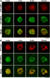
Temporal localization of LIMK1 and SSH1L in SDF-1α–stimulated Jurkat cells. (A) Localization of CFP-LIMK1 (red) and YFP-SSH1L (green) in fixed Jurkat cells unstimulated (0 min) or stimulated for 1, 5, and 20 min with 5 nM SDF-1α. Merged images are shown in the bottom panels. (B) Localization of YFP-actin (red) and CFP-SSH1L (green) in fixed Jurkat cells unstimulated (0 min) or stimulated for 1, 5, and 20 min with 5 nM SDF-1α. Merged images are shown in the bottom panels. Bars, 5 μm.
Figure 3.
Figure 3.
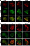
Cofilin, but not P-cofilin, accumulates in the SDF-1α–induced lamellipodial membrane protrusion in Jurkat cells. Jurkat cells were left unstimulated (0 min) or were stimulated with 5 nM SDF-1α for the indicated periods of time. Cells were costained with anti–β-actin mAb (red) and anticofilin (A) or anti–P-cofilin (B) pAbs (green). Merged images are shown in the bottom panels. Bars, 5 μm.
Figure 4.
Figure 4.
Effect of SSH1L, LIMK1, or cofilin siRNA on SDF-1α–induced T cell chemotaxis and chemokinesis. Jurkat cells were transfected with siRNA plasmids for GFP (control), SSH1L, LIMK1, cofilin, or empty vector (−) as indicated. Chemotactic responses toward 5 nM SDF-1α and chemokinetic responses in the presence of 5 nM SDF-1α were determined in the Transwell culture chambers, as described in Materials and methods. The data are expressed as the means ± SEM (error bars) of three independent experiments. *, P < 0.05; **, P < 0.01; ***, P < 0.005, compared with cells transfected with the empty vector.
Figure 5.
Figure 5.
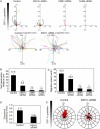
Effect of SSH1L, LIMK1, or cofilin siRNA on SDF-1α–induced T cell chemotaxis in Dunn chambers. Jurkat cells were transfected with siRNA plasmids for SSH1L, LIMK1, cofilin, or empty plasmid (control) and were analyzed for their ability to migrate in an SDF-1α gradient in the Dunn chamber during a 50-min period. (A) The migration paths of 30 randomly chosen cells were traced for 50 min. The intersection of the x and y axes was taken to be the starting point of each cell path, whereas the source of SDF-1α was at the top. Magnified views of the paths of control cells and SSH1L siRNA cells are also shown. (B) The net translocation distance (straight distance from the start to the end point) of each cell over the 50-min period is shown as the mean ± SEM (error bars) of the paths of 50 randomly chosen cells. *, P < 0.01 compared with control cells. (C) The migration speed (total length of the migration path per hour) of each cell is shown as the mean ± SEM of the paths of 50 randomly chosen cells. *, P < 0.05; **, P < 0.01 compared with control cells. (D) The directional persistency index (the ratio of the net translocation distance to the cumulative length of migration path) of control and SSH1L siRNA cells. (E) Circular histograms showing the percentage of cells whose final position was located within each of 18 equal sectors (20°). The source of SDF-1α was at the top. Data from control and SSH1L siRNA cells are shown.
Figure 6.
Figure 6.
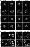
Effect of cofilin, LIMK1, or SSH1L siRNA on F-actin assembly and membrane protrusion formation before and after SDF-1α stimulation. (A) Time-lapse fluorescence analysis. Jurkat cells cotransfected with YFP-actin and siRNA plasmids for mutated SSH1L (control), cofilin, LIMK1, or SSH1L were analyzed by time-lapse fluorescence microscopy, making use of YFP fluorescence. Numbers indicate the times after SDF-1α stimulation. See Videos 4–7 (available at http://www.jcb.org/cgi/content/full/jcb.200504029/DC1). (B) Jurkat cells transfected with siRNA plasmids were left unstimulated (top) or stimulated for 5 min with SDF-1α (bottom) and were fixed and stained with rhodamine-phalloidin to visualize F-actin. Bars, 10 μm.
Figure 7.
Figure 7.
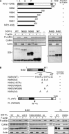
Trp-458 is critical for the F-actin–mediated activation of SSH1L. (A) The role of amino acids 457–461 in F-actin–mediated SSH1L activation. Schematic structures of SSH1L and deletion mutants are shown. The conserved regions in the SSH family are indicated by the A, B, P (phosphatase), and S (Ser-rich) domains. Wild-type (WT) and deletion mutants of (myc + His)-tagged SSH1L were expressed in 293T cells, immunoprecipitated with an anti-myc antibody, and subjected to in vitro phosphatase assays using cofilin-(His)6 as a substrate in the presence or absence of F-actin. P-cofilin levels were measured by Pro-Q staining. Total cofilin and actin were measured by Coomassie blue staining. The expression of SSH1L mutants was analyzed by immunoblotting with the anti-myc antibody. *, Ig heavy chain. (B) Trp-458 is required for F-actin–mediated SSH1L activation. Point mutants of N461 and full-length (FL) SSH1L were subjected to in vitro cofilin phosphatase assays as described in A. Arrow indicates the replacement of Trp-458 with Ala.
Figure 8.
Figure 8.
F-actin–mediated activation of SSH1L is critical for polarized lamellipodium formation and chemotaxis. (A) Expression of siRNA-resistant (sr) SSH1L(WT), but not sr-C393S, sr-W458A, or NP mutant, rescues the inhibitory effect of SSH1L siRNA on T cell chemotaxis. Jurkat cells were cotransfected with SSH1L siRNA plasmids together with expression plasmids for sr-SSH1L(WT), sr-C393S, sr-W458A, or NP mutants cultured for 60 h and were subjected to Transwell culture chamber assays as described in Fig. 4. *, P < 0.005; **, P < 0.05, compared with cells transfected with empty vector. Error bars represent SEM. (B) Expression of sr-SSH1L(WT), but not sr-C393S or sr-W458A mutant, recovers the SDF-1α–induced polarized F-actin assembly in SSH1L siRNA cells. Jurkat cells transfected as in A were cultured for 60 h, stimulated with 5 nM SDF-1α for 5 min, and stained with rhodamine-phalloidin for F-actin as described in Fig. 6 B. Bar, 5 μm. (C) Quantitative analysis of cell morphology changes. Jurkat cells transfected with SSH1L siRNA plasmids together with the indicated sr-SSH1L expression plasmids were cultured for 60 h, stimulated with 5 nM SDF-1α for 5 min, and stained as in B. Cells were categorized into three classes, as shown on the right: class 1 (round cells without a lamellipodium), class 2 (cells with a single lamellipodium), and class 3 (cells with multiple lamellipodia around the cells). The percentages of cells in each class are shown as the means of triplicate experiments (200–300 cells were counted in each experiment).
Figure 9.
Figure 9.
A model for the LIMK1- and SSH1L-mediated spatiotemporal regulation of cofilin activity during SDF-1α–induced polarized F-actin assembly and cell migration. The unstimulated Jurkat cell has a round, symmetrical shape. Exposure of the cell to SDF-1α induces the activation of LIMK1 through Rac and leads to a transient increase in P-cofilin levels, which is required for the formation of F-actin–rich lamellipodial protrusions in the initial stages of cell response. SSH1L translocates to the lamellipodia and is activated by associating with F-actin. Because SSH1L knockdown cells retain multiple protrusions during the cell stimulation, SSH1L is required for the conversion of multiple protrusions to the single lamellipodium. In later stages, SSH1L locally stimulates cofilin activation and actin filament turnover in the lamellipodium in the front of the cell, whereas LIMK1 is diffusely distributed in the cell and may help to stabilize actin filaments in the rear of the cell.

Similar articles

Cited by

References

    1. Allen, W.E., D. Zicha, A.J. Ridley, and G.E. Jones. 1998. A role for Cdc42 in macrophage chemotaxis. J. Cell Biol. 141:1147–1157. - PMC - PubMed
    1. Arakawa, Y., H. Bito, T. Furuyashiki, T. Tsuji, S. Takemoto-Kimura, K. Kimura, K. Nozaki, N. Hashimoto, and S. Narumiya. 2003. Control of axon elongation via an SDF-1α/Rho/mDia pathway in cultured cerebellar granule neurons. J. Cell Biol. 161:381–391. - PMC - PubMed
    1. Arber, S., F.A. Barbayannis, H. Hanser, C. Schneider, C.A. Stanyon, O. Bernard, and P. Caroni. 1998. Regulation of actin dynamics through phosphorylation of cofilin by LIM-kinase. Nature. 393:805–809. - PubMed
    1. Bailly, M., and G.E. Jones. 2003. Polarised migration: cofilin holds the front. Curr. Biol. 13:R128–R130. - PubMed
    1. Bamburg, J.R. 1999. Proteins of the ADF/cofilin family: essential regulators of actin dynamics. Annu. Rev. Cell Dev. Biol. 15:185–230. - PubMed

Publication types

MeSH terms