. 2005 Nov;25(21):9189-97.
doi: 10.1128/MCB.25.21.9189-9197.2005.

Long-chain base kinase Lcb4 Is anchored to the membrane through its palmitoylation by Akr1

Affiliations

Long-chain base kinase Lcb4 Is anchored to the membrane through its palmitoylation by Akr1

Akio Kihara et al. Mol Cell Biol. 2005 Nov.

Abstract

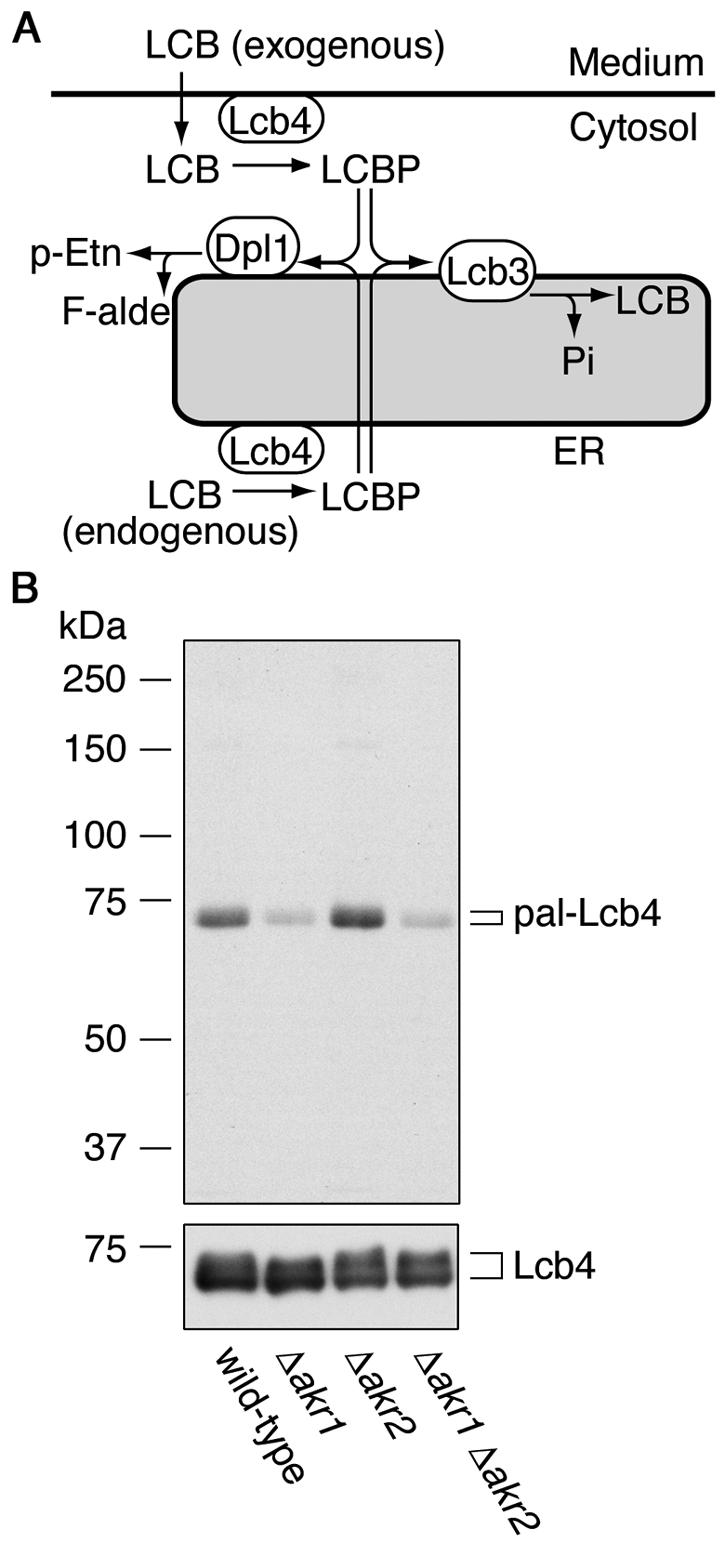
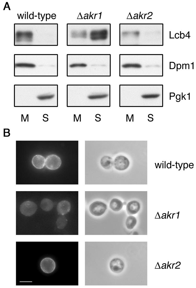
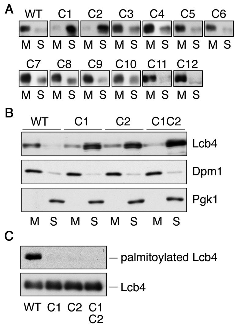
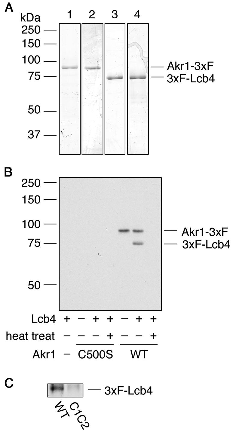
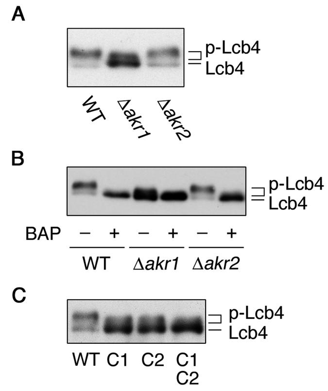
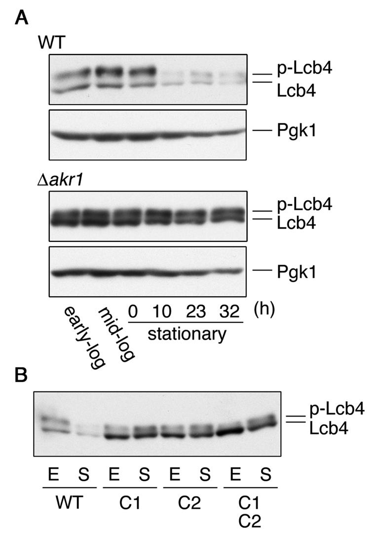
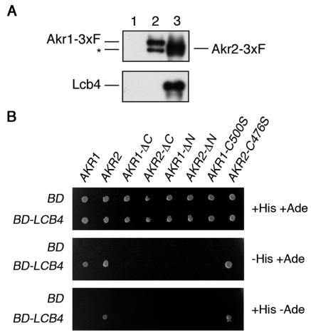
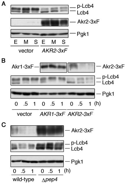
Sphingoid long-chain base kinase Lcb4 catalyzes the production of the bioactive lipid molecules the long-chain base 1-phosphates. Although Lcb4 has no apparent transmembrane-spanning domain, it is tightly associated with the membrane. Here, we demonstrate that Lcb4 is modified by palmitoylation. This modification was greatly reduced in mutants for AKR1, which was recently identified as encoding a protein acyltransferase. In vitro experiments revealed that Akr1 indeed acts as a protein acyltransferase for Lcb4. Studies using site-directed mutagenesis indicated that Cys-43 and Cys-46 are palmitoylated. The loss of palmitoylation on Lcb4 caused several effects, including mislocalization of the protein to the cytosol, reduced phosphorylation, and loss of downregulation during the stationary phase. Although Akr2 is highly homologous to Akr1, the deletion of AKR2 did not result in any remarkable phenotypes. However, overproduction of Akr2 resulted in reduced amounts of Lcb4. We demonstrated that Akr2 is an unstable protein and is degraded in the vacuole. Akr2 exhibits high affinity for Lcb4, and in Akr2-overproducing cells this interaction caused unusual delivery of Lcb4 to the vacuole and degradation.


Figures

FIG. 1.
FIG. 1.
Lcb4 is palmitoylated and Akr1 is involved. (A) Pathways for LCB and LCBP metabolism. Exogenous LCB is imported into the cells and then converted to LCBP by the kinase Lcb4. LCBP is then degraded in the ER via one of two pathways: conversion to long-chain fatty aldehyde (F-alde) and phosphoethanolamine (p-Etn) by the lyase Dpl1 or conversion to LCB by the phosphatase Lcb3. (B) KA311A (wild type), KHY689 (Δakr1), KHY672 (Δakr2), and KHY690 (Δakr1 Δakr2) cells, each harboring pAK514 (GAL1 promoter-LCB4), were grown at 30°C in SC medium lacking uracil. Lcb4 expression was induced by using YPGal medium and, after 1 h, cells were labeled with 0.3 mCi of [3H]palmitic acid for 2 h at 30°C. Lcb4 was immunoprecipitated from total cell lysates (equal in radioactivity) with anti-Lcb4 antibodies and separated by SDS-PAGE, followed by autoradiography (upper panel) or by immunoblotting with anti-Lcb4 antibodies (lower panel). pal-Lcb4, palmitoylated Lcb4.
FIG. 2.
FIG. 2.
Akr1 is required for the membrane association of Lcb4. (A) Total lysates were prepared from KHY195 (wild-type), KHY212 (Δakr1), and KHY199 (Δakr2) cells. Membrane (M) and soluble (S) proteins were separated by centrifugation at 100,000 × g for 1 h and then by SDS-PAGE. Lcb4, Dpm1 (a membrane protein marker), and Pgk1 (a soluble protein marker) were then detected by immunoblotting. (B) SEY6210 (wild-type), KHY211 (Δakr1), and KHY197 (Δakr2) cells, each bearing pWK79 (LCB4), were fixed by formaldehyde, converted to spheroplasts, permeabilized with 0.1% Triton X-100, and stained with anti-Lcb4 antibodies (left panels). Phase-contrast images are also shown (right panels). Bar, 5 μm.
FIG. 3.
FIG. 3.
Cys-43 and Cys-46 residues of Lcb4 are palmitoylated. (A) Total lysates prepared from SIY03 (Δlcb4) cells bearing pWK78 (LCB4), pFK18 (LCB4-C1), pFK19 (LCB4-C2), pFK20 (LCB4-C3), pFK21 (LCB4-C4), pFK22 (LCB4-C5), pFK23 (LCB4-C6), pFK24 (LCB4-C7), pFK25 (LCB4-C8), pFK26 (LCB4-C9), pFK27 (LCB4-C10), pFK28 (LCB4-C11), or pFK29 (LCB4-C12) were centrifuged at 100,000 × g for 1 h. Proteins in the resulting pellet (membrane fraction; M) and supernatant (soluble fraction; S) were separated by SDS-PAGE, followed by immunoblotting with anti-Lcb4. (B) Proteins were separated as in panel A by using total lysates prepared from TS11 (Δlcb4 Δpep4) cells bearing pWK78, pFK18, pFK19, or pFK33 (LCB4-C1C2). Lcb4, Dpm1, and Pgk1 were detected by immunoblotting. (C) FKY146 (Δlcb4 Δpep4) cells harboring pAK514 (GAL1 promoter-LCB4), pFK72 (LCB4-C1), pFK73 (LCB4-C2), or pFK74 (LCB4-C1C2) were labeled with 0.3 mCi of [3H]palmitic acid for 2 h at 30°C. Lcb4 was immunoprecipitated from total cell lysates (equal in radioactivity) by using anti-Lcb4 antibodies and separated by SDS-PAGE, followed by autoradiography (upper panel) or immunoblotting with anti-Lcb4 antibodies (lower panel). WT, wild type.
FIG. 4.
FIG. 4.
Akr1 palmitoylates Lcb4 in vitro. (A) Akr1-3xFLAG (lane 1), Akr1-C500S-3xFLAG (lane 2), 3xFLAG-Lcb4 (lane 3), and 3xFLAG-Lcb4-C1C2 (lane 4) were purified by using anti-FLAG M2 agarose. Purified proteins (0.5 μg) were separated by SDS-PAGE and stained with Coomassie brilliant blue. (B) Reaction mixtures containing [3H]palmitoyl-CoA, and purified proteins (0.1 μg of Akr1-3xFLAG and/or 0.24 μg of 3xFLAG-Lcb4) were incubated at 30°C for 1 h. As indicated, Akr1-3xFLAG protein was heat inactivated at 100°C for 15 min. (C) Akr1-3xFLAG (0.5 μg) was incubated with 3xFLAG-Lcb4 (1 μg) or 3xFLAG-Lcb4-C1C2 (1 μg) in the presence of [3H]palmitoyl-CoA at 37°C for 1 h. Proteins were separated by SDS-PAGE, followed by autoradiography. 3xF, 3xFLAG; WT, wild type.
FIG. 5.
FIG. 5.
Palmitoylation is required for the phosphorylation of Lcb4. (A) Total lysates were prepared from SEY6210 (wild type), KHY211 (Δakr1), and KHY197 (Δakr2) cells. Proteins were separated by SDS-PAGE and detected by immunoblotting with anti-Lcb4 antibodies. (B) Total lysates prepared as in panel A were treated with or without bacterial alkaline phosphatase (BAP) and separated by SDS-PAGE, followed by immunoblotting with anti-Lcb4 antibodies. (C) Total lysates prepared from TS11 (Δlcb4 Δpep4) cells harboring pWK78 (LCB4), pFK18 (LCB4-C1), pFK19 (LCB4-C2), or pFK33 (LCB4-C1C2) were separated by SDS-PAGE, followed by immunoblotting with anti-Lcb4 antibodies. WT, wild type, p-Lcb4, hyperphosphorylated Lcb4.
FIG. 6.
FIG. 6.
Palmitoylation is essential for the downregulation of Lcb4 during stationary phase. (A) SEY6210 (wild-type) and KHY211 (Δakr1) cells grown overnight were diluted into fresh YPD medium at a density of A600 = 0.3 and incubated at 30°C. Cells were harvested in early log phase, mid-log phase, and 0, 10, 23, or 32 h after reaching stationary phase. Total lysates were prepared from cells (ca. 2 × 10⁷), and proteins (12 μg) were separated by SDS-PAGE and subjected to immunoblotting with anti-Lcb4 or anti-Pgk1 antibodies. (B) SIY03 (Δlcb4) cells bearing pWK78 (LCB4), pFK18 (LCB4-C1), pFK19 (LCB4-C2), or pFK33 (LCB4-C1C2) were harvested in early log phase (E) or 10 h after reaching stationary phase (S). Total cell lysates were prepared, and proteins (15 μg) were separated by SDS-PAGE, followed by immunoblotting with anti-Lcb4 antibodies. WT, wild type; p-Lcb4, hyperphosphorylated Lcb4.
FIG. 7.
FIG. 7.
Akr2 interacts with Lcb4. (A) Total cell lysates prepared from KHY195 (Δpep4)/pWK62 (LCB4)/pRS423 (vector) (lane 1), KHY195/pWK62/pAK456 (AKR1-3xFLAG) (lane 2), and KHY195/pWK62/pAK457 (AKR2-3xFLAG) (lane 3) cells were solubilized with 1% Triton X-100. Proteins were immunoprecipitated by using anti-FLAG M2 affinity gels and separated by SDS-PAGE, followed by immunoblotting with anti-FLAG (M2) or anti-Lcb4 antibodies. The asterisk indicates the degradation product of Akr1-3xFLAG. (B) Plasmids containing GLA4 AD fused with the indicated genes were introduced into PJ69-4A cells harboring pGBDU-C1 (GAL4 BD) or pWK05 (GAL4 BD-LCB4). Cells (ca. 3 × 10⁵) were plated on leucine-free SC medium lacking uracil (top panel), uracil and histidine (middle panel), or uracil and adenine (bottom panel), followed by incubation at 30°C. 3xF, 3xFLAG.
FIG. 8.
FIG. 8.
Overproduction of Akr2 causes destabilization of Lcb4. (A) SEY6210 cells bearing pRS423 (vector) or pAK457 (AKR2-3xFLAG) were harvested in early log phase (E), in mid-log phase (M), or at the beginning of stationary phase (S). Total lysates were prepared from cells (ca. 2 × 10⁷), and proteins (9 μg) were separated by SDS-PAGE and immunoblotted with anti-Lcb4, anti-FLAG M2, and anti-Pgk1 antibodies. (B) SEY6210 cells bearing pRS423, pAK456 (AKR1-3xFLAG), or pAK457, grown to early log phase, were treated with cycloheximide (10 μg/ml). Cells were collected at the indicated time point after treatment, and total cell lysates were prepared. Proteins (10 μg) were separated by SDS-PAGE, followed by immunoblotting with anti-Lcb4, anti-FLAG M2, and anti-Pgk1 antibodies. (C) SEY6210 (wild-type) cells bearing pAK457 and TVY1 (Δpep4) cells bearing pAK457 were treated with cycloheximide and processed as in panel B. 3xF, 3xFLAG; p-Lcb4, hyperphosphorylated Lcb4.

Similar articles

Cited by

References

    1. Abeliovich, H., T. Darsow, and S. D. Emr. 1999. Cytoplasm to vacuole trafficking of aminopeptidase I requires a t-SNARE-Sec1p complex composed of Tlg2p and Vps45p. EMBO J. 18:6005-6016. - PMC - PubMed
    1. Babu, P., R. J. Deschenes, and L. C. Robinson. 2004. Akr1p-dependent palmitoylation of Yck2p yeast casein kinase 1 is necessary and sufficient for plasma membrane targeting. J. Biol. Chem. 279:27138-27147. - PubMed
    1. Birchwood, C. J., J. D. Saba, R. C. Dickson, and K. W. Cunningham. 2001. Calcium influx and signaling in yeast stimulated by intracellular sphingosine 1-phosphate accumulation. J. Biol. Chem. 276:11712-11718. - PubMed
    1. Casey, P. J. 1995. Protein lipidation in cell signaling. Science 268:221-225. - PubMed
    1. Christianson, T. W., R. S. Sikorski, M. Dante, and P. Hieter. 1992. Multifunctional yeast high-copy-number shuttle vectors. Gene 110:119-122. - PubMed

Publication types

MeSH terms

Substances