. 2005 Nov;79(21):13735-46.
doi: 10.1128/JVI.79.21.13735-13746.2005.

Human immunodeficiency virus type 1 Vpr interacts with antiapoptotic mitochondrial protein HAX-1

Affiliations

Human immunodeficiency virus type 1 Vpr interacts with antiapoptotic mitochondrial protein HAX-1

Venkat S R K Yedavalli et al. J Virol. 2005 Nov.

Abstract

Human immunodeficiency virus type 1 viral protein R (Vpr) is required for viral pathogenesis and has been implicated in T-cell apoptosis through its activation of caspase 3 and caspase 9 and perturbation of mitochondrial membrane potential. To understand better Vpr-mitochondria interaction, we report here the identification of antiapoptotic mitochondrial protein HAX-1 as a novel Vpr target. We show that Vpr and HAX-1 physically associate with each other. Overexpression of Vpr in cells dislocates HAX-1 from its normal residence in mitochondria and creates mitochondrion instability and cell death. Conversely, overexpression of HAX-1 suppressed the proapoptotic activity of Vpr.


Figures

FIG. 1.
FIG. 1.
Interaction between Vpr and HAX-1. (A) Vpr and HAX-1 interact in yeast two-hybrid assay. The L40 yeast expressing both LexA BD and GAL4AD hybrid proteins was analyzed on selective plates containing 2 mM concentration of 3-amino-(1,2,4) triazole. Growth in the absence of His with a positive β-galactosidase signal are indications of the interaction between the hybrid proteins. LexA-lamin fusion protein was used as a negative control. (B) HAX-1 and Vpr interact in vitro. Bacterially expressed purified GST-Vpr was used for in vitro binding assay with [35S]methionine-labeled in vitro-translated HAX-1 protein. HAX-1 bound GST-Vpr (lane 3) but not GST-alone (lane 2). (C) Interaction between Vpr and HAX-1 in human cells. HAX-1 interacts with Vpr expressed from HIV-1 in MT4 cells infected with infectious HIV-1 NL4-3. MT4 cells were harvested 4 days postinfection by centrifugation and were lysed in RIPA buffer. Cell lysates were subjected to sonication and coimmunoprecipitation assays with anti-Vpr or with irrelevant control antibody (isotype control). The immunoprecipitates were resolved by SDS-PAGE, transferred to membrane, and probed with mouse monoclonal anti-HAX-1 antibody. The top panel shows coimmunoprecipitation of HAX-1 with Vpr. The middle panel shows Western blotting of endogenous HAX-1. The bottom panel shows expression of Vpr from pNL4-3.
FIG. 2.
FIG. 2.
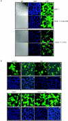
Vpr-induced cell death is suppressed by HAX-1 overexpression. (A) HAX-1 influences cellular apoptosis. Expression of DN HAX-1 induces cell death in HeLa cells. pcDNA HAX-1 WT, pcDNA HAX-1(1-141), and pcDNA HAX-1(142-279) were cotransfected with pCMV-GFP into HeLa cells. At 36 to 48 h posttransfection, cells were fixed and stained with the nuclear stain DAPI. The transfected GFP-expressing cells were examined by microscopy for differential interference contrast (DIC), DAPI, and green fluorescence. pcDNA HAX-1(1-141) expression in transfected cells resulted in significant cell death (compare subpanels 9 and 12 with subpanels 3 and 6). Apoptotic cells were detected as GFP-positive cells with nuclear condensation and fragmentation. (B) HAX-1 suppressed Vpr-induced cell death. HeLa cells were transfected with HAX-1 and Vpr as indicated along with cytomegalovirus (CMV)-GFP as a marker for transfected cells. Overexpression of HAX-1 suppressed Vpr-induced cell death (compare subpanels 2 to 4 with subpanels 6 to 8). (C) Western blot analysis of the expression of Vpr and HAX-1 in transfected HeLa cells. Cell lysates prepared from transfections above (Fig. 2B) were analyzed by blotting for expression of the indicated proteins from transiently transfected plasmids. The top row shows detection of transfected HAX-1 with a light exposure of film. The middle row shows detection of Flag-tagged transfected Vpr. The bottom row shows β-actin signals as loading controls. (D) Graphic representation of suppression cell death induced by HIV-1 Vpr. Cell viability of transfected cells was observed 36 to 48 h posttransfection by counting the number of live green (total cells) and apoptotic green (apoptotic cells) fluorescent cells under the microscope. Values are representative of three independent assays.
FIG. 2.
FIG. 2.
Vpr-induced cell death is suppressed by HAX-1 overexpression. (A) HAX-1 influences cellular apoptosis. Expression of DN HAX-1 induces cell death in HeLa cells. pcDNA HAX-1 WT, pcDNA HAX-1(1-141), and pcDNA HAX-1(142-279) were cotransfected with pCMV-GFP into HeLa cells. At 36 to 48 h posttransfection, cells were fixed and stained with the nuclear stain DAPI. The transfected GFP-expressing cells were examined by microscopy for differential interference contrast (DIC), DAPI, and green fluorescence. pcDNA HAX-1(1-141) expression in transfected cells resulted in significant cell death (compare subpanels 9 and 12 with subpanels 3 and 6). Apoptotic cells were detected as GFP-positive cells with nuclear condensation and fragmentation. (B) HAX-1 suppressed Vpr-induced cell death. HeLa cells were transfected with HAX-1 and Vpr as indicated along with cytomegalovirus (CMV)-GFP as a marker for transfected cells. Overexpression of HAX-1 suppressed Vpr-induced cell death (compare subpanels 2 to 4 with subpanels 6 to 8). (C) Western blot analysis of the expression of Vpr and HAX-1 in transfected HeLa cells. Cell lysates prepared from transfections above (Fig. 2B) were analyzed by blotting for expression of the indicated proteins from transiently transfected plasmids. The top row shows detection of transfected HAX-1 with a light exposure of film. The middle row shows detection of Flag-tagged transfected Vpr. The bottom row shows β-actin signals as loading controls. (D) Graphic representation of suppression cell death induced by HIV-1 Vpr. Cell viability of transfected cells was observed 36 to 48 h posttransfection by counting the number of live green (total cells) and apoptotic green (apoptotic cells) fluorescent cells under the microscope. Values are representative of three independent assays.
FIG. 3.
FIG. 3.
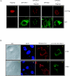
Localization of Vpr and HAX-1 in HeLa cells. Transfected cells were visualized 36 to 48 h after transfection. (A) Flag-Vpr and GFP-HAX-1 colocalize. Flag-Vpr is primarily nuclear (subpanel 1); GFP-HAX-1 protein is mainly mitochondrial (subpanel 2). The cotransfected GFP-HAX-1 and Flag-Vpr colocalize outside of the nucleus and the mitochondria (subpanels 3 and 4). In contrast, Flag-Vpr and GFP-Bcl2 do not colocalize (subpanels 5 to 7). (B) HAX-1 localizes to extra mitochondrial sites in the presence of Vpr. HeLa cells transfected with HAX-1 alone (subpanels 1 to 4) and HAX-1 plus Vpr (subpanels 5 to 8) were stained with Mitotracker before fixation and were probed with anti-HA antibody-anti-mouse Alexa 488. HAX-1 in the absence of Vpr colocalized with Mitotracker-stained mitochondria but in the presence of Vpr formed cytoplasmic bodies that are not stained by Mitotracker.
FIG. 4.
FIG. 4.
HAX-1 interaction with the C terminus of Vpr correlated with suppression of apoptosis. (A) Confocal examination of distribution of Vpr mutants in HeLa cells in the presence or absence of HAX-1. Staining patterns of YFP-VprS79A and YFP-Vpr(1-70) are shown. (B) Mapping of the Vpr region required for HAX-1 interaction. HeLa cells transiently expressing the indicated protein(s) were immunoprecipitated with mouse monoclonal antibody to HAX-1. The immunoprecipitates were subjected to SDS-PAGE, transferred to membrane, and probed with rabbit polyclonal anti-YFP. The top panel shows that YFP-Vpr WT coimmunoprecipitated with HA-HAX-1, whereas YFP alone, YFP-Vpr(1-70), and YFP-Vpr(1-51) did not. The middle panel shows equal expression levels of all YFP-tagged proteins. The bottom panel verified for equal expression of transfected HA-HAX-1. (C) Schematic representations of Vpr mutants, their interactions with HAX-1, and their apoptosis-inducing capacity.
FIG. 5.
FIG. 5.
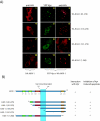
Mapping of amino acids 118 to 141 of HAX-1 as a region of interaction with Vpr. (A) Confocal examination of expression and distribution of HAX-1 in HeLa cells. Fluorescent images indicate that the various HAX-1 deletion mutants are either capable or not capable of sequestering Vpr. (B) Diagrammatic summary of HAX-1 deletion mutant correlating the ability to sequester Vpr with ability to halt Vpr-induced apoptosis. Bcl2 homology domains 1 and 2 (BH1 and -2, green), PEST sequence (PEST, red), and transmembrane domain (TMD, brown) are indicated.

Similar articles

Cited by

References

    1. Ayyavoo, V., A. Mahboubi, S. Mahalingam, R. Ramalingam, S. Kudchodkar, W. V. Williams, D. R. Green, and D. B. Weiner. 1997. HIV-1 Vpr suppresses immune activation and apoptosis through regulation of nuclear factor κB. Nat. Med. 3:1117-1123. - PubMed
    1. Belzacq, A. S., H. L. Vieira, F. Verrier, G. Vandecasteele, I. Cohen, M. C. Prevost, E. Larquet, F. Pariselli, P. X. Petit, A. Kahn, R. Rizzuto, C. Brenner, and G. Kroemer. 2003. Bcl-2 and Bax modulate adenine nucleotide translocase activity. Cancer Res. 63:541-546. - PubMed
    1. Bouhamdan, M., S. Benichou, F. Rey, J. M. Navarro, I. Agostini, B. Spire, J. Camonis, G. Slupphaug, R. Vigne, R. Benarous, and J. Sire. 1996. Human immunodeficiency virus type 1 Vpr protein binds to the uracil DNA glycosylase DNA repair enzyme. J. Virol. 70:697-704. - PMC - PubMed
    1. Boya, P., A. L. Pauleau, D. Poncet, R. A. Gonzalez-Polo, N. Zamzami, and G. Kroemer. 2004. Viral proteins targeting mitochondria: controlling cell death. Biochim. Biophys. Acta 1659:178-189. - PubMed
    1. Brenner, C., H. Cadiou, H. L. Vieira, N. Zamzami, I. Marzo, Z. Xie, B. Leber, D. Andrews, H. Duclohier, J. C. Reed, and G. Kroemer. 2000. Bcl-2 and Bax regulate the channel activity of the mitochondrial adenine nucleotide translocator. Oncogene 19:329-336. - PubMed

Publication types

Substances