. 2006 Feb 15;394(Pt 1):227-36.
doi: 10.1042/BJ20050605.

The N-terminal domain of the human eIF2beta subunit and the CK2 phosphorylation sites are required for its function

Affiliations

The N-terminal domain of the human eIF2beta subunit and the CK2 phosphorylation sites are required for its function

Franc Llorens et al. Biochem J. .

Abstract

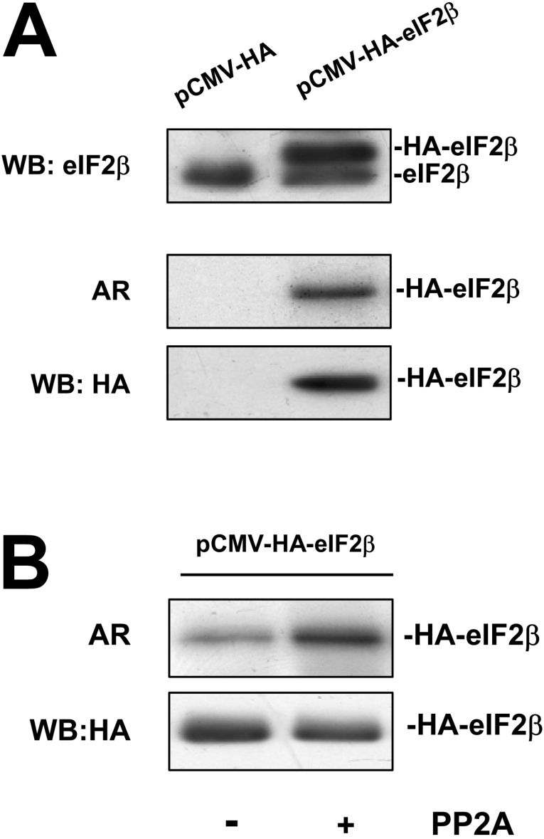
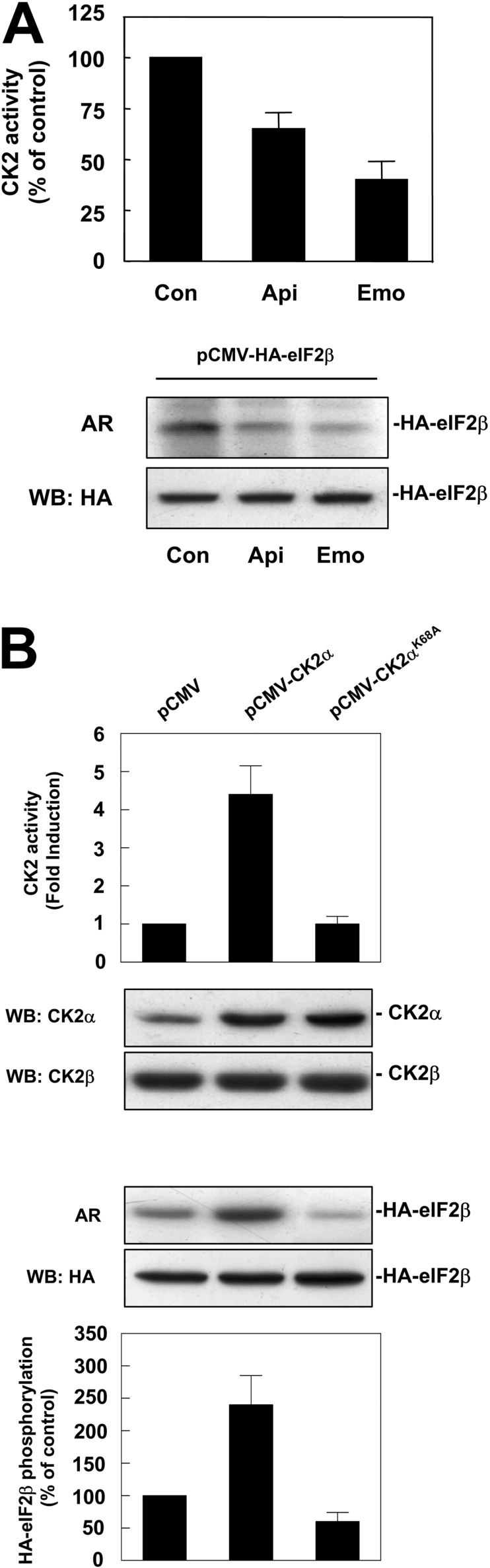
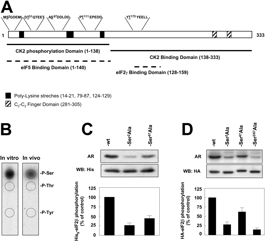
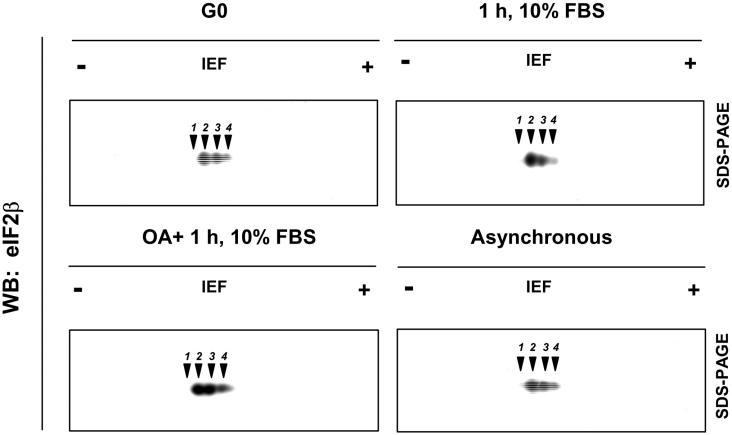
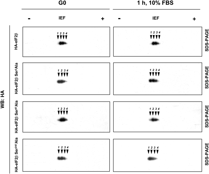
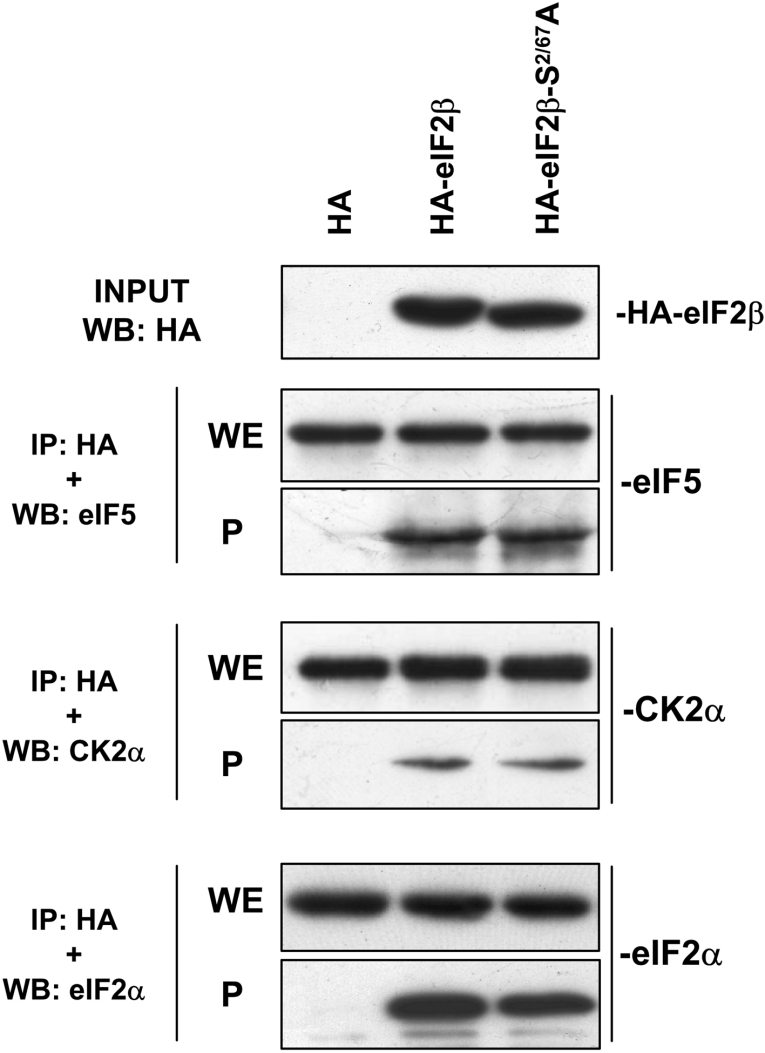
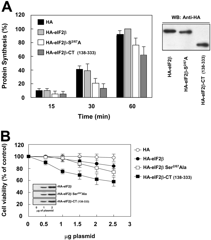
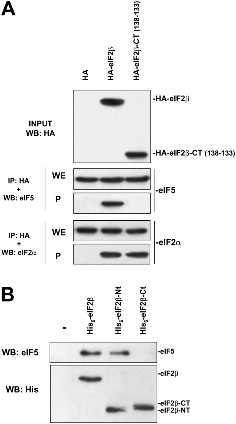
CK2 (protein kinase CK2) is known to phosphorylate eIF2 (eukaryotic translation initiation factor 2) in vitro; however, its implication in this process in living cells has remained to be confirmed. The combined use of chemical inhibitors (emodin and apigenin) of CK2 together with transfection experiments with the wild-type of the K68A kinase-dead mutant form of CK2alpha evidenced the direct involvement of this protein kinase in eIF2beta phosphorylation in cultured HeLa cells. Transfection of HeLa cells with human wild-type eIF2beta or its phosphorylation site mutants showed Ser2 as the main site for constitutive eIF2beta phosphorylation, whereas phosphorylation at Ser67 seems more restricted. In vitro phosphorylation of eIF2beta also pointed to Ser2 as a preferred site for CK2 phosphorylation. Overexpression of the eIF2beta S2/67A mutant slowed down the rate of protein synthesis stimulated by serum, although less markedly than the overexpression of the Delta2-138 N-terminal-truncated form of eIF2beta (eIF2beta-CT). Mutation at Ser2 and Ser67 did not affect eIF2beta integrating into the eIF2 trimer or being able to complex with eIF5 and CK2alpha. The eIF2beta-CT form was also incorporated into the eIF2 trimer but did not bind to eIF5. Overexpression of eIF2beta slightly decreased HeLa cell viability, an effect that was more evident when overexpressing the eIF2beta S2/67A mutant. Cell death was particularly marked when overexpressing the eIF2beta-CT form, being detectable at doses where eIF2beta and eIF2beta S2/67A were ineffective. These results suggest that Ser2 and Ser67 contribute to the important role of the N-terminal region of eIF2beta for its function in mammals.


Figures

Figure 1
Figure 1. HA–eIF2β is constitutively phosphorylated in HeLa cells
(A) HeLa cells were transfected with either pCMV-HA or pCMV-HA-eIF2β. Cell lysates were analysed by Western blotting with anti-eIF2β antibody. In a parallel experiment, HeLa cells were labelled with [32P]Pi and cell lysates were immunoprecipitated with anti-HA antibody and subjected to SDS/PAGE followed by autoradiography (AR) and Western blotting against anti-HA antibody. (B) HeLa cells were transfected with pCMV-HA-eIF2β and cell lysates were immunoprecipitated with anti-HA antibody. Immunoprecipitates were left untreated or dephosphorylated with PP2A catalytic subunit and re-phosphorylated with CK2 holoenzyme. Samples were subjected to SDS/PAGE followed by autoradiography (AR) and Western blotting against HA antibody to ensure equal amount of HA–eIF2β in the assay.
Figure 2
Figure 2. HA–eIF2β is phosphorylated by CK2 in HeLa cells
(A) pCMV-HA-eIF2β transfected HeLa cells were labelled with [32P]Pi in the absence or presence of CK2 inhibitors apigenin and emodin (40 μM) during the labelling time as indicated. An aliquot of cell lysate was tested for CK2 activity over CK2-specific peptide substrate RRRAADSDDDDD to monitor CK2 inhibition on the assay (upper panel). Samples were immunoprecipitated with anti-HA antibody and subjected to SDS/PAGE followed by autoradiography (AR) and Western blotting against HA antibody to ensure equal amount of HA–eIF2β in the assay (lower panels). (B) HeLa cells were transfected with pCMV, pCMV-CK2α or pCMV-CK2αK68A and cell lysates were assayed for CK2 activity over CK2-specific peptide substrate RRRAADSDDDDD and subjected to Western blotting against CK2α and CK2β antibodies (upper panels). In a parallel experiment, HA–eIF2β was co-transfected with the CK2α plasmids indicated above and HeLa cells were labelled with [32P]Pi. Cell lysates were immunoprecipitated with anti-HA antibody and subjected to SDS/PAGE followed by autoradiography (AR) and Western blotting against HA antibody to ensure equal amount of HA–eIF2β in the assay. Quantification of autoradiography is also shown in the graph (lower panels).
Figure 3
Figure 3. eIF2β is phosphorylated in vitro and in vivo on Ser2 and Ser67
(A) Domain structure of eIF2β. Potential CK2 phosphorylation sequences are indicated over the sequence of the open box representing the polypeptide chain, and phosphorylation sites are underlined. Black boxes indicate polylysine stretches and shadowed boxes the C2-C2 finger domain. Black lines indicate the characterized domains for interaction and phosphorylation by CK2. Discontinuous lines indicate the location in human eIF2β for binding to eIF5 and eIF2γ extrapolated from the data reported on yeast. (B) Phospho amino acid analysis of (left panel) His6–eIF2β phosphorylated by CK2 holoenzyme resolved by SDS/PAGE and transferred on to PVDF membrane and (right panel) HA–eIF2β from HeLa cells labelled with [32P]Pi, immunoprecipitated with anti-HA, resolved by SDS/PAGE and transferred on to PVDF membrane. In both cases, radioactive bands were excised from the membrane and the samples were analysed by one-dimensional high-voltage electrophoresis on a TLC cellulose plate and autoradiography. P-Ser, P-Thr and P-Tyr denote the positions of phosphoserine, phosphothreonine and phosphotyrosine respectively as determined by ninhydrin staining of standards. (C) Densitometry of the autoradiography of in vitro phosphorylation of His6–eIF2β, His6–eIF2βS2A and His6–eIF2βS67A by CK2 holoenzyme. Western blotting with anti-His6 was carried out to ensure equal amount of protein in the assay. (D) In vivo phosphorylation of HA–eIF2β, HA–eIF2βS2A, HA–eIF2βS67A and HA–eIF2βS2/67A. Transfected HeLa cells were labelled with [32P]Pi and cell lysates were immunoprecipitated with anti-HA antibody and subjected to SDS/PAGE followed by autoradiography and Western blotting against HA antibody to ensure equal amount of HA–eIF2β in the assay. Densitometries of the autoradiography are shown.
Figure 4
Figure 4. Phosphorylation of eIF2β in HeLa cells is affected by serum and okadaic acid
HeLa cells were serum-starved for 24 h with 0.5% serum and left untreated (G0), re-stimulated for 1 h with 10% serum (1 h, 10% FBS), re-stimulated for 1 h with 10% serum in the presence of okadaic acid (1 μM) (OA+1 h, 10% FBS) or left in 10% FBS as the normal growth conditions (Asynchronous). Equal amounts of cell extract protein were precipitated in 10% trichloroacetic acid, subjected to two-dimensional electrophoresis as described in the Experimental section and developed with anti-eIF2β antibodies. Spot localization is indicated by numbers over arrowheads. The experiment was performed three times with similar results. WB, Western blot; IEF, isoelectric focusing.
Figure 5
Figure 5. Ser2 is constitutively phosphorylated in human eIF2β
HA–eIF2β, HA–eIF2βS2A, HA–eIF2βS67A and HA–eIF2βS2/67A transfected HeLa cells were serum-starved for 24 h with 0.5% serum and left untreated (G0) or re-stimulated for 1 h with 10% serum (1 h, 10% FBS). Equal amounts of cell extracts were precipitated in 10% trichloroacetic acid, subjected to two-dimensional electrophoresis as described in the Experimental section and developed with anti-HA antibodies. Spot localization is indicated by numbers over arrowheads. The experiment was performed three times with similar results. WB, Western blot; IEF, isoelectric focusing.
Figure 6
Figure 6. HA–eIF2β and CK2 phosphorylation mutant HA–eIF2βS2/67A associates with eIF5, CK2α and eIF2α
HeLa cells were transfected with pCMV-HA, pCMV-HA-eIF2β and pCMV-HA-eIF2βS2/67A and cell lysates were subjected to immunoprecipitation with anti-HA antibody. Immunoprecipitates (P) and an aliquot of the whole cell extract (WE) were subjected to SDS/PAGE, transferred on to PVDF membranes and developed with antibodies against eIF5, CK2α and eIF2α as indicated. The top panel indicates the input used for the assay. IP, immunoprecipitation.
Figure 7
Figure 7. Mutation of CK2 phosphorylation sites on HA–eIF2β impairs protein synthesis at a high rate but has minor effects on cell viability
(A) HeLa cells were transfected with pCMV-HA, pCMV-HA-eIF2β, pCMV-HA-eIF2βS2/67A and pCMV-HA-eIF2β(138-333) and serum-starved for 24 h. Then cells were re-stimulated with 10% FBS in the presence of [35S]methionine/cysteine for the indicated times. Cell extracts were analysed for radioactivity incorporation as indicated in the Experimental section. Radioactivity was expressed taking the maximum value as 100%. An aliquot of samples were subjected to Western blotting with anti-HA antibody to ensure equal expression levels of transfected proteins on the assay. (B) HeLa cells were transfected with increasing amounts of pCMV-HA, pCMV-HA-eIF2β, pCMV-HA-eIF2βS2/67A or pCMV-HA-eIF2β (138–333), and after 48 h of transfection, cell viability was analysed using the MTT [3-(4,5-dimethylthiazol-2-yl)-2,5-diphenyl-2H-tetrazolium bromide] assay. Cell viability was expressed taking the maximum value as 100%.
Figure 8
Figure 8. N-terminal deletion mutant HA–eIF2β (HA–eIF2β 138–333) does no associate with eIF5
(A) HeLa cells were transfected with pCMV-HA, pCMV-HA-eIF2β and pCMV-HA-eIF2β (138–333) and cell lysates were subjected to immunoprecipitation with anti-HA antibody. Immunoprecipitates (P) and an aliquot of the whole cell extract (WE) were subjected to SDS/PAGE, transferred on to PVDF membranes and developed with antibodies against eIF5 and eIF2α as indicated. The top panel indicates the input used for the assay. (B) His6–eIF2β, His6–eIF2βCT (138–333) or His6–eIF2βNT (1–137) was incubated with 500 μg of HeLa cell extract. A slurry of Ni-NTA–agarose (1:1; 20 μl) was added to recover recombinant proteins and eIF5 bound to His6-tagged proteins was analysed by SDS/PAGE and Western blotting with anti-eIF5 antibody (upper panel). An aliquot of the experiment was analysed by SDS/PAGE and Western blotting using anti-His6 antibody to ensure equal amount of recombinant protein in the assay (lower panel).

Similar articles

Cited by

References

    1. Preiss T., Hentze M. W. Starting the protein synthesis: eukaryotic translation initiation. BioEssays. 2003;25:1201–1211. - PubMed
    1. Kapp L. D., Lorsh J. R. The molecular mechanisms of eukaryotic translation. Annu. Rev. Biochem. 2004;73:657–704. - PubMed
    1. Kimball S. R. Eukaryotic initiation factor eIF2. Int. J. Biochem. Cell Biol. 1999;31:25–29. - PubMed
    1. Krishnamoorthy T., Pavitt G. D., Zhang F., Dever T. E., Hinnebusch A. G. Tight binding of the phosphorylated α subunit of initiation factor 2 (eIF2α) to the regulatory subunits of guanine nucleotide exchange factor eIF2B is required for inhibition of translation initiation. Mol. Cell. Biol. 2001;21:5018–5030. - PMC - PubMed
    1. Thompson G. M., Pacheco E., Melo E. O., Castilho B. A. Conserved sequences in the β subunit of archaeal and eukaryal translation initiation factor 2 (eIF2), absent from eIF5, mediate interaction with eIF2γ. Biochem. J. 2000;347:703–709. - PMC - PubMed

Publication types

Substances