

A requirement for sustained ERK signaling during thymocyte positive selection in vivo
- PMID: 16174747
- PMCID: PMC1224638
- DOI: 10.1073/pnas.0505110102
A requirement for sustained ERK signaling during thymocyte positive selection in vivo
Abstract
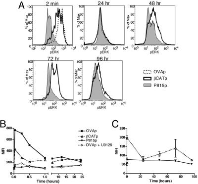
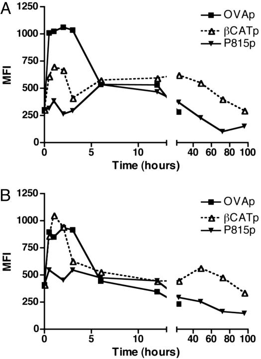
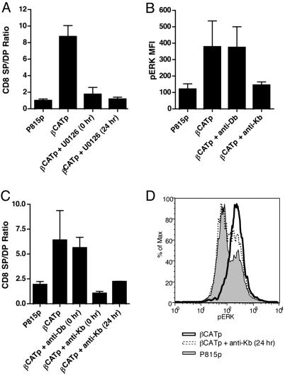
It is unknown how the contrasting events of positive and negative selection can lead to the distinct biological outcomes of life or death. An increasing body of evidence suggests that the duration of extracellular signal-regulated kinase (ERK) signaling plays a role in thymocyte selection. However, it remains unclear what the kinetics of ERK activation are during positive selection in vivo. In this study, we examined the magnitude and duration of ERK signaling in intact murine thymic tissues cultured under conditions of negative or positive selection. We found that negative selection induced a rapid and robust ERK activation that is associated with death, whereas positive selection stimulated a lower intensity and brief ERK activation that quickly declined and then gradually increased and was sustained over several days. The expression pattern of Egr-1 (early growth response-1), a downstream ERK effector, correlates with the biphasic kinetics of ERK during positive selection. Id3 (inhibitor of differentiation/DNA binding 3) also exhibits biphasic kinetics but appeared to be independent of ERK signaling. Furthermore, inhibitors of T cell receptor ligation and ERK activation block maturation of CD8 single-positive thymocytes even when added after 24 h. These results demonstrate that the in vivo duration of ERK signaling must be sustained to support positive selection.
Figures


Similar articles
-
Ternary complex factor SAP-1 is required for Erk-mediated thymocyte positive selection.Nat Immunol. 2004 Mar;5(3):289-98. doi: 10.1038/ni1038. Epub 2004 Feb 8. Nat Immunol. 2004. PMID: 14770179
-
Early growth response (Egr)-1 gene induction in the thymus in response to TCR ligation during early steps in positive selection is not required for CD8 lineage commitment.J Immunol. 2000 Sep 1;165(5):2444-50. doi: 10.4049/jimmunol.165.5.2444. J Immunol. 2000. PMID: 10946269
-
Degree of ERK activation influences both positive and negative thymocyte selection.Eur J Immunol. 2000 Apr;30(4):1060-8. doi: 10.1002/(SICI)1521-4141(200004)30:4<1060::AID-IMMU1060>3.0.CO;2-2. Eur J Immunol. 2000. PMID: 10760794
-
Regulation of T cell development in the thymus.Immunol Res. 2000;21(2-3):71-81. doi: 10.1385/IR:21:2-3:71. Immunol Res. 2000. PMID: 10852104 Review.
-
Real-time measurement of signaling and motility during T cell development in the thymus.Semin Immunol. 2005 Dec;17(6):411-20. doi: 10.1016/j.smim.2005.09.004. Epub 2005 Oct 25. Semin Immunol. 2005. PMID: 16256363 Review.
Cited by
-
Noncanonical mode of ERK action controls alternative αβ and γδ T cell lineage fates.Immunity. 2014 Dec 18;41(6):934-46. doi: 10.1016/j.immuni.2014.10.021. Epub 2014 Nov 28. Immunity. 2014. PMID: 25526308 Free PMC article.
-
Vive la peptide différence!Nat Immunol. 2016 Jul 19;17(8):896-8. doi: 10.1038/ni.3507. Nat Immunol. 2016. PMID: 27434001 No abstract available.
-
A stepwise and digital pattern of RSK phosphorylation determines the outcome of thymic selection.iScience. 2023 Aug 9;26(9):107552. doi: 10.1016/j.isci.2023.107552. eCollection 2023 Sep 15. iScience. 2023. PMID: 37646020 Free PMC article.
-
B-Raf-mediated signaling pathway regulates T cell development.Eur J Immunol. 2008 Feb;38(2):518-27. doi: 10.1002/eji.200737430. Eur J Immunol. 2008. PMID: 18228248 Free PMC article.
-
Inherent low Erk and p38 activity reduce Fas Ligand expression and degranulation in T helper 17 cells leading to activation induced cell death resistance.Oncotarget. 2016 Aug 23;7(34):54339-54359. doi: 10.18632/oncotarget.10913. Oncotarget. 2016. PMID: 27486885 Free PMC article.
References
-
- Starr, T. K., Jameson, S. C. & Hogquist, K. A. (2003) Annu. Rev. Immunol. 21, 139-176. - PubMed
-
- Sugawara, T., Moriguchi, T., Nishida, E. & Takahama, Y. (1998) Immunity 9, 565-574. - PubMed
-
- Gong, Q., Cheng, A. M., Akk, A. M., Alberola-Ila, J., Gong, G., Pawson, T. & Chan, A. C. (2001) Nat. Immunol. 2, 29-36. - PubMed
Publication types
MeSH terms
Substances
Grants and funding
LinkOut - more resources
Full Text Sources
Molecular Biology Databases
Research Materials
Miscellaneous
