Review
. 2005 Sep;69(3):462-500.
doi: 10.1128/MMBR.69.3.462-500.2005.

Molecular biology of pseudorabies virus: impact on neurovirology and veterinary medicine

Affiliations
Review

Molecular biology of pseudorabies virus: impact on neurovirology and veterinary medicine

Lisa E Pomeranz et al. Microbiol Mol Biol Rev. 2005 Sep.

Abstract

Pseudorabies virus (PRV) is a herpesvirus of swine, a member of the Alphaherpesvirinae subfamily, and the etiological agent of Aujeszky's disease. This review describes the contributions of PRV research to herpesvirus biology, neurobiology, and viral pathogenesis by focusing on (i) the molecular biology of PRV, (ii) model systems to study PRV pathogenesis and neurovirulence, (iii) PRV transsynaptic tracing of neuronal circuits, and (iv) veterinary aspects of pseudorabies disease. The structure of the enveloped infectious particle, the content of the viral DNA genome, and a step-by-step overview of the viral replication cycle are presented. PRV infection is initiated by binding to cellular receptors to allow penetration into the cell. After reaching the nucleus, the viral genome directs a regulated gene expression cascade that culminates with viral DNA replication and production of new virion constituents. Finally, progeny virions self-assemble and exit the host cells. Animal models and neuronal culture systems developed for the study of PRV pathogenesis and neurovirulence are discussed. PRV serves asa self-perpetuating transsynaptic tracer of neuronal circuitry, and we detail the original studies of PRV circuitry mapping, the biology underlying this application, and the development of the next generation of tracer viruses. The basic veterinary aspects of pseudorabies management and disease in swine are discussed. PRV infection progresses from acute infection of the respiratory epithelium to latent infection in the peripheral nervous system. Sporadic reactivation from latency can transmit PRV to new hosts. The successful management of PRV disease has relied on vaccination, prevention, and testing.


Figures

FIG. 1.
FIG. 1.
Structure of the PRV virion. PRV virions are composed of four structural elements. The double-stranded DNA genome is housed in an icosahedral capsid. The tegument is a collection of approximately 12 proteins organized into at least two layers, one which interacts with envelope proteins and one that is closely associated with the capsid. The envelope is a lipid bilayer infused with transmembrane proteins, many of which are modified by glycosylation. Listed are proteins thought to be components of the virion; however, not all proteins are represented in the cartoon.
FIG.2.
FIG.2.
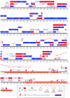
Linear map of the PRV genome: predicted gene and transcript organization. The PRV genome consists of a long and a short unique segment, named UL and US, respectively. The US region is flanked by the inverted repeats IRS and TRS. The predicted locations of core and accessory genes, transcripts, DNA repeats, splice sites, and the origin of replication are indicated. (Modified from reference .)
FIG. 3.
FIG. 3.
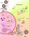
Replication cycle of PRV. 1. Entry begins with attachment or binding of the virus particle to the cell surface. In PRV, this initial binding step is an interaction between gC in the virion envelope and heparan sulfate on the surface of the cell. 2. The next steps of entry require gD, gB, gH, and gL. In PRV, although gD is not essential for membrane fusion or cell-cell spread, gD interacts with the cellular herpesvirus entry mediator (HVEM) and is required for entry of extracellular virus (penetration). 3. After fusion of the virion envelope with the cell membrane, the capsid and tegument proteins are released into the cell. The viral tegument proteins begin takeover of the host cell protein synthesis machinery immediately after entering the cell. 4. The capsid and tightly bound inner tegument proteins are transported along microtubules to the cell nucleus. 5. The VP16 tegument protein localizes to the nucleus independent of the capsid and transactivates cellular RNA polymerase II transcription of the only immediate-early protein of PRV, the HSV ICP4 homolog IE180. 6. IE180 protein expressed in the cytoplasm is transported back to the nucleus. 7. There, it transactivates RNA polymerase II transcription of the early genes. 8. Early proteins fall into two main categories. The first category comprises 15 proteins involved in viral DNA synthesis. 9. Seven of these proteins (UL5, UL8, UL9/OBP, UL29/ssDNABP, UL30/DNA Pol, UL42/Pap, and UL52) (shown in blue) are essential for replication of the viral DNA. DNA replication occurs by a rolling-circle mechanism. 10. The second category comprises three proteins thought to act as transactivators of transcription (EP0, US1, and UL54). 11. Onset of DNA synthesis signals the start of the late stage of the PRV replication cycle and synthesis of true late proteins. 12. The capsid proteins are transported to the nucleus, where they assemble around a scaffold composed of the product of the UL26 and UL26.5 genes. 13. The mature capsid is composed of five proteins (UL19/VP5, UL18/VP23, UL25, UL38, and UL35). The product of the UL6 gene acts as a portal for insertion of the genomic DNAinto the capsid. UL32, UL33, UL15 (Ex2), and UL17 are all involved in cleavage and packaging of the viral DNA. 14. During primary envelopment, the fully assembled nucleocapsid buds out of the nucleus, temporarily entering the perinuclear space. This process involves the products of the UL31 and UL34 genes along with the US3 kinase. 15 and 16. The nucleocapsid (15) loses its primary envelope and (16) gains its final envelope by associating with tegument and envelope proteins and budding into the trans-Golgi apparatus. 17. The mature virus is brought to the cell surface within a sorting compartment/vesicle derived from the envelopment compartment.
FIG. 4.
FIG. 4.
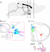
Stomach injection model. A. Sagittal view of the rat brain. S1, S2, and S3 refer to the levels of coronal sections depicted in panels B, C, D, and E. B. Innervation of smooth muscle of the ventral stomach. The area boxed in red is magnified to the right. Motor neurons from the dorsal motor nucleus of the vagus send projections through the vagus nerve to the ventral wall of the stomach. Sensory innervation of the ventral stomach through the left nodose ganglion is shown in pink. Neurons in the dorsal motor nucleus of the vagus exhibit anti-PRV immune reactivity 30 h after stomach injection of wild-type PRV-Becker (64, 338). PRV travels retrogradely to second-order neurons in the medial nucleus of the solitary tract between 50 and 60 h postinjection and to third-order neurons in the area postrema between 60 and 70 h postinjection. Labeling of neurons within the left nodose ganglia can be observed by 45 and 50 h postinjection. C. No cross talk with tongue or esophageal innervation after stomach injection. Injection of PRV-Becker into the ventrolateral musculature of the tongue (pathway shown in green) results in a very different pattern of infection from injection into the stomach (shown in blue) (64). After transport through the hypoglossal nerve, PRV immune reactivity can be seen in the hypoglossal nucleus (XII) 30 h postinjection. By about 52 h postinjection, PRV infection can be observed in second-order neurons in the spinal trigeminal nucleus (pars oralis and pars interpolaris) and the ventrolateral brainstem tegumentum and monoaminergic cell groups (not shown). Injection into the smooth muscle of the esophagus (shown in orange) produced labeling in the dorsal nucleus ambiguus. By 48 hours postinfection, labeling was detected in small bipolar neurons of the nucleus centralis of the medial NTS. The segregation of labeled structures following injection of stomach and esophagus is significant because the axons of these efferent circuits travel together in the vagus nerve, yet PRV infection is absent from the nucleus ambiguous following stomach injection and absent from the dorsal motor nucleus of the vagus followingesophageal injection. D. PRV requires an intact circuit for spread in the nervous system. In addition to surgical severance of the left vagus nerve which eliminates PRV transport to the left dorsal motor nucleus of the vagus, further proof that PRV neuronal spread requires intact, synaptically connected neurons is provided by tracing studies that span progressing developmental stages (339). PRV-Bartha immune reactivity in the central nervous system was examined 2.5 days after injection into the stomachs of newborn rats. Rats injected on postnatal day 1 (P1) exhibited PRV immune reactivity in the dorsal motor nucleus of the vagus, medial nucleus of the solitary tract, area postrema, and paraventricular nucleus of the hypothalamus by 2.5 days postinjection. No animals in the P1 group exhibited anti-PRV labeling in the central nucleus of the amygdala, lateral hypothalamic area, bed nucleus of the stria terminalis, insular cortex, or medial prefrontal cortex with the exception of one rat with six labeled neurons in the central nucleus of the amygdala. Rats injected at later developmental stages exhibit progressively more viral penetrance into the central nervous system; 2.5 days postinjection, P4 rats exhibit labeling of all structures observed in the P1 group plus extensive labeling in the central nucleus of the amygdala, lateral hypothalamic area, and bed nucleus of the stria terminalis. Only rats injected on P8 exhibit infection of neurons in the insular cortex and medial prefrontal cortex. E. Comparison of anterograde- and retrograde-defective alphaherpesviruses. In adult rats, stomach injection of PRV-Bartha results in retrograde-only transport of viral infection (pathway and PRV-immune reactive structures shown in orange) from the dorsal motor nucleus of the vagus to medial nucleus of the solitary tract to area postrema (340). Anti-PRV immunoreactivity 4 to 5 days postinjection does not change after elimination of anterograde transport by surgically severing the axons of pseudounipolar neurons projecting from the nodose ganglia (shown in pink; see panel B). HSV-H129 has a retrograde spread defect. HSV-H129 can spread retrogradelyin first-order neurons but only anterograde spread is observed in second-order pathways. Although HSV immune reactive structures appear similar to those infected by PRV after injection into the ventral stomach (HSV-H129, intact compare with PRV-Bartha), vagal deafferentation (illustrated by a red X on the sensory pathway from the nodose ganglia) eliminates infection of the medial nucleus of the solitary tract and area postrema (HSV-H129, vagal deafferentation). Abbreviations: AMBd, dorsal nucleus ambiguus; AP, area postrema; BNST, bed nucleus of the stria terminalis; CeA, central nucleus of the amygdala; DMV, dorsal motor nucleus of the vagus; IC, insular cortex; LHA, lateral hypothalamic area; mNST, medial nucleus of the solitary tract; mPFC, medial prefrontal cortex; nCen, nucleus centralis of the medial solitary nucleus; NG, nodose ganglion; PVN, paraventricular nucleus of the hypothalamus. (Figure modified from reference with permission of the publisher.)
FIG. 4.
FIG. 4.
Stomach injection model. A. Sagittal view of the rat brain. S1, S2, and S3 refer to the levels of coronal sections depicted in panels B, C, D, and E. B. Innervation of smooth muscle of the ventral stomach. The area boxed in red is magnified to the right. Motor neurons from the dorsal motor nucleus of the vagus send projections through the vagus nerve to the ventral wall of the stomach. Sensory innervation of the ventral stomach through the left nodose ganglion is shown in pink. Neurons in the dorsal motor nucleus of the vagus exhibit anti-PRV immune reactivity 30 h after stomach injection of wild-type PRV-Becker (64, 338). PRV travels retrogradely to second-order neurons in the medial nucleus of the solitary tract between 50 and 60 h postinjection and to third-order neurons in the area postrema between 60 and 70 h postinjection. Labeling of neurons within the left nodose ganglia can be observed by 45 and 50 h postinjection. C. No cross talk with tongue or esophageal innervation after stomach injection. Injection of PRV-Becker into the ventrolateral musculature of the tongue (pathway shown in green) results in a very different pattern of infection from injection into the stomach (shown in blue) (64). After transport through the hypoglossal nerve, PRV immune reactivity can be seen in the hypoglossal nucleus (XII) 30 h postinjection. By about 52 h postinjection, PRV infection can be observed in second-order neurons in the spinal trigeminal nucleus (pars oralis and pars interpolaris) and the ventrolateral brainstem tegumentum and monoaminergic cell groups (not shown). Injection into the smooth muscle of the esophagus (shown in orange) produced labeling in the dorsal nucleus ambiguus. By 48 hours postinfection, labeling was detected in small bipolar neurons of the nucleus centralis of the medial NTS. The segregation of labeled structures following injection of stomach and esophagus is significant because the axons of these efferent circuits travel together in the vagus nerve, yet PRV infection is absent from the nucleus ambiguous following stomach injection and absent from the dorsal motor nucleus of the vagus followingesophageal injection. D. PRV requires an intact circuit for spread in the nervous system. In addition to surgical severance of the left vagus nerve which eliminates PRV transport to the left dorsal motor nucleus of the vagus, further proof that PRV neuronal spread requires intact, synaptically connected neurons is provided by tracing studies that span progressing developmental stages (339). PRV-Bartha immune reactivity in the central nervous system was examined 2.5 days after injection into the stomachs of newborn rats. Rats injected on postnatal day 1 (P1) exhibited PRV immune reactivity in the dorsal motor nucleus of the vagus, medial nucleus of the solitary tract, area postrema, and paraventricular nucleus of the hypothalamus by 2.5 days postinjection. No animals in the P1 group exhibited anti-PRV labeling in the central nucleus of the amygdala, lateral hypothalamic area, bed nucleus of the stria terminalis, insular cortex, or medial prefrontal cortex with the exception of one rat with six labeled neurons in the central nucleus of the amygdala. Rats injected at later developmental stages exhibit progressively more viral penetrance into the central nervous system; 2.5 days postinjection, P4 rats exhibit labeling of all structures observed in the P1 group plus extensive labeling in the central nucleus of the amygdala, lateral hypothalamic area, and bed nucleus of the stria terminalis. Only rats injected on P8 exhibit infection of neurons in the insular cortex and medial prefrontal cortex. E. Comparison of anterograde- and retrograde-defective alphaherpesviruses. In adult rats, stomach injection of PRV-Bartha results in retrograde-only transport of viral infection (pathway and PRV-immune reactive structures shown in orange) from the dorsal motor nucleus of the vagus to medial nucleus of the solitary tract to area postrema (340). Anti-PRV immunoreactivity 4 to 5 days postinjection does not change after elimination of anterograde transport by surgically severing the axons of pseudounipolar neurons projecting from the nodose ganglia (shown in pink; see panel B). HSV-H129 has a retrograde spread defect. HSV-H129 can spread retrogradelyin first-order neurons but only anterograde spread is observed in second-order pathways. Although HSV immune reactive structures appear similar to those infected by PRV after injection into the ventral stomach (HSV-H129, intact compare with PRV-Bartha), vagal deafferentation (illustrated by a red X on the sensory pathway from the nodose ganglia) eliminates infection of the medial nucleus of the solitary tract and area postrema (HSV-H129, vagal deafferentation). Abbreviations: AMBd, dorsal nucleus ambiguus; AP, area postrema; BNST, bed nucleus of the stria terminalis; CeA, central nucleus of the amygdala; DMV, dorsal motor nucleus of the vagus; IC, insular cortex; LHA, lateral hypothalamic area; mNST, medial nucleus of the solitary tract; mPFC, medial prefrontal cortex; nCen, nucleus centralis of the medial solitary nucleus; NG, nodose ganglion; PVN, paraventricular nucleus of the hypothalamus. (Figure modified from reference with permission of the publisher.)
FIG. 5.
FIG. 5.
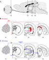
Rat eye infection model: comparison of wild-type (PRV-Becker) and attenuated (PRV-Bartha) neuronal spreads. A. Sagittal view of the rat brain. E1, E2, E3, and E4 refer to the level of coronal sections depicted in panels B and C. B. Spread of PRV-Becker (shown in red) after virus injection into the vitreous humor of the rat eye. PRV-Becker spreads from infected retinal ganglion cells (level E1) through the optic nerve to second-order neurons in the supracharismatic nucleus (level E2), dorsal and ventral aspects of the geniculate nuclei (dorsal aspect of the lateral geniculate nucleus and ventral aspect of the lateral geniculate nucleus) and intergeniculate nucleus (level E3), and the superior colliculus of thevisual cortex (level E4). Only first-order projections are shown. C. Transport of PRV-Bartha (shown in blue) is restricted to retrograde-only pathways. Although cells of the retinal ganglia (level E1) are infected with PRV-Bartha, this virus is restricted from anterograde spread through the optic nerve to retinorecipient neurons. Instead, retrograde spread of infection to first-order neurons in the ciliary ganglion leads to transport through the oculomotor nerve to second-order neurons in the Edinger-Westphal nucleus (level E4). Infection of neurons in the olivary pretectal nucleus, intergeniculate nucleus (level E3) and supracharismatic nucleus (level E2) is also by retrograde axonal transport of virus as shown (369). Abbreviations: CG, ciliary ganglion; dLGN, dorsal aspect of the lateral geniculate nucleus; EW, Edinger-Westphal nucleus; IGL, intergeniculate nucleus; OPN, olivary pretectal nucleus; SC, superior colliculus; SCN, suprachiasmatic nucleus; vLGN, ventral aspect of the lateral geniculate nucleus. (Figure modified from reference with permission of the publisher.)

Similar articles

Cited by

References

    1. Abmayr, S. M., J. L. Workman, and R. G. Roeder. 1988. The pseudorabies immediate-early protein stimulates in vitro transcription by facilitating TFIID: promoter interactions. Genes Dev. 2:542-553. - PubMed
    1. Adams, R., C. Cunningham, M. D. Davison, C. A. MacLean, and A. J. Davison. 1998. Characterization of the protein encoded by gene UL49A of herpes simplex virus type 1. J. Gen. Virol. 79:813-823. - PubMed
    1. Adelman, K., B. Salmon, and J. D. Baines. 2001. Herpes simplex virus DNA packaging sequences adopt novel structures that are specifically recognized by a component of the cleavage and packaging machinery. Proc. Natl. Acad. Sci. USA 98:3086-3091. - PMC - PubMed
    1. Aleman, N., M. I. Quiroga, M. Lopez-Pena, S. Vazquez, F. H. Guerrero, and J. M. Nieto. 2003. L-particle production during primary replication of pseudorabies virus in the nasal mucosa of swine. J. Virol. 77:5657-5667. - PMC - PubMed
    1. Ambagala, A. P., R. S. Gopinath, and S. Srikumaran. 2003. Inhibition of TAP by pseudorabies virus is independent of its vhs activity. Virus Res. 96:37-48. - PubMed

Publication types

MeSH terms

LinkOut - more resources