. 2005 Sep;79(18):11914-24.
doi: 10.1128/JVI.79.18.11914-11924.2005.

The human herpesvirus 6 G protein-coupled receptor homolog U51 positively regulates virus replication and enhances cell-cell fusion in vitro

Affiliations

The human herpesvirus 6 G protein-coupled receptor homolog U51 positively regulates virus replication and enhances cell-cell fusion in vitro

Zhu Zhen et al. J Virol. 2005 Sep.

Abstract

Human herpesvirus 6 (HHV-6) is a ubiquitous T-lymphotropic betaherpesvirus that encodes two G protein-coupled receptor homologs, U12 and U51. HHV-6A U51 has been reported to bind to CC chemokines including RANTES, but the biological function of U51 remains uncertain. In this report, we stably expressed short interfering RNAs (siRNAs) specific for U51 in human T cells and then infected these cells with HHV-6. Viral DNA replication was reduced 50-fold by the U51 siRNA, and virally induced cytopathic effects were also inhibited. In contrast, viral replication and syncytium formation were unaltered in cells that expressed a scrambled derivative of the siRNA or an irrelevant siRNA and were restored to normal when a human codon-optimized derivative of U51 was introduced into cells containing the U51 siRNA. To examine the mechanism whereby U51 might contribute to viral replication, we explored the signaling characteristics of U51. None of the chemokines and opioids tested was able to induce G protein coupling by U51, and no evidence for opioid ligand binding by U51 was obtained. The effect of U51 on cell-cell fusion was also evaluated; these studies showed that U51 enhanced cell fusion mediated by the G protein of vesicular stomatitis virus. However, a U51-specific antiserum had no virus-neutralizing activity, suggesting that U51 may not be involved in the initial interaction between the virus particle and host cell. Overall, these studies suggest that HHV-6 U51 is a positive regulator of virus replication in vitro, perhaps because it may promote membrane fusion and facilitates cell-cell spread of this highly cell-associated virus.


Figures

FIG. 1.
FIG. 1.
Design and screening of siRNAs targeting U51 and gB. (A) Four different siRNAs against HHV-6AU51 were designed to target distinct regions of the U51 open reading frame (ORF); numbers refer to the first nucleotide position of each siRNA relative to the predicted translational start codon of the U51 ORF. HEK293 cells were cotransfected with an expression vector carrying an SV5 epitope-tagged, wild-type version of the HHV-6A U51 ORF (HHV-6A U51nco) plus either empty pcDNA3 plasmid, empty pSuppressorRetro (pSR) vector (si Vec), or pSR constructs containing the siRNA-carrying inserts indicated in the figure; the lane labeled “siNeg.Ctrl.” corresponds to a pSR construct that contains an siRNA of irrelevant sequence which has no homology in the human genome. The U51 expression construct and the various siRNA-carrying plasmids were added to cells at a 1:6 molar ratio and formulated with Lipofectamine-2000 reagent (Invitrogen). At 48 h posttransfection, cell lysates were prepared and analyzed by Western blotting with an SV5 epitope-specific antibody (upper panel); the band detected corresponds to a protein of approximately 30-kDa molecular mass (as expected for U51). The blot was then stripped and reprobed with a β-tubulin antibody to confirm equal loading (lower panel). (B) Two different siRNA constructs against HHV-6A gB were designed and tested in a similar manner as for siU51. In this experiment, the gB expression plasmid vector pDisplay has an HA epitope tag. The blot shown was probed with an anti-HA antibody; the blot was then stripped and reprobed with a β-tubulin antibody to confirm equal loading (lower panel).
FIG. 2.
FIG. 2.
Suppression of U51 mRNA expression in virus-infected cell lines stably expressing siRNA-U51 (si6U51-812, si6U51-130). Stable SupT1 cells expressing the indicated siRNAs were generated following appropriate drug selection of cells transduced with corresponding retroviral vectors (pSuppressorRetro; Imgenex). The siRNA-expressing cells were then infected with HHV-6A (strain U1102) at an MOI of 0.1 TCID50/cell, and total cellular RNA was extracted 24 h thereafter. Quantitative RT-PCR analysis was then performed to assess levels of mRNA corresponding to U51. mRNA levels were normalized to GAPDH mRNA for each sample. Results represent mean values from a single experiment that was performed in triplicate (three independent infections); error bars correspond to the standard error of these mean values. There is a statistically significant difference in U51 mRNA levels in the siU51-SupT1 stable cell sample versus control SupT1 cells that were also infected with HHV-6 (P < 0.001; two-tailed t test).
FIG. 3.
FIG. 3.
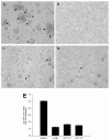
Effect of U51 knockdown on virus replication and syncytium formation. SupT1 cells stably expressing siRNA targeting U51, gB, or an irrelevant sequence (siNeg.Ctrl.) were infected with HHV-6A strain U1102 at an MOI of 0.1 TCID50/cell. Virally induced cytopathic effects were then examined in the cultures at 6 days postinfection. The photomicrographs shown were taken on an Olympus IX81 microscope under bright-field illumination; final magnification is 10×. The various panels correspond, respectively, to (A) SupT1 cells expressing an irrelevant siRNA (siNeg.Ctrl.) or (B) a gB-specific siRNA (si6gB) as well as two different clonal SupT1 cell sublines, each of which expresses a U51-specific siRNA, (C) si6U51-812 and (D) si6U51-130. It can be readily appreciated that virally induced syncytium formation was greatly reduced in the SupT1 cells that expressed either the gB-specific siRNA or the two U51-specific siRNAs. (E) Cell-free supernatants were collected from virus-infected SupT1 cultures at 6 days postinfection, and virus genomic DNA in the supernatant was measured by quantitative DNA PCR analysis using primers and TaqMan probes specific for the HHV-6 U38 gene. The data shown are from the same samples as in panels A to D; the results are representative of three separate experiments. The viral DNA copy number in SupT1 cells stably expressing either si6AgB or si6U51 were both significantly different from the viral DNA copy number in control SupT1 cells that were also infected with HHV-6 (P < 0.05 for each pairwise comparison between the three experimental cell lines and the control SupT1 cells). The detection sensitivity of the assay is about 10 copies.
FIG. 4.
FIG. 4.
Expression of a codon-optimized form of U51 can restore virus replication in SupT1 cells that express a U51-specific siRNA. SupT1 cells stably expressing an siRNA targeting U51 (si6U51-812) were transduced with a retrovirus vector that carried a human codon-optimized (CO) derivative of the HHV-6A U51 ORF. This CO version of the U51 ORF carries an mRNA that is significantly different from the wild-type U51 mRNA at the nucleotide level, and as a result it is resistant to inhibition by the U51 siRNA. The SupT1(si6U51-812) cells and their CO-U51-transduced counterparts were then infected with HHV-6A strain U1102 at an MOI of 0.1 TCID50/cell. Virally induced cytopathic effects were then examined in the cultures at 6 days postinfection, as described in the legend to Fig. 3. The various panels correspond, respectively, to SupT1 si6U51-812 cells transduced with the empty retrovirus vector (A), uninfected SupT1 cells (B), or CO-U51-encoding retrovirus vector encoding U51 from HHV-6A (C) or HHV-6B (D). It can be readily appreciated that virally induced syncytium formation was restored in the SupT1(si6U51-812) cells upon coexpression of CO-U51. (E) Cell-free supernatants were collected from virus-infected SupT1 cultures at 6 days postinfection, and virus genomic DNA in the supernatant was measured by quantitative DNA PCR analysis, as described in the legend to Fig. 3E. The various lanes indicate control si6U51-812-expressing SupT1 cells and their CO-U51-transduced counterparts. The results are representative of three separate experiments. The viral DNA copy number in siU51-SupT1 stable cells transduced with either 6AU51co or 6BU51co were both significantly different from the viral DNA copy number in control siU51-SupT1 cells that were also infected with HHV-6 (P < 0.05 in both cases; two-tailed t test).
FIG. 4.
FIG. 4.
Expression of a codon-optimized form of U51 can restore virus replication in SupT1 cells that express a U51-specific siRNA. SupT1 cells stably expressing an siRNA targeting U51 (si6U51-812) were transduced with a retrovirus vector that carried a human codon-optimized (CO) derivative of the HHV-6A U51 ORF. This CO version of the U51 ORF carries an mRNA that is significantly different from the wild-type U51 mRNA at the nucleotide level, and as a result it is resistant to inhibition by the U51 siRNA. The SupT1(si6U51-812) cells and their CO-U51-transduced counterparts were then infected with HHV-6A strain U1102 at an MOI of 0.1 TCID50/cell. Virally induced cytopathic effects were then examined in the cultures at 6 days postinfection, as described in the legend to Fig. 3. The various panels correspond, respectively, to SupT1 si6U51-812 cells transduced with the empty retrovirus vector (A), uninfected SupT1 cells (B), or CO-U51-encoding retrovirus vector encoding U51 from HHV-6A (C) or HHV-6B (D). It can be readily appreciated that virally induced syncytium formation was restored in the SupT1(si6U51-812) cells upon coexpression of CO-U51. (E) Cell-free supernatants were collected from virus-infected SupT1 cultures at 6 days postinfection, and virus genomic DNA in the supernatant was measured by quantitative DNA PCR analysis, as described in the legend to Fig. 3E. The various lanes indicate control si6U51-812-expressing SupT1 cells and their CO-U51-transduced counterparts. The results are representative of three separate experiments. The viral DNA copy number in siU51-SupT1 stable cells transduced with either 6AU51co or 6BU51co were both significantly different from the viral DNA copy number in control siU51-SupT1 cells that were also infected with HHV-6 (P < 0.05 in both cases; two-tailed t test).
FIG. 5.
FIG. 5.
Virus infectivity was unaffected by an antibody specific for U51. Two-hundred microliters of an HHV-6A virus stock (strain U1102) was preincubated with either 5 μl of human plasma (“baby plasma”) or 6 μg of affinity-purified rabbit antisera specific for HHV-6B U51 (anti-6B U51) or HHV-7 U51 (anti-7 U51) for 1 h at 37°C. The virus-antiserum mixture was then added to SupT1 cells (approximate MOI of 0.1 TCID50/cell). Six days later, cell-free culture supernatants were collected and viral genomic DNA was measured by a quantitative PCR assay as previously described. The results are representative of three separate experiments. As expected, the human plasma efficiently neutralized HHV-6A infectivity (P < 0.05; two-tailed t test); in contrast, the U51-specific antisera had no such effect (P = 0.117; two-tailed t test).
FIG. 6.
FIG. 6.
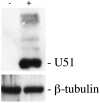
Tet-inducible overexpression of HHV-6B U51 in stably transduced HEK293 cells. HEK293 cells were cotransfected with the regulatory plasmid pcDNA6/TR and the inducible expression vector pcDNA4/TO (Invitrogen), which contained an insert sequence corresponding to the wild-type (non-codon-optimized) HHV-6B U51 sequence, with an added N-terminal SV5 epitope tag. Positive cell colonies were selected in the presence of 2 μg/ml blasticidin and 60 μg/ml zeocin for 3 weeks. Cell lysates from those positive cells, either in the absence of tetracycline treatment (“-”) or following induction with 1 μg/ml tetracycline for 24 h (“+”), were prepared and analyzed by Western blot using an SV5-specific antibody (upper panel) or a β-tubulin antibody (lower panel). Protein expression for a representative cell clone is shown; this clone was used in the subsequent [35S]GTPγS binding assay (Table 1).
FIG. 7.
FIG. 7.
U51 enhances cell-cell fusion in the presence of VSV-G in vitro. Equal numbers of HEK293 cells were transfected either with a HIV-1 Tat expressing plasmid (pcTat) (50) or with a plasmid containing a luciferase reporter gene under the transcriptional control of the HIV-1 LTR (17). All of the cells were also transfected with plasmid expression vectors encoding the following proteins: none (pcDNA3 lane), VSV-G alone (VSV-G lane) or VSV-G plus HCMV US28, HHV-6A U51 (6AU51CO), or the rat kappa opioid receptor (KOR), which was included as a negative control. The pcTat and LTRluc cell pools were then trypsinized 4 h posttransfection, mixed, and allowed to re-adhere to tissue culture plastic; 44 h later, luciferase activity was measured. The experiment shown is representative of three independent experiments. Shown are the mean relative light units (RLU) and standard deviations for three replicate samples obtained. As previously reported, HCMV US28 enhanced cell fusion initiated by US28 (39) (P < 0.05). HHV-6A U51 had a similar, though slightly less pronounced, effect (P < 0.05), while KOR had no such effect (P = 0.431; two-tailed t test).

Similar articles

Cited by

References

    1. Ablashi, D. V., N. Balachandran, S. F. Josephs, C. L. Hung, G. R. Krueger, B. Kramarsky, S. Z. Salahuddin, and R. C. Gallo. 1991. Genomic polymorphism, growth properties, and immunologic variations in human herpesvirus-6 isolates. Virology 184:545-552. - PubMed
    1. Alkhatib, G., C. Combadiere, C. C. Broder, Y. Feng, P. E. Kennedy, P. M. Murphy, and E. A. Berger. 1996. CC CKR5: a RANTES, MIP-1alpha, MIP-1beta receptor as a fusion cofactor for macrophage-tropic HIV-1. Science 272:1955-1958. - PubMed
    1. Arvanitakis, L., E. Geras-Raaka, A. Varma, M. C. Gershengorn, and E. Cesarman. 1997. Human herpesvirus KSHV encodes a constitutively active G-protein-coupled receptor linked to cell proliferation. Nature 385:347-350. - PubMed
    1. Aubin, J. T., H. Collandre, D. Candotti, D. Ingrand, C. Rouzioux, M. Burgard, S. Richard, J. M. Huraux, and H. Agut. 1991. Several groups among human herpesvirus 6 strains can be distinguished by Southern blotting and polymerase chain reaction. J. Clin. Microbiol. 29:367-372. - PMC - PubMed
    1. Beisser, P. S., G. Grauls, C. A. Bruggeman, and C. Vink. 1999. Deletion of the R78 G protein-coupled receptor gene from rat cytomegalovirus results in an attenuated, syncytium-inducing mutant strain. J. Virol. 73:7218-7230. - PMC - PubMed

Publication types

MeSH terms

LinkOut - more resources