. 2005 Sep;25(18):8344-55.
doi: 10.1128/MCB.25.18.8344-8355.2005.

Direct modulation of RNA polymerase core functions by basal transcription factors

Affiliations

Direct modulation of RNA polymerase core functions by basal transcription factors

Finn Werner et al. Mol Cell Biol. 2005 Sep.

Abstract

Archaeal RNA polymerases (RNAPs) are recruited to promoters through the joint action of three basal transcription factors: TATA-binding protein, TFB (archaeal homolog of TFIIB), and TFE (archaeal homolog of TFIIE). Our results demonstrate several new insights into the mechanisms of TFB and TFE during the transcription cycle. (i) The N-terminal Zn ribbon of TFB displays a surprising degree of redundancy for the recruitment of RNAP during transcription initiation in the archaeal system. (ii) The B-finger domain of TFB participates in transcription initiation events by stimulating abortive and productive transcription in a recruitment-independent function. TFB thus combines physical recruitment of the RNAP with an active role in influencing the catalytic properties of RNAP during transcription initiation. (iii) TFB mutations are complemented by TFE, thereby demonstrating that both factors act synergistically during transcription initiation. (iv) An additional function of TFE is to dynamically alter the nucleic acid-binding properties of RNAP by stabilizing the initiation complex and destabilizing elongation complexes.


Figures
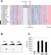
FIG. 1.
FIG. 1.
The RNA polymerase dock domain is involved in promoter-directed transcription. (A) Sequence alignment of euryarchaeal and eukaryotic dock domains. Euryarchaeal sequences (vertical white bar) and eukaryotic sequences (black bar) are identified to the left of the species. The sequences of Methanocaldococcus jannaschii, Methanococcus maripaludis, Methanococcus vannielii, Methanothermobacter thermautotrophicus (M. thermauto.), Pyrococcus furiosus, Pyrococcus horikoshii, Thermococcus celer, Halobacter salinarum, Methanococcoides burtonii, Methanosarcina barkeri, Methanosarcina acetivorans, Methanosarcina mazei, Archaeoglobus fulgidus, Methanopyrus kandleri, Saccharomyces cerevisiae, Drosophila melanogaster, Mus musculus, and Homo sapiens are shown. The dock domain is located between the conserved sequence blocks C and D (12). The Methanocaldococcus jannaschii A′ residue R388 (mjA′-R388) and K396 residue and the Saccharomyces cerevisiae RPB1 R412 residue (scRPB1-R412) are indicated. The alignment was performed using MultAlin (14). All invariant residues are shown in red, and highly conserved residues are shown in blue. Gaps introduced to maximize alignment are indicated by dashes. (B) The dock mutant M. jannaschii RNAP variants are defective in promoter-directed transcription assays (reaction mixtures contain 0.1 μg TBP; 0.1 μg TFB; and 0.1, 0.3, or 1.0 μg RNAP). (C) The dock mutant M. jannaschii RNAP variants display a comparable amount of activity in nonspecific transcription assays (reaction mixtures contain 0.5 μg RNAP). neg. ctrl., negative control.
FIG. 2.
FIG. 2.
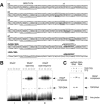
Deletion analysis of Methanocaldococcus jannaschii TFB. (A) M. jannaschii TFB is comprised of four distinct regions: N-terminal Zn ribbon (red), B-finger (yellow), flexible linker (green), and C-terminal core domain (blue). The various domain deletion variants used in this study are shown. The B-finger sequences of M. jannaschii TFB (mjTFB) and Saccharomyces cerevisiae TFIIB (scTFIIB) are shown at the bottom of the panel. (B) The N-terminal fragments TFB-Zn and TFB-Zn-B-L are capable of competing for full-length TFB in promoter-directed transcription assays (reaction mixtures contain 0.1 μg TBP, 0.1 μg TFB, 1.0 μg RNAP, and 0.5 or 1.0 μg TFB-Zn or TFB-Zn-B-L). (C) Activity of the TFB deletion variants in promoter-directed transcription assays (reaction mixtures contain 0.1 μg TBP, 0.1 μg TFB or TFB variants, and 1.0 μg RNAP). One lane contains no TFB variant (−). (D) Activity of the TFB B-finger mutants in promoter-directed transcription assays (reaction mixtures contain 0.1 μg TBP, 0.1 μg TFB or TFB variants, 1.0 μg RNAP, and 10 or 200 μM NTP). wt, wild type.
FIG. 3.
FIG. 3.
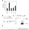
The TFB B-finger is stimulating RNAP in a recruitment-independent manner. (A) M. jannaschii TFB stimulates RNAP activity in a nonspecific transcription assay. This effect is dependent on the B-finger domain but independent of the core domain (reaction mixtures contain 1 μg TFB or TFB variants and 1.0 μg RNAP). (B) Nonspecific abortive initiation assay with RNAP in the presence (+) or absence (−) of TFB using one of the three dinucleotide substrates (ApG, ApA, or UpG) (reaction mixtures contain 1.0 μg TFB or TFB-ΔB and 2.0 μg RNAP). 32P-labeled RNA 9-mer marker was run in one lane. (C) Factor-independent transcription initiation using the 3′-tailed template is stimulated by TFB (reaction mixtures contain 1.0 μg TFB or TFB-ΔB and 2.0 μg RNAP). nt, nucleotides. (D) RNAP forms heparin-stable complexes with the 3′-tailed template in the presence (+) of TFB, and this effect is dependent on the B-finger (reaction mixtures contain 0.5 μg TFB or TFB-ΔB and 2.0 μg RNAP). (E) Sequence of the 3′-tailed template based on the SSV6 promoter; the transcription start site of the double-stranded promoter is indicated by bold type.
FIG. 4.
FIG. 4.
TFE complements TFB mutations in vitro. (A) Promoter-directed transcription assays using RNAP, TBP, full-length TFB, TFB-ΔZn, and TFB-ΔZn/R92E in combination with TFE (reaction mixtures contain 0.1 μg TBP, 0.1 μg TFB, 0.25 or 0.5 μg TFE, and 1.0 μg RNAP). (B) Electrophoretic mobility shift assays using various combinations of RNAP, TBP, and TFE together with full-length and mutant variants of TFB as indicated (reaction mixtures contain 0.5 μg TBP, 0.5 μg TFB or TFB variant, 0.5 μg TFE, and 2.0 μg RNAP). The presence of TFE causes a minor decrease in the mobility of the preinitiation complexes. wt, wild type. (C) In the presence (+) of TFB and TFE, RNAP is capable of forming heparin-stable complexes with the template strand (tDNA) but not with the nontemplate strand (nDNA) of the SSV T6 promoter. This interaction is dependent on the TFB B-finger. Both single-stranded DNA probes tDNA and nDNA form a nonspecific shift (marked with an asterisk in panel C) that is presumably due to a stable secondary structure not to be confused with the TBP/DNA shift in panel B. Reaction mixtures contain 0.5 μg TFB or TFB-ΔB, 0.5 μg TFE, and 2.0 μg RNAP.
FIG. 5.
FIG. 5.
RNAP binding to heteroduplex DNA and DNA/RNA-hybrid templates. (A) Sequences of nucleic acid templates based on the SSV T6 promoter used for EMSAs. The B-recognition element (BRE)/TATA elements are underlined, the transcription start site is indicated by +1 and bold type, the heteroduplex mutations are boxed and in italic type, and RNA sequences are indicated by bold italic type. wt, wild type. (B) RNAP was used in EMSAs with various combinations of TBP, TFB, and TFB on the homoduplex and heteroduplex templates m1, m3, m6, and m7 (reaction mixtures contain 0.5 μg TBP, 0.5 μg TFB, 0.5 μg TFE, and 2.0 μg RNAP). wt, wild type. (C) RNAP forms stable complexes with DNA/RNA-hybrid probes (reaction mixtures contain 0.5 μg TBP, 0.5 μg TFB, 0.5 μg TFE, and 2.0 μg RNAP).
FIG. 6.
FIG. 6.
Modulation of RNAP core functions by TFB and TFE during transcription initiation. (A) RNAP is recruited to the TBP/TFB/DNA complex through interactions between the TFB Zn ribbon and the RNAP dock domain. In this closed complex, RNAP makes only superficial contacts with promoter DNA. F/E, RNAP subunits F and E. (B) During open complex formation, the DNA strands are melted and the template strand is inserted into the active site where it is stabilized by the TFB B-finger. TFE furthermore stabilizes this complex by influencing the position of the RNAP clamp by subunits F and E and possibly by interacting directly with the nontemplate strand. (C) The abortive initiation phase is characterized by the production of short RNA transcripts. This step is stimulated by TFB presumably because the TFB B-finger facilitates a DNA/rNTP or even DNA/RNA/rNTP configuration that is conducive to catalysis (indicated by the red-orange flash). (D) During the escape of the RNAP from the promoter, basal transcription factors TBP, TFB, and TFE are shed and the RNA transcript is directed towards RNAP subunits F and E in the elongation complex.

Similar articles

Cited by

References

    1. Barberis, A., C. W. Muller, S. C. Harrison, and M. Ptashne. 1993. Delineation of two functional regions of transcription factor TFIIB. Proc. Natl. Acad. Sci. USA 90:5628-5632. - PMC - PubMed
    1. Bartlett, M. S., M. Thomm, and E. P. Geiduschek. 2004. Topography of the euryarchaeal transcription initiation complex. J. Biol. Chem. 279:894-903. - PubMed
    1. Bell, S. D., and S. P. Jackson. 2000. The role of transcription factor B in transcription initiation and promoter clearance in the archaeon Sulfolobus acidocaldarius. J. Biol. Chem. 275:12934-12940. - PubMed
    1. Bell, S. D., and S. P. Jackson. 2001. Mechanism and regulation of transcription in archaea. Curr. Opin. Microbiol. 4:208-213. - PubMed
    1. Bell, S. D., A. B. Brinkman, J. van der Oost, and S. P. Jackson. 2001. The archaeal TFIIEalpha homologue facilitates transcription initiation by enhancing TATA-box recognition. EMBO Rep. 2:133-138. - PMC - PubMed

Publication types

LinkOut - more resources