Comparative Study
. 2005 Sep 15;19(18):2122-37.
doi: 10.1101/gad.1339905. Epub 2005 Aug 30.

p53 isoforms can regulate p53 transcriptional activity

Affiliations
Comparative Study

p53 isoforms can regulate p53 transcriptional activity

Jean-Christophe Bourdon et al. Genes Dev. .

Abstract

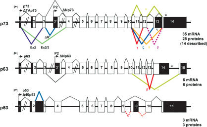
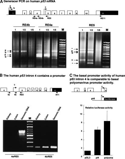
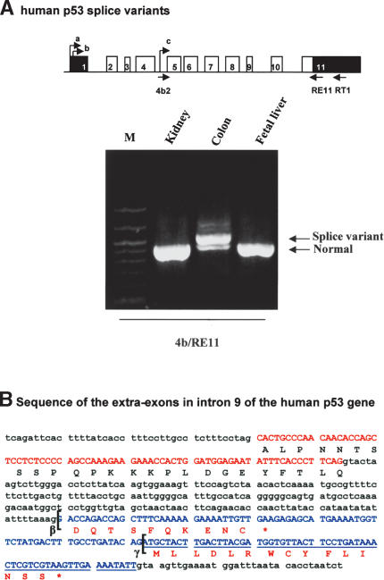
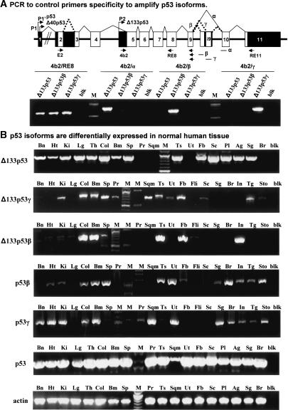
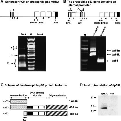
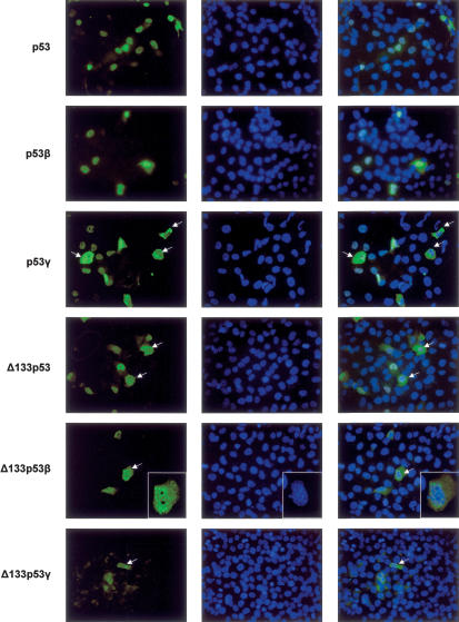
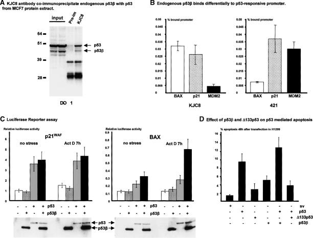
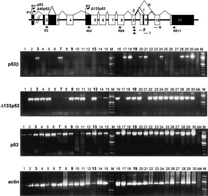
The recently discovered p53-related genes, p73 and p63, express multiple splice variants and N-terminally truncated forms initiated from an alternative promoter in intron 3. To date, no alternative promoter and multiple splice variants have been described for the p53 gene. In this study, we show that p53 has a gene structure similar to the p73 and p63 genes. The human p53 gene contains an alternative promoter and transcribes multiple splice variants. We show that p53 variants are expressed in normal human tissue in a tissue-dependent manner. We determine that the alternative promoter is conserved through evolution from Drosophila to man, suggesting that the p53 family gene structure plays an essential role in the multiple activities of the p53 family members. Consistent with this hypothesis, p53 variants are differentially expressed in human breast tumors compared with normal breast tissue. We establish that p53beta can bind differentially to promoters and can enhance p53 target gene expression in a promoter-dependent manner, while Delta133p53 is dominant-negative toward full-length p53, inhibiting p53-mediated apoptosis. The differential expression of the p53 isoforms in human tumors may explain the difficulties in linking p53 status to the biological properties and drug sensitivity of human cancer.


Figures

Figure 1.
Figure 1.
Structure of the human p53, p63, and p73 genes. p63 and p73 express multiple splice variants and contain an internal promoter, while no internal promoter or multiple splice variants have been described for p53. (Black) Noncoding sequence; (white) coding sequence.
Figure 2.
Figure 2.
The human p53 gene contains an internal promoter. (A) GeneRacer PCR on human p53 gene. Schema of the human p53 gene: (black) noncoding sequence; (white) coding sequence. Total RNA extract from normal human colon was treated for GeneRacer PCR according to the manufacturer's protocol to allow specific amplification of capped mRNA. The reverse transcription was performed by using primer RE11. A first PCR was performed using the GeneRacer primer-1 and one of the p53-specific primers (RE4a or RE4b or RE5). One microliter of the first PCR diluted (1/2, 1/5) or neat (1) was used to perform a semi-nested PCR using GeneRacer primer-2 and the appropriate p53-specific primer (RE4b or RE4a or RE5). The product of the second PCR was analyzed on 1% agarose gel, and the DNA was stained with ethidium bromide. After cloning and sequencing, bands a and b were found to contain sequences from exon 1 of human p53 while band c contained sequences from intron 4 of the human p53 gene. () Nonspecific bands. (B) The human p53 intron 4 contains a promoter. The RT-PCR was performed from total RNA extracted from kidney and colon human tissues using primer RT1. The PCR was performed using the primer RE5 with the primers 4a or 4b. PCR from human genomic DNA was performed using the same primers to control experimental conditions. (C) Luciferase reporter assay. The basal promoter activity of intron 4 of the human p53 gene is comparable to basal polyomavirus promoter activity. The human p53 intron 4 (pi4) was cloned upstream of the luciferase gene in the promoterless plasmid pGL3 basic and transfected with the Renilla luciferase expression vector into H1299 cells. The Renilla vector is used as an internal control of transfection efficiency. Cells were harvested 24 h after transfection, and the luciferase and Renilla activities were analyzed simultaneously. Luciferase activity was normalized to Renilla activity. Results shown are the average of four independent experiments performed in triplicate. SDs are indicated as error bars.
Figure 3.
Figure 3.
Intron 9 of the human p53 gene can be alternatively spliced. (A) RT-PCR. Human p53 splice variants. The reverse transcriptions of total RNA extracted from normal human tissue (colon, kidney, and fetal liver) were performed using primer RT1. Two successive PCRs were performed, the first using the primers 4b and RE11, followed by a second PCR using primers 4b2 and RE11. The arrows indicate the fragments corresponding to p53 variants initiated in intron 4. (B) Sequence of the extra-exons in intron 9 of the human p53 gene. Schema of the human p53 gene with the alternative splices (β and γ): (black) noncoding sequence; (white) coding sequence. Sequences of exon 9 and intron 9: (red) sequence of exon 9; (blue) sequence of the extra-exons in intron 9. The brackets indicate the splice sites giving p53β and p53γ protein isoforms.
Figure 4.
Figure 4.
Schemas of the human p53 gene structure and p53 protein isoforms. (A) Schema of the human p53 gene structure. (B) Schema of the p53 protein isoforms theoretically encoded by the human p53 gene.
Figure 5.
Figure 5.
Expression of the p53 isoforms in normal human tissue. (A) PCR to control primers' specificity to amplify p53 isoforms. Primers were designed to be amplified specifically by normal splicing, splice β, and splice γ and were assessed by PCR on cloned cDNA of Δ133p53, Δ133p53β, and Δ133p53γ. (B) p53 isoforms are differentially expressed in normal human tissue. p53 mRNA variants were amplified specifically by PCR from total RNA extracted from normal human tissue (Clontech). (Bn) Brain; (Ht) heart; (Ki) kidney; (Lg) lung; (Th) thymus; (Col) colon; (Bm) bone marrow; (Sp) spleen; (Pr) prostate; (Ts) testis; (Sqm) skeletal muscle; (Ut) uterus; (Fb) fetal brain; (Sc) spinal cord; (Pl) placenta; (Ag) adrenal gland; (Sg) salivary gland; (Br) breast; (Fli) fetal liver; (In) intestine; (Tg) thyroid gland; (Sto) stomach; (blk) blank. Actin is shown as a control.
Figure 6.
Figure 6.
The Drosophila p53 gene contains an internal promoter. (A) GeneRacer PCR on Drosophila p53 mRNA. Schema of the Drosophila p53 gene: (black) noncoding sequence; (white) coding sequence. Total RNA extract from Drosophila embryos was treated according to the manufacturer's protocol to allow amplification of capped mRNA. The reverse transcription was performed by using primer DRE8. The first PCR was performed using the GeneRacer primer-1 and the Drosophila p53-specific primer DRE51. One microliter of the first PCR was used to perform a nested PCR using the GeneRacer primer-2 and the p53-specific primer DRE52. The product of the second PCR was analyzed on a 1% agarose gel, and the DNA was stained with ethidium bromide. After cloning and sequencing, the bands (1) contained sequences from two additional exons (exon A and B) upstream of the previously described exon 1 of the Drosophila p53 gene. The upper band contained a splice variant. Band 2 contains sequences from the previously described exon 1 of Drosophila p53. () Nonspecific bands. (B) The Drosophila p53 gene contains an internal promoter. The reverse transcription was performed using primer DRE8 on total Drosophila embryo RNA extracts. A first PCR was performed using primers derived from exon A (DFA) or from exon 1 (DF1) and the reverse primer DRE51 followed by a semi-nested PCR using primer DFA or DF1 with primer DRE52. The arrows indicate the PCR fragments corresponding to Drosophila p53. Fragment dp53n corresponds to the Drosophila p53 mRNA not spliced at intron B and initiated at the first site (1) of initiation of transcription. Fragment dp53L contains the Drosophila p53 mRNA initiated at the first site (1) of initiation of transcription with splicing of intron B. Fragment dp53 contains the Drosophila p53 mRNA initiated at the second site of transcription initiation. (C) Schema of the Drosophila p53 protein isoforms. The Drosophila p53 gene can encode, theoretically, three different protein isoforms. The splice form dp53n may encode a truncated form of Drosophila p53 containing only the N-terminal end (123 amino acids). The form dp53L may encode a Drosophila p53 of 495 amino acids containing a large N-terminal domain. The form dp53 encodes the published Drosophila p53 protein. (D) In vitro translation of dp53L. The dp53L cDNA was fused to a Flag-tag and cloned in a T7-driven promoter expression vector. dp53L protein was produced by in vitro translation using a reticulocytes lysates kit and revealed by Western blot using anti-Flag antibody.
Figure 7.
Figure 7.
Endogenous p53 isoforms are expressed at the protein level. (A) Transfection of p53 isoforms in H1299. A p53 variant can lead to p53 protein isoform expression. H1299 cells, devoid of p53 expression, were transfected with p53 cDNA variants cloned in mammalian expression vectors as indicated. Proteins were extracted 24 h after transfection, and analyzed by Western blot using either sheep polyclonal (sp) anti-human p53 or mouse monoclonal (DO-12) anti-human p53 (Vojtesek et al. 1995). (B) Endogenous p53 protein isoforms can be detected in human cell lines. Proteins from human cell lines expressing wild-type p53 (293) or mutant p53 (T47D, SW620, HT-29), or devoid of p53 expression (H1299, Saos-2) were extracted as described in Materials and Methods. Ten micrograms of protein extract were analyzed by Western blot. p53 expression was revealed using DO-12 mouse monoclonal antibody. (C) Anti-p53β antibody (KJC8) is specific for p53β and Δ133p53β. H1299 cells, devoid of p53 expression, were transfected with p53, p53β, Δ133p53β, or empty expression vectors. Protein extracts were analyzed by Western blot using either CM1 rabbit polyclonal anti-human p53 or peptide affinity-purified KJC8 rabbit polyclonal anti-p53β antibody. (D) U2OS cells express endogenous p53β and Δ133p53β protein isoforms. U2OS cells were transfected with 400 nM final concentration of small interference RNA oligonucleotides specific for p53 (Smartpool p53siRNA; Dharmacon) or control (Smartpool controlsiRNA; Dharmacon). Cells were harvested 96 h after transfection, and proteins were analyzed by Western blot using CM1 rabbit polyclonal anti-human p53 or KJC8 rabbit polyclonal anti-p53β. The anti-actin antibody was used to control loading and transfer efficiency. (E) p53β and Δ133p53β are not accumulated in MCF7 tumor cells in response to Actinomycin-D treatment. MCF7 cells expressing wild-type p53 were untreated or treated with 60 ng/mL of the DNA-damaging drug Actinomycin-D (Act-D), a potent p53 inducer. Proteins were extracted 6 h after treatment and analyzed (5 μg) by Western blot using either DO-1 mouse monoclonal anti-human p53 or peptide affinity-purified KJC8 rabbit polyclonal anti-p53β antibody. The anti-actin antibody was used to control loading and transfer efficiency.
Figure 8.
Figure 8.
Subcellular localization of the p53 protein isoforms. H1299 cells transfected with p53, p53β, p53γ, or Δ133p53 expression vectors were stained by indirect immunofluorescence (FITC) using rabbit polyclonal CM1 anti-human p53 antibody. H1299 cells transfected with Δ133p53β expression vector were stained by indirect immunofluorescence (FITC) using rabbit polyclonal KJC8 anti-p53β antibody. H1299 cells transfected with Δ133p53γ expression vector were stained by indirect immunofluorescence (FITC) using mouse monoclonal DO-12 anti-p53 antibody. DNA was stained by DAPI. The merge is shown. The cells with a particular staining are indicated with an arrow.
Figure 9.
Figure 9.
p53β binds the p53-responsive promoter and enhances p53 transcriptional activity on the BAX promoter but not on the p21 promoter. (A) KJC8 antibody coimmunoprecipitates endogenous p53β with p53 from MCF7 protein extract. Protein from MCF7 cells cultured under standard cell culture conditions were extracted and treated for immunoprecipitation as described in Materials and Methods. Proteins (100 μg) were immunoprecipitated with KJC8 antibody or with preimmune serum, and p53 content was revealed after Western blot using mouse monoclonal antibody (DO-1) anti-human p53. Ten percent of the input was loaded as a control. (B) Endogenous p53β binds differentially to the p53-responsive promoter. Protein from MCF7 cells cultured under standard cell culture conditions were extracted and treated for ChIP assay as described in Materials and Methods. Extracts were immunoprecipitated with KJC8 antibodyb mouse monoclonal 421 anti-p53 antibody, or nonrelated rabbit polyclonal antibody as a negative control. The amounts of BAX and p21 promoter contained in the input or immunoprecipitated with each of the antibodies were quantified by real-time PCR as described (Kaeser and Iggo 2002). The amount of promoter nonspecifically immunoprecipitated with the nonrelated rabbit polyclonal antibody was subtracted from the amount of promoter immunoprecipitated with KJC8 or 421 antibody. The results are expressed as the percentage of promoter specifically immunoprecipitated by KJC8 or 421 antibody compared with the total amount of promoter contained in the input. The results shown are the average of four independent experiments. (C) Luciferase reporter assay. p53β enhances p53 transcriptional activity on the BAX promoter but not the p21 promoter. H1299 cells (20,000 cells/well) were seeded on a 24-well plate. Cells were cotransfected in duplicate with the plasmids bax-luc or p21-luc containing the natural promoters driving the luciferase reporter gene, with a trace amount of pSVrenilla as an internal control, and 25 ng/well of p53 and/or 50 ng/well of p53β as indicated. Cells were treated 18 h after transfection with 60 ng/mL of Actinomycin D (ActD) for 6 h. The amount of SV-40 promoter was maintained constant in each transfection by adding empty SV-40 expression vector. Cells were harvested 24 h after transfection. Luciferase activity was normalized to Renilla activity. The results shown are the average of four independent experiments performed in duplicate. SDs are indicated as error bars. To take account of the transfection efficiency, luciferase assay protein extracts were analyzed by Western blot after normalization for Renilla activity. p53 and p53β expressions were revealed using DO-1 monoclonal antibodies. (D) Effect of p53β and Δ133p53 on p53-mediated apoptosis. H1299 cells were transfected with 5 μg/mL of the indicated expression vector or empty SV-40 expression vector (sv). In the experiments of coexpression of p53 with p53 isoforms, H1299 cells were cotransfected with 5 μg/mL of p53 expression vector and 5 μg/mL of the indicated p53 isoform expression vectors. The amount of SV-40 promoter was maintained constant at 10 μg/mL in each transfection by adding empty SV-40 expression vector. The DNA content of each transfected population was determined by flow cytometry analysis. The percentage of sub-G1 DNA content represents the percentage of apoptotic cells 48 h after transfection. The histogram represents the average of three independent transfections. SDs are reported as error bars.
Figure 10.
Figure 10.
p53 mRNA variants are differentially expressed in tumors from patient to patient. Total RNA from 30 breast tumors were provided by Tayside Tumour Bank. Reverse transcription was performed as described in Materials and Methods using primer RT1 and 400 ng of total RNA. The p53 cDNAs were amplified by PCR using appropriate primers as described in Material and Methods and on schema of the p53 gene structure. p53γ, Δ133p53β, and Δ133p53γ mRNAs were not detected (data not shown). Actin was used a control. Tumors expressing mutant p53 are marked in bold: tumor 3 (mutant p53 K132 → R), tumor 7 (mutant p53 R248 → Q), tumor 9 (mutant p53 K132 → N), tumor 13 (mutant p53 V216 → M), and tumor 19 (mutant p53 R248 → Q). All the other tumors express wild-type p53.

Comment in

Similar articles

Cited by

References

    1. Almog N., Goldfinger, N., and Rotter, V. 2000. p53-dependent apoptosis is regulated by a C-terminally alternatively spliced form of murine p53. Oncogene 19: 3395-3403. - PubMed
    1. Bates G.J., Nicol, S.M., Wilson, B.J., Jacobs, A.M., Bourdon, J.C., Wardrop, J., Gregory, D.J., Lane, D.P., Perkins, N.D., and Fuller-Pace, F.V. 2005. The DEAD box protein p68: A novel transcriptional coactivator of the p53 tumour suppressor. EMBO J. 24: 543-553. [Epub 2005 January 20.] - PMC - PubMed
    1. Benard J., Douc-Rasy, S., and Ahomadegbe, J.C. 2003. TP53 family members and human cancers. Hum. Mutat. 21: 182-191. - PubMed
    1. Bienz-Tadmor B., Zakut-Houri, R., Libresco, S., Givol, D., and Oren, M. 1985. The 5′ region of the p53 gene: Evolutionary conservation and evidence for a negative regulatory element. EMBO J. 4: 3209-3213. - PMC - PubMed
    1. Bourdon J.-C., Renzing, J., Robertson, P.L., Fernandes, K.N., and Lane, D.P. 2002. Scotin, a novel p53-inducible proapoptotic protein located in the ER and the nuclear membrane. J. Cell. Biol. 158: 235-246. [Epub 2002 July 22.] - PMC - PubMed

Publication types

MeSH terms