. 2005 Aug 29;170(5):837-45.
doi: 10.1083/jcb.200503125. Epub 2005 Aug 22.

PTP-1B is an essential positive regulator of platelet integrin signaling

Affiliations

PTP-1B is an essential positive regulator of platelet integrin signaling

Elena Garcia Arias-Salgado et al. J Cell Biol. .

Abstract

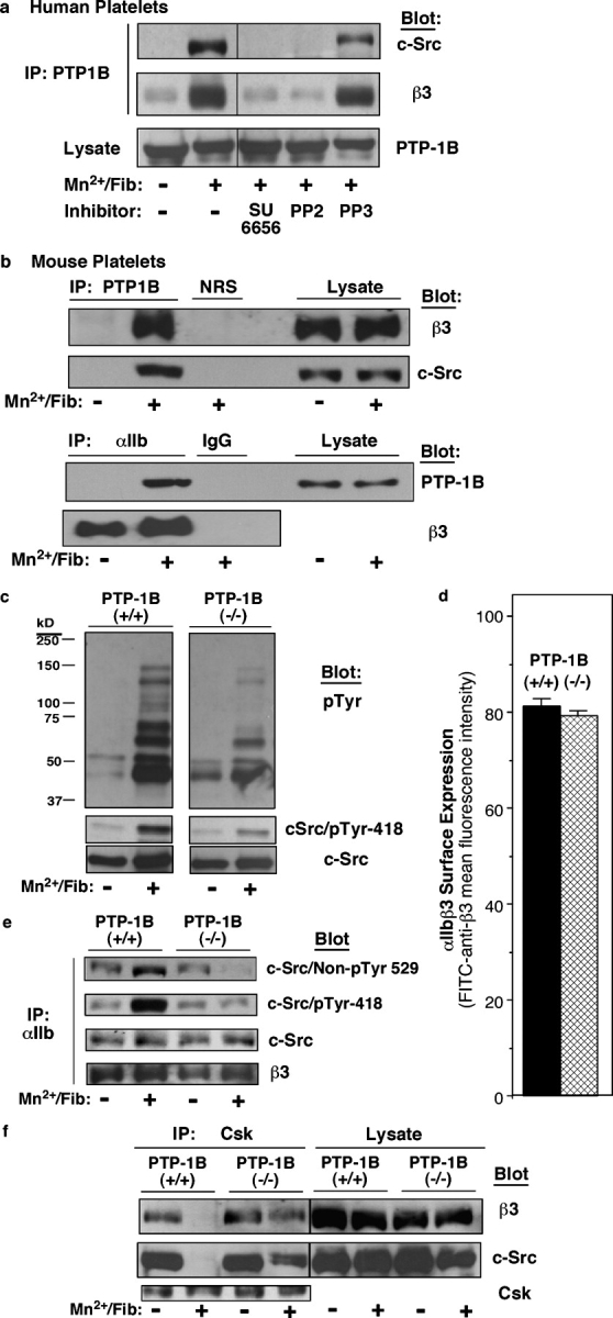
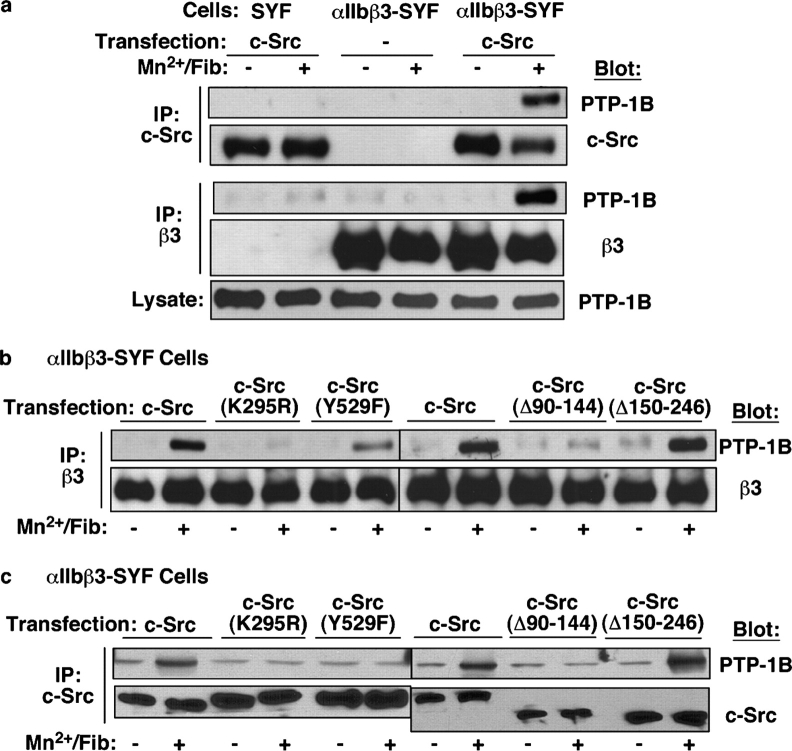
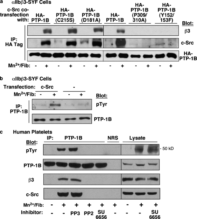
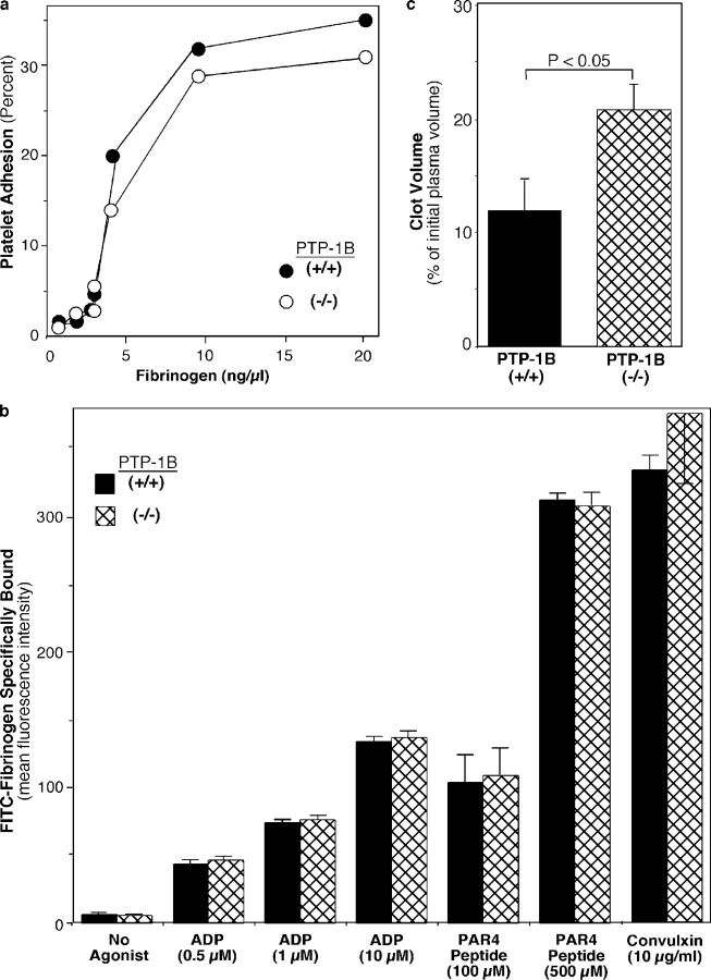
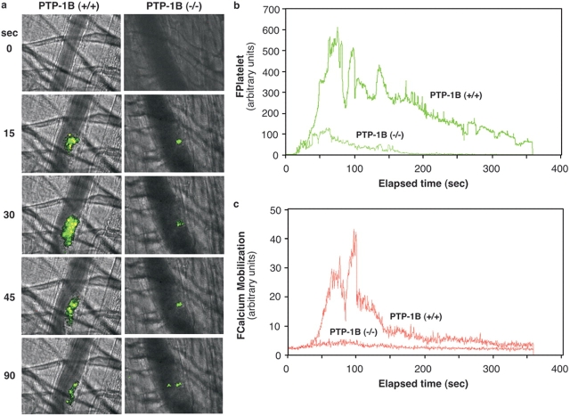
Outside-in integrin alphaIIbbeta3 signaling is required for normal platelet thrombus formation and is triggered by c-Src activation through an unknown mechanism. In this study, we demonstrate an essential role for protein-tyrosine phosphatase (PTP)-1B in this process. In resting platelets, c-Src forms a complex with alphaIIbbeta3 and Csk, which phosphorylates c-Src tyrosine 529 to maintain c-Src autoinhibition. Fibrinogen binding to alphaIIbbeta3 triggers PTP-1B recruitment to the alphaIIbbeta3-c-Src-Csk complex in a manner that is dependent on c-Src and specific tyrosine (tyrosine 152 and 153) and proline (proline 309 and 310) residues in PTP-1B. Studies of PTP-1B-deficient mouse platelets indicate that PTP-1B is required for fibrinogen-dependent Csk dissociation from alphaIIbbeta3, dephosphorylation of c-Src tyrosine 529, and c-Src activation. Furthermore, PTP-1B-deficient platelets are defective in outside-in alphaIIbbeta3 signaling in vitro as manifested by poor spreading on fibrinogen and decreased clot retraction, and they exhibit ineffective Ca2+ signaling and thrombus formation in vivo. Thus, PTP-1B is an essential positive regulator of the initiation of outside-in alphaIIbbeta3 signaling in platelets.


Figures

Figure 1.
Figure 1.
Interactions between PTP-1B, αIIbβ3, and c-Src in platelets. (a) Washed human platelets were incubated for 15 min at RT with 250 μg/ml fibrinogen in the presence or absence of 0.5 mM MnCl2. Some samples were preincubated for 15 min with 2 μM SU6656, 5 μM PP2, or 5 μM PP3; the latter is an inactive congener of PP2. Clarified lysates were immunoprecipitated (IP) and probed on immunoblots as indicated. Vertical lines in the blots indicate grouping of images from different parts of the same gel. (b) Washed mouse platelets were incubated with MnCl2 and fibrinogen, and immunoblots of immunoprecipitates were probed as in a. Control immunoprecipitations used normal rabbit serum (NRS) or rat IgG (IgG). (c) Role of PTP-1B in platelet tyrosine phosphorylation. Fibrinogen binding to PTP-1B+/+ and PTP-1B−/− platelets was induced as in b. Lysates were immunoblotted with antibodies to phosphotyrosine (pTyr) or c-Src phosphotyrosine 418 and reprobed with antibodies to c-Src. (d) αIIbβ3 surface expression in PTP-1B+/+ (black bar) and PTP-1B−/− (hatched bar) platelets was quantified by flow cytometry. Mean fluorescence intensities are depicted in arbitrary units, and error bars represent means ± SEM of three experiments. (e) PTP-1B is required for activation of integrin-associated c-Src. Fibrinogen binding to PTP-1B+/+ and PTP-1B−/− platelets was induced as in b, and αIIbβ3 immunoprecipitates were probed on immunoblots as indicated. (f) PTP-1B is required for dissociation of Csk from the αIIbβ3–c-Src complex. Fibrinogen binding to PTP-1B+/+ and PTP-1B−/− platelets was induced as in b, and Csk immunoprecipitates were probed on immunoblots as indicated. Each immunoblot panel is representative of three to five independent experiments.
Figure 2.
Figure 2.
Structural features of c-Src that are required for interactions with PTP-1B and αIIbβ3. (a) SYF cells or SYF cells stably expressing human αIIbβ3 (αIIbβ3-SYF) were transiently transfected with wild-type c-Src or empty vector. After 48 h, transfected cells were incubated at 37°C for 15 min in the presence or absence of 1 mM MnCl2 and 250 μg/ml fibrinogen. Clarified lysates were immunoprecipitated with antibodies to c-Src or β3, and immunoprecipitates were probed on immunoblots as indicated. Lysates were probed for PTP-1B as a loading control. (b and c) αIIbβ3-SYF cells were transfected with wild-type c-Src or an indicated c-Src mutant. After 48 h, transfected cells were incubated in the presence or absence of MnCl2 and fibrinogen as in a. Clarified lysates were immunoprecipitated with antibodies to β3 (b) or c-Src (c) and with the precipitates probed on immunoblots. Data are from a single experiment that was representative of three that were performed.
Figure 3.
Figure 3.
Structural features of PTP-1B that are required for interactions with αIIbβ3 and c-Src. (a) αIIbβ3-SYF cells were transiently cotransfected with c-Src and wild-type or mutant forms of HA-tagged human PTP-1B. After 48 h, transfected cells were incubated with or without MnCl2 and fibrinogen as described in Fig. 2. Clarified lysates were immunoprecipitated with antibodies to the HA tag, and precipitates were probed on immunoblots as indicated. (b and c) PTP-1B is tyrosine phosphorylated in response to fibrinogen binding. αIIbβ3-SYF cells transfected with c-Src and empty vector (b) or human platelets (c) were incubated with or without 0.5 mM MnCl2 and 250 μg/ml fibrinogen for 10 min. Some platelet samples were preincubated for 15 min with c-Src inhibitors (5 μM PP2 or 2 μM SU6656) or 5 μM PP3 as a control. Clarified lysates were immunoprecipitated with an antibody to PTP-1B, and immunoprecipitates and lysates were probed on immunoblots. Data are from a single experiment that was representative of three that were performed. NRS, normal rabbit serum.
Figure 4.
Figure 4.
PTP-1B / platelets are defective in αIIbβ3-dependent spreading on fibrinogen. (a) Platelets from PTP-1B+/+ and PTP-1B−/− mice were plated on fibrinogen-coated coverslips for 40 min at RT in the presence or absence of 100 μM ADP. Adherent cells were fixed, permeabilized, and stained with rhodamine-phalloidin (F-actin, red) and antiphosphotyrosine antibodies (green). Images were acquired with a confocal fluorescence microscope. Bar, 10 μm. (b) Platelet surface areas from at least 25 images were analyzed and depicted in the left panel as means ± SEM. The right panel depicts the percentage of platelets containing one or more filopodia and/or lamellipodia. At least 80 cells each were analyzed.
Figure 5.
Figure 5.
Role of PTP-1B in the interaction of platelets with fibrinogen and fibrin. (a) Platelet adhesion. Washed platelets (1.5 × 10⁶ in 50 μl incubation buffer) were incubated in fibrinogen-coated microtiter wells for 1 h at RT, and platelet adhesion was quantified. Platelets from at least four mice were used to generate duplicate points at each fibrinogen concentration. (b) Soluble fibrinogen binding. Platelets were incubated at RT for 20 min with 150 μg/ml FITC-fibrinogen in the presence or absence of ADP, convulxin, or PAR4 receptor–activating peptide (AYPGKF; Faruqi et al., 2000). Fibrinogen binding was analyzed by flow cytometry. Data are the means ± SEM of quadruplicate determinations from an experiment that was representative of three that were performed. (c) Fibrin clot retraction was assessed 2 h after the addition of thrombin and CaCl2 to platelet-rich plasma. Clot volumes, expressed as a percentage of the initial volume of platelet-rich plasma, were significantly greater in PTP-1B−/− than in PTP+/+ samples, indicating less clot retraction. Data represent means ± SEM of seven experiments.
Figure 6.
Figure 6.
Defective thrombus formation in PTP-1B / mice. PTP-1B+/+ and PTP-1B−/− platelets were labeled ex vivo with Fura 2-AM and were reinfused into PTP-1B+/+ and PTP-1B−/− recipient mice, respectively. Then, vessel walls of arterioles in recipient cremaster muscles were subjected to laser injury, and the accumulation of fluorescent platelets into developing thrombi was assessed. (a) Representative composite brightfield and fluorescence images of Fura 2–labeled platelets up to 90 s after laser injury of an arteriole. Green represents labeled platelets and yellow represents cytoplasmic free calcium. Blood flow is from bottom to top. See Videos 1 and 2 (available at http://www.jcb.org/cgi/content/full/jcb.200503125/DC1) for examples of thrombus formation in PTP-1B+/+ and PTP-1B−/− mice, respectively. (b) Accumulation of fluorescent platelets into the developing thrombus. Fluorescent signal was detected at 510 nm after excitation at 380 nm. FPlatelet is defined as the integrated fluorescence intensity associated with platelets. (c) Calcium mobilization within fluorescent platelets of the developing thrombus. Fluorescent signal was detected at 510 nm after excitation at 340 nm. FCalcium mobilization is defined as the integrated fluorescence intensity associated with calcium mobilization. Each curve in b and c is a composite of 18 independent thrombi generated in three mice (six thrombi per mouse). To analyze these data, all 18 curves were plotted versus time, and median values were determined at each time point and depicted in the figure.

Similar articles

Cited by

References

    1. Arias-Salgado, E.G., S. Lizano, S. Sarker, J.S. Brugge, M.H. Ginsberg, and S.J. Shattil. 2003. Src kinase activation by a novel and direct interaction with the integrin β cytoplasmic domain. Proc. Natl. Acad. Sci. USA. 100:13298–13302. - PMC - PubMed
    1. Arregui, C.O., J. Balsamo, and J. Lilien. 1998. Impaired integrin-mediated adhesion and signaling in fibroblasts expressing a dominant-negative mutant PTP1B. J. Cell Biol. 143:861–873. - PMC - PubMed
    1. Brunton, V.G., I.R.J. MacPherson, and M.C. Frame. 2004. Cell adhesion receptors, tyrosine kinases and actin modulators: a complex three-way circuitry. Biochim. Biophys. Acta. 1692:121–144. - PubMed
    1. Buensuceso, C., M. De Virgilio, and S.J. Shattil. 2003. Detection of integrin αIIbβ3 clustering in living cells. J. Biol. Chem. 278:15217–15224. - PubMed
    1. Byzova, T.V., R. Rabbani, S. D'Souza, and E.F. Plow. 1998. Role of integrin αVβ3 in vascular biology. Thromb. Haemost. 80:726–734. - PubMed

Publication types

Substances