. 2005 Sep;79(17):10852-63.
doi: 10.1128/JVI.79.17.10852-10863.2005.

Key Golgi factors for structural and functional maturation of bunyamwera virus

Affiliations

Key Golgi factors for structural and functional maturation of bunyamwera virus

Reyes R Novoa et al. J Virol. 2005 Sep.

Abstract

Several complex enveloped viruses assemble in the membranes of the secretory pathway, such as the Golgi apparatus. Among them, bunyaviruses form immature viral particles that change their structure in a trans-Golgi-dependent manner. To identify key Golgi factors for viral structural maturation, we have purified and characterized the three viral forms assembled in infected cells, two intracellular intermediates and the extracellular mature virion. The first viral form is a pleomorphic structure with fully endo-beta-N-acetylglucosaminidase H (Endo-H)-sensitive, nonsialylated glycoproteins. The second viral intermediate is a structure with hexagonal and pentagonal contours and partially Endo-H-resistant glycoproteins. Sialic acid is incorporated into the small glycoprotein of this second viral form. Growing the virus in glycosylation-deficient cells confirmed that acquisition of Endo-H resistance but not sialylation is critical for the trans-Golgi-dependent structural maturation and release of mature viruses. Conformational changes in viral glycoproteins triggered by changes in sugar composition would then induce the assembly of a compact viral particle of angular contours. These structures would be competent for the second maturation step, taking place during exit from cells, that originates fully infectious virions.


Figures
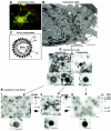
FIG. 1.
FIG. 1.
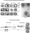
Isolation of three viral forms from Bunyamwera virus-infected BHK-21 cells. (A) At 8 h postinfection, confocal microscopy shows the large perinuclear structure (arrow), known as the viral factory (vf), where viral proteins (here Gc is labeled in red) and mitochondria (green) are recruited. Labeling was performed as described in Materials and Methods. Small red dots seen on the cell periphery correspond to secretory vesicles filled with viruses. (B) Electron microscopy shows the viral factory around the nucleus (N) as associations of mitochondria (mi) and membranes (arrows). In these factories viral particles are assembled by recruitment of the structural components represented in C: an envelope with spikes (made of Gc and Gn glycoproteins) and an internal core containing three ribonucleoproteins (RNPs) of RNA, nucleocapsid protein (N), and RNA polymerase (L). (D) Viral particles with three different morphologies are seen in thin sections of infected cells: intracellular viruses on the left correspond either to type I (annular structures that correspond to immature precursors) or type II (an intermediate dense form thatassembles in a trans-Golgi-dependent manner) morphology. Extracellular virions (E) on the right are also dense particles but with a more defined coat of spikes (arrowheads). Higher-magnification fields on the bottom show the structural characteristics of the viral particles in more detail. (E) Isolation of these three viral forms gave homogeneous populations of type I (left), type II (middle), and extracellular (right) viruses. Viral structural proteins were separated by SDS-PAGE and analyzed by Coomassie blue staining (CB) and Western blotting (WB). The asterisk on the gel for extracellular viruses marks a band of seralbumin identified by MALDI peptide mass fingerprinting. Thin sections of purified viruses (high-magnification EM fields on the bottom) show the internal structure of the three purified viral forms. Bars: 1 μm in B, 100 nm in D and E.
FIG. 2.
FIG. 2.
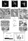
Structural and biochemical characterization of isolated viral forms. (A) EM shows the characteristics of type I (upper line), type II (middle), and extracellular (bottom) viruses. Contour and surface analyses by negative staining and freeze-etching, respectively (two columns on the left), show that type I virus is pleomorphic with a rounded contour and a smooth surface, while type II virus has acquired a more compact, angular contour. In addition, E virions have neat surface spikes. Immunogold labeling with anti-Gc and anti-Gn antibodies (two columns on the right) shows full recognition of Gc on E viruses, while Gn was detected on type I viruses exclusively. Values for infectivity (expressed as number of infectious particles/total number of particles) are indicated on the right. The large EM field shows extracellular virions treated with 1 M sucrose. Particles are opened in one spot and are releasing the RNPs (arrows), leaving a thick, geometrical envelope (arrowheads). (B) Posttranslational modifications in bunyavirus structural proteins were analyzed in isolated viral forms. Sensitivity to endoglycosidase H (on the left) distinguishes immature from mature glycoproteins. Total sensitivity is seen as a decrease in molecular weight for the corresponding band after Endo-H digestion, SDS-PAGE, and Western blot, while partial sensitivity is shown as an increase in band width by Western blot due to the generation of two or more bands after digestion (see Coomassie blue for Gc of E viruses on the right). Both Gc and Gn glycoproteins were totally sensitive to Endo-H in type I virus while sensitivity was partial in type II and E viruses. The presence of sialic acid is shown on the right. Lectin binding to proteins transferred to nitrocellulose showed reactivity in Gn of type II and extracellular viruses, but no reaction in the position corresponding to Gc. Binding of a conjugate of Triticum vulgaris agglutinin and 10-nm gold particles showed strong exposure of sialic acid on the surface of type II and E viruses, while type I virus was totally devoid of labeling. Bars: 100 nm.
FIG. 3.
FIG. 3.
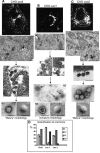
Viral forms produced at 14 h postinfection in BHK-21 cells, BHK-21 cells treated with the drug megalomycin (MGM), and BHK Ricr 14 cells. (A) Characteristic viral factory induced by Bunyamwera virus in BHK-21 cells, as seen by immunofluorescence (top), is seen as accumulation of Gc protein close to the nucleus (arrow). EM fields of these areas show numerous type II viruses at 14 h postinfection, as well as typical extracellular (E) virions, with dense internal structure. Geometrical contours, as seen by negative staining (NS) and freeze-etching (FE),are typical of a mature, functional morphology. (B) In the presence of the drug megalomycin, infection progresses, as indicated by immunofluorescence of Gc (top). As seen by EM, type I virus-like particles are the only morphology assembled and accumulated in these cells. No extracellular forms are detected. (C) The GlcNAc-TI-deficient BHK Ricr 14 cell line also supports virus growth, as seen by immunofluorescence of Gc (top), but only type I-like viruses accumulate intracellularly and exit the cells (central EM fields). Their morphology is undistinguishable from that corresponding to immature type I viruses assembled in normally infected BHK-21 cells (Fig. 1) and from the viral particles accumulated in megalomycin-treated BHK-21 cells shown in B. Extracellular “immature” viruses are designated E*. As indicated at the bottom, infectivity in supernatants of Ricr 14 cells is significantly lower compared to that of supernatants from infected BHK-21 cells at the same times postinfection. (D) Endo-H sensitivity analyzed in cell monolayers showed partial sensitivity in glycoprotein Gc of infected BHK-21 cells while fully sensitive Gc is detected in infected Ricr14 cells. (E) Quantification of viral forms (expressed as the percentage of viruses with a particular morphology after counting more than 500 viruses for each cell line in thin sections and TEM) showed the presence of numerous mature virions in BHK-21 cells, while immature viruses accumulate and are secreted from BHK Ricr 14 cells. Bars: 150 nm.
FIG.4.
FIG.4.
Viral forms assembled in CHO pro5 cells and two derived glycosylation-deficient cell lines, Lec1 and Lec2. (A) Perinuclear accumulation of viral proteins is seen in infected CHO pro5 cells as seen by immunofluorescence of Gc (top). EM fields show the two typical intracellular morphologies (I and II) as well as extracellular virions (E) with a dense interior. Negative staining (NS) and freeze-etching (FE) show compact particles with rough surfaces, but the layer of spikes is not clearly seen. (B) The N-acetylglucosaminyltransferase I-deficient CHO Lec1 cell line also accumulates viral proteins in the perinuclear area, rendering a Golgi-like pattern by immunofluorescence of Gc (top). A few dots representing secretory vesicles with viruses are observed. At the EM level, type I-like annular intracellular viruses accumulated inside infected cells (middle panel). These immature forms are able to exit the cells and they exhibit a pleomorphic, round morphology (E*). These particles are labile and easily disrupted, as observed by negative staining (arrows). Freeze-etching shows a smooth surface for these particles. (C) The sialylation-deficient CHO Lec2 cell line exhibits patterns similar to the parental cell line by both immunofluorescence and TEM (top). Intracellular viruses correspond to both type I and type II morphologies, while only normal extracellular virions (E), with compact and dense internal structure, are detected. (D) Quantification of viral forms assembled in CHO pro5, Lec1, and Lec2 cells shows similar patterns for CHO pro5 and Lec2 cells, while Lec1 accumulates immature forms at both intracellular and extracellular locations. Quantification was done on thin sections of infected cells and is represented as the percentage of each morphology after counting more than 500 viral particles for each cell line. Bars: 150 nm.
FIG. 5.
FIG. 5.
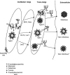
Proposal for the structural changes taking place during the generation of the three viral forms, and the potential molecular changes involved, associated with modifications of viral glycoproteins in the trans-Golgi subcompartment, according to the results obtained in this study. The shape and final state of sugar chains are indicated for the three viral forms. The asterisk marks the viral form released by Lec2 cells that exhibits a normal shape but an abnormal surface of spikes and low infectivity values.

Similar articles

Cited by

References

    1. Allison, S. L., Y. J. Tao, G. O'Riordain, C. W. Mandl, S. C. Harrison, and F. X. Heinz. 2003. Two disinct size classes of immature and mature subviral particles from tick-borne encephalitis virus. J. Virol. 77:11357-11366. - PMC - PubMed
    1. Altan-Bonnet, N., R. Sougrat, and J. Lippincott-Schwartz. 2004. Molecular basis for Golgi maintenance and biogenesis. Curr. Opin. Cell Biol. 16:364-372. - PubMed
    1. Baumeister, W. 2002. Electron tomography: towards visualizing the molecular organization of the cytoplasm. Curr. Opin. Struct. Biol. 12:679-684. - PubMed
    1. Bijlmakers, M. J., and M. Marsh. 2003. The on-off story of protein palmitoylation. Trends Cell Biol. 13:32-42. - PubMed
    1. Bonay, P., S. Munro, M. Fresno, and B. Alarcon. 1996. Intra-Golgi transport inhibition by megalomycin. J. Biol. Chem. 271:3719-3726. - PubMed

Publication types

LinkOut - more resources