. 2005 Dec;53(12):1525-37.
doi: 10.1369/jhc.5A6630.2005. Epub 2005 Aug 8.

Osteoclast responses to lipopolysaccharide, parathyroid hormone and bisphosphonates in neonatal murine calvaria analyzed by laser scanning confocal microscopy

Affiliations

Osteoclast responses to lipopolysaccharide, parathyroid hormone and bisphosphonates in neonatal murine calvaria analyzed by laser scanning confocal microscopy

Keiko Suzuki et al. J Histochem Cytochem. 2005 Dec.

Abstract

Because the development and activity of osteoclasts in bone remodeling is critically dependent on cell-cell and cell-matrix interactions, we used laser confocal microscopy to study the response of osteoclasts to lipopolysaccharide (LPS; 10 microg/ml), parathyroid hormone (PTH; 10(-8) M), and bisphosphonates (BPs; 1-25 microM clodronate or 0.1-2.5 microM risedronate) in cultured neonatal calvaria. Following treatment with LPS or PTH (<48 hr), osteopontin (OPN) and the alphavbeta3 integrin were found colocalized with the actin ring in the sealing zone of actively resorbing osteoclasts. In contrast, non-resorbing osteoclasts in BP-treated cultures showed morphological abnormalities, including retraction of pseudopods and vacuolization of cytoplasm. In the combined presence of LPS and BP, bone-resorbing osteoclasts were smaller and the sealing zone diffuse, reflecting reduced actin, OPN, and beta3 integrin staining. Depth analyses of calvaria showed that the area of resorbed bone was filled with proliferating osteoblastic cells that stained for alkaline phosphatase, collagen type I, and bone sialoprotein, regardless of the presence of BPs. These studies show that confocal microscopy of neonatal calvaria in culture can be used to assess the cytological relationships between osteoclasts and osteoblastic cells in response to agents that regulate bone remodeling in situ, avoiding systemic effects that can compromise in vivo studies and artifacts associated with studies of isolated osteoclasts.


Figures
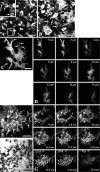
Figure 1.
Figure 1.
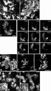
Tartrate-resistant acid phosphatase (TRACP) staining of non-resorbing and resorbing osteoclasts in cultured calvaria. Laser scanning confocal microscopy of neonatal mouse calvaria stained for TRACP activity after culturing for 24 hr (C, D) or 48 hr (A, B, E-G) in the absence (A-D) or presence (E-G) of 10−8 M parathyroid hormone (PTH). The pictures were created by estimating the regions of best focus in the z-stacks made of 11 and 15 images taken at 0.5-μm intervals in A and B, respectively (MetaMorph). Arrows in the picture at higher magnification in A indicate round prefusion osteoclasts and in B indicate long pseudopods extending from the osteoclasts. (C) Picture created by estimating the regions of best focus in a z-stack made of 40 images taken at 0.4-μm intervals in the TRACP-stained calvaria. Arrows indicate long pseudopods. (D) Serial optical sections of a single osteoclast taken at 2-μm intervals from upper (periosteal) surface (0 μm) to the lower (endosteal) surface (16 μm) of the calvaria. (E) Picture created by estimating the regions of best focus in a z-stack made of 37 images. Arrows indicate the sealing zone surrounding resorption areas, also shown using Nomarsky optics (F), associated with resorbing osteoclasts in TRACP-stained calvaria cultured in the presence of 10−8 M PTH. (G) Serial optical sections taken at 2.8-μm intervals from upper surface (0 μm) to lower surface (22.4 μm) of the calvaria. Arrowheads indicate the edge of attachment zone at 2.8 μm. Bars: A, B = 50 μm; C = 30 μm; E, F = 200 μm.
Figure 2.
Figure 2.
Effects of bisphosphonates (BPs) on the differentiation and activation of osteoclasts. Laser scanning confocal microscopy of neonatal mouse calvaria stained for TRACP activity after culturing for 24 hr (A-D, H) or 48 hr (I) in the presence of BP alone (A-D) or in combination with 10−8 M PTH (H) or 10 μg/ml LPS (I). (A, B) Calvaria were cultured in the presence of 1 μM clodronate alone. The pictures were created by estimating the regions of best focus in the z-stacks made of 44 and 51 images taken at 0.7- and 0.5-μm intervals in A and B, respectively (MetaMorph). Arrows in A indicate cell processes. Arrowheads in the picture at higher magnification in B indicate cytoplasmic vacuolization. (C, D) Calvaria were cultured in the presence of 2.5 μM risedronate alone. (C) Picture created by estimating the regions of best focus in a z-stack made of 43 images taken at 0.5-μm intervals in the TRACP-stained calvaria. Arrowheads indicate cytoplasmic vacuolization. (D) Serial optical sections taken from upper surface (0 μm) to lower surface (16 μm) of the calvarium. (E-G) Preosteoclasts derived from mouse bone marrow cells were incubated for 3 days in the absence (E) or presence (F) of 25 μM clodronate or 2.5 μM risedronate (G) and stained with Alexa Fluor 488-conjugated phalloidin for 30 min. Arrows, dotted arrows, and arrowheads indicate pseudopods extending from the osteoclasts, retracted pseudopods, and cytoplasmic vacuolization, respectively. (H, I) Calvaria were cultured in the presence of 10−8 M PTH and 25 μM clodronate (H) or 10 μg/ml lipopolysaccharides (LPS) and 2.5 μM risedronate (I). The pictures were created by estimating the regions of best focus in the z-stacks made of 26 and 19 images taken at 0.6-μm intervals in H and I, respectively (MetaMorph). Arrowheads indicate the cytoplasmic vacuolization. Bars: A, B, H, I = 50 μm; C, E-G = 30 μm.
Figure 3.
Figure 3.
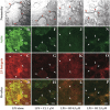
Effects of BPs on the colocalization of actin and β3 integrin in sealing zone of resorbing osteoclasts in calvaria cultured for 48 hr in the presence of LPS. Laser scanning confocal microscopy of neonatal mouse calvaria stained for F-actin (Alexa Fluor 488-conjugated phalloidin) and β3 integrin (hamster anti-mouse β3 integrin antibody followed by Alexa Fluor 594-conjugated anti-hamster IgG) after culturing for 48 hr in the presence of 10 μg/ml LPS alone (A-D) or in combination with 1 μM clodronate (E-H), 0.5 μM risedronate (I-L), or 2.5 μM risedronate (M-P). Arrows indicate the edge of resorption lacunae in the Nomarsky image and corresponding sites in the fluorescent images (actin: B, F, J, N; β3 integrin: C, G, K, O). Bars = 50 μm.
Figure 4.
Figure 4.
Effects of BP on the colocalization of actin and osteopontin (OPN) in sealing zone of resorbing osteoclasts in calvaria cultured for 48 hr in the presence of LPS. Laser scanning confocal microscopy of neonatal mouse calvaria stained for F-actin (Alexa Fluor 488-conjugated phalloidin) and OPN (LF123 antisera followed by Texas Red conjugated anti-rabbit IgG) after culturing for 48 hr in the presence of 10 μg/ml LPS alone (A-D) or in combination with 2.5 μM risedronate (E-J). Arrows indicate the edge of resorption lacunae in the Nomarsky image and corresponding sites in the fluorescent images (actin: B, F; OPN: C, G). Bars = 50 μm.
Figure 5.
Figure 5.
Cell proliferation observed after bone resorption induced by LPS. (A-D) Optical sections of calvaria cultured in the presence of LPS taken at the focal plane 16.1 μm above the level shown in Figure 3B. The sealing zone of osteoclasts observed in LPS-treated calvaria in Figure 3B was superimposed in white (C) on the merged image of actin (A) and β3 integrin (B). (D) Merged image at higher magnification. Bars: A, B = 50 μm; D = 20 μm.
Figure 6.
Figure 6.
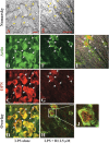
Calvaria cultured for 48 hr with vehicle alone or 10 μg/ml LPS and 2.5 μM risedronate (Nomarsky images; A and E, respectively) were double stained for alkaline phosphatase activity (B, F, I) and actin (C, G, J) using ELF 97 phosphatase substrate and Alexa Fluor 594-conjugated phalloidin, respectively. Depth analyses in the LPS and 2.5-μM risedronate group were made on the blue (F-H) and pink (I-K) lines separately. Asterisks in F and I indicate the resorption lacunae. The horizontal green lines in XZ scans in B and D are due to autofluorescence derived from a glass coverslip. Bars = 100 μm. Depth in the XZ scans is 100 and 70 μm in B-D and F-K, respectively.
Figure 7.
Figure 7.
Calvaria cultured for 48 hr with vehicle alone or 10 μg/ml LPS alone (Nomarsky images; A and E, respectively) were double stained with rabbit anti-rat collagen type I antibody (B, F) or rabbit anti-rat bone sialoprotein (BSP) antibody (I, J) followed by Alexa Fluor 488-conjugated anti-rabbit IgG and Alizarin Red S for mineral. XZ (depth)-scans were made on the blue horizontal lines in the Nomarsky images. Bars = 100 μm. Depth in the XZ scans is 100 μm.
Figure 8.
Figure 8.
Calvaria cultured for 48 hr under various conditions, i.e., in the presence of vehicle (A-D), 1 μM clodronate alone (E-H), 10 μg/ml LPS alone (I-L) or in combination with 1 μM clodronate (M-P) or 0.1 μM risedronate (Q-T). Calvaria were stained for actin with Alexa Fluor 488-conjugated phalloidin (B, F, J, N, R) and for mineral using Alizarin Red S (C, G, K, O, S). Top panels (A, E, I, M, Q) show the Nomarsky images where white areas indicate the resorption lacuna. XZ (depth)-scans of actin and mineral staining made on the blue horizontal line in the corresponding Nomarsky images. Bars = 100 μm. Depth in the XZ scans is 100 μm.

Similar articles

Cited by

References

    1. Andersson G, Johansson EK. (1996) Adhesion of human myelomonocytic (HL-60) cells induced by 1, 25-dihydroxyvitamin D3 and phorbol myristate acetate is dependent on osteopontin synthesis and the alpha v beta 3 integrin. Connect Tissue Res 35: 163–171 - PubMed
    1. Burdi AR. (1965) Toluidine Blue-Alizarin Red S staining of cartilage and bone in whole-mount skeletons in vitro. Stain Technol 40: 45–48 - PubMed
    1. Cecchini MG, Fleisch H. (1990) Bisphosphonates in vitro specifically inhibit, among the hematopoietic series, the development of the mouse mononuclear phagocyte lineage. J Bone Miner Res 5: 1019–1027 - PubMed
    1. Chellaiah MA, Hruska KA. (2003) The integrin alpha(v)beta(3) and CD44 regulate the actions of osteopontin on osteoclast motility. Calcif Tissue Int 72: 197–205 - PubMed
    1. Chellaiah MA, Kizer N, Biswas R, Alvarez U, Strauss-Schoenberger J, Rifas L, Rittling SR, et al. (2003) Osteopontin deficiency produces osteoclast dysfunction due to reduced CD44 surface expression. Mol Biol Cell 14: 173–189 - PMC - PubMed

Publication types

MeSH terms